GreenLight Laser Vaporisation for Benign Prostate Enlargement

A Modern, Low-Bleeding Treatment for an Enlarged Prostate

Benign prostate enlargement, also called benign prostatic hyperplasia (BPH), is extremely common as men get older. As the prostate enlarges around the urethra, it can gradually restrict urinary flow, rather like slowly tightening a collar around the urinary pipe.

Symptoms may include:

  • A slow or weak urinary stream
  • Difficulty starting urination
  • Straining to urinate
  • Intermittent or stop-start flow
  • A feeling that the bladder has not emptied properly
  • Urinary frequency and urgency
  • Getting up several times at night to urinate
  • Episodes of urinary retention

Many men can initially be managed with lifestyle modification or medication. When symptoms become troublesome, complications develop, or medication is no longer effective or desirable, surgery may be considered.

One well-established minimally invasive surgical option is GreenLight laser photoselective vaporisation of the prostate (PVP).


What Is GreenLight Laser Prostate Surgery?

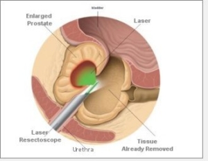
GreenLight PVP is an endoscopic procedure performed through the urethra. There are no abdominal incisions.

A telescope is passed through the urethra to the prostate. A specialised laser fibre is then used to progressively vaporise the obstructing prostate tissue and create a wider channel through which urine can flow.

Unlike TURP, where pieces of prostate tissue are physically cut away, GreenLight treatment predominantly converts the obstructing tissue into vapour while simultaneously sealing blood vessels.

The result is a combination of tissue removal and excellent haemostasis.


What Laser Is Used?

The GreenLight system uses a 532-nanometre green laser.

Modern GreenLight systems commonly use a lithium triborate (LBO) crystal to generate the 532 nm wavelength, with the contemporary high-powered platform delivering up to 180 watts.

Earlier systems included:

Generation Laser Maximum power
Early GreenLight KTP 80 W
GreenLight HPS LBO 120 W
GreenLight XPS LBO 180 W

The current European Association of Urology guideline describes the 180 W system as the contemporary standard platform for GreenLight PVP.


Why Is the Laser Green?

The colour is not merely decorative.

The 532 nm wavelength is strongly absorbed by haemoglobin, the pigment contained within red blood cells. It is much less strongly absorbed by water.

This property allows the laser energy to be preferentially absorbed by the vascular prostate tissue.

The laser has a relatively shallow tissue penetration of approximately 0.8 mm, producing rapid vaporisation while creating a limited zone of coagulation underneath the treated surface.

This gives GreenLight its two particularly useful characteristics:

Vaporisation + haemostasis

As prostate tissue is vaporised, small blood vessels are simultaneously coagulated.

This is why the operative field can remain remarkably clear and why GreenLight surgery generally produces less bleeding than traditional TURP.


Who May Benefit from GreenLight Laser Surgery?

GreenLight PVP may be considered for men with moderate to severe urinary symptoms caused by benign prostatic obstruction, particularly when conservative or medical treatment has not provided adequate relief.

Surgery may also be recommended when BPH results in complications such as:

  • Recurrent urinary retention
  • Dependence on a urinary catheter
  • Recurrent urinary tract infections associated with obstruction
  • Bladder stones
  • Recurrent visible haematuria attributable to BPH
  • Progressive bladder dysfunction
  • Significant residual urine
  • Upper urinary tract deterioration or renal impairment secondary to obstruction

Surgery is also reasonable when symptoms remain sufficiently troublesome despite medication, or when a patient does not wish to continue long-term medical therapy.


What Size Prostate Can Be Treated?

GreenLight PVP is particularly well established for prostates approximately 30–80 mL in volume.

The 2026 European Association of Urology guidelines recommend 532 nm GreenLight PVP as an alternative to TURP for men with moderate-to-severe LUTS and benign prostatic obstruction in this prostate-size range.

Larger prostates can certainly be treated by experienced GreenLight surgeons, but the operation becomes progressively more time-consuming as prostate volume increases.

Importantly, evidence supporting PVP in prostates above approximately 100 mL is less robust than the evidence supporting anatomical enucleation procedures such as HoLEP.

The AUA guideline similarly notes that PVP may be less effective in very large prostates and that retreatment becomes an increasingly important consideration.

For a very large prostate, HoLEP or robotic-assisted simple prostatectomy/enucleation may therefore provide more complete adenoma removal.


What Happens During GreenLight PVP?

The operation is usually performed under general or spinal anaesthesia.

A telescope is passed through the penis and urethra until the obstructing prostate is visualised.

The laser fibre is introduced through the telescope.

The surgeon progressively vaporises the obstructing prostate tissue, generally working from the bladder neck towards the apex of the prostate while carefully protecting the urinary sphincter.

The goal is not necessarily to remove the entire prostate.

Instead, the obstructing transition-zone tissue is removed sufficiently to create a wide, low-resistance urinary channel.

A urinary catheter is usually placed at the end of the operation.

Because bleeding is generally limited, catheterisation and hospitalisation can often be shorter than following conventional TURP.

Some appropriately selected patients can undergo the procedure as day surgery.


Advantages of GreenLight Laser Surgery

Less bleeding

This is arguably GreenLight’s greatest strength.

Because the 532 nm wavelength is absorbed by haemoglobin, tissue vaporisation and coagulation occur simultaneously.

Compared with TURP, studies have demonstrated lower transfusion rates and less bleeding.

Particularly useful when bleeding risk matters

GreenLight may be attractive in older patients or men with cardiovascular disease who require antiplatelet or anticoagulant medication.

The EAU guidelines specifically recognise GreenLight PVP as an option in selected patients receiving anticoagulant or antiplatelet therapy, although individual management of these medications still needs to be determined before surgery.

Shorter catheterisation

Many patients can have their catheter removed relatively quickly after surgery.

Shorter hospital stay

GreenLight surgery is frequently suitable for overnight or even day-case treatment in appropriately selected patients.

No external incision

The entire procedure is performed through the urethra.

No TUR syndrome

GreenLight PVP uses saline irrigation and does not carry the classical dilutional hyponatraemia or “TUR syndrome” associated with older monopolar TURP techniques.

Effective symptom improvement

GreenLight PVP can produce substantial improvements in:

  • Urinary flow
  • Bladder emptying
  • IPSS symptom scores
  • Quality of life

Randomised trials of contemporary GreenLight PVP have demonstrated symptom and flow improvements broadly comparable with TURP over short- to medium-term follow-up.


What Are the Side Effects?

Most men experience some temporary urinary irritation during the recovery period.

Burning or stinging when urinating

This is common initially and usually improves as the prostatic urethra heals.

Urinary frequency and urgency

The bladder may remain irritable for several weeks.

It is important to remember that removing the obstruction does not instantly reset a bladder that may have been struggling against that obstruction for many years.

Blood in the urine

Small amounts of bleeding can occur intermittently during healing, despite the excellent haemostatic properties of the laser.

Temporary difficulty urinating

Occasionally swelling after surgery means that the catheter needs to remain in place longer or needs to be reinserted.

Urinary infection

As with any endoscopic urinary procedure, urinary infection can occur.


Retrograde Ejaculation

One of the most important issues to discuss before surgery is ejaculation.

Following conventional GreenLight PVP, semen may travel backwards into the bladder rather than forwards through the penis during orgasm.

This is called retrograde ejaculation.

The orgasm usually remains present, but little or no semen may be produced.

This is particularly important for younger men concerned about fertility or preservation of ejaculation.

GreenLight should therefore not automatically be described as an “ejaculation-preserving” operation.


Does GreenLight Cause Erectile Dysfunction?

For most men, erectile function is preserved.

Current evidence does not demonstrate a major difference in erectile-function outcomes between GreenLight PVP and conventional TURP.

Sexual function, however, is influenced by many factors including age, vascular health, diabetes, medications and pre-existing erectile function.


Less Common Complications

Potential complications include:

  • Significant bleeding
  • Urinary tract infection
  • Temporary urinary retention
  • Urethral stricture
  • Bladder-neck contracture
  • Temporary urinary incontinence
  • Rare persistent urinary incontinence
  • Persistent urgency or overactive bladder symptoms
  • Incomplete removal of obstructing tissue
  • Recurrent prostate enlargement
  • Requirement for further prostate surgery

One Important Limitation: There Is Usually No Tissue for Pathology

This is an important difference between GreenLight PVP and TURP or HoLEP.

With TURP, prostate chips are removed.

With HoLEP, the enucleated adenoma is morcellated and retrieved.

With GreenLight PVP, much of the treated tissue is vaporised.

Consequently, there may be little or no prostate tissue available for histological examination.

For this reason, appropriate assessment for prostate cancer should be undertaken before GreenLight surgery when clinically indicated, using PSA, examination, MRI and/or prostate biopsy where appropriate.


GreenLight vs TURP vs HoLEP vs Robotic-Assisted Simple Prostatectomy

There is no single “best” prostate operation for every man.

The appropriate procedure depends on:

prostate size + prostate anatomy + bleeding risk + bladder function + patient priorities + surgeon expertise.

Feature GreenLight PVP TURP HoLEP Robotic Simple Prostatectomy / Enucleation
Approach Transurethral Transurethral Transurethral Abdominal robotic
Energy 532 nm laser Electrical 2,140 nm holmium laser Robotic dissection ± energy
Tissue treatment Vaporisation Resection Anatomical enucleation Anatomical enucleation
Tissue for pathology Limited/none Yes Yes Yes
Bleeding Very low Low-moderate Very low Low-moderate
Best established size ~30–80 mL ~30–80 mL Virtually size-independent Large/very large prostates
Large prostate >100 mL Possible, but less ideal Less attractive Excellent option Excellent option
Catheter duration Usually short Short Usually short Usually longer
Hospital stay Short Short Short Generally longer
Anticoagulation advantage Good Less favourable Good Less favourable
Learning curve Moderate Familiar technique Significant Significant
Durability in very large glands Less certain Good Excellent Excellent
Incisions None None None Abdominal port incisions

GreenLight vs TURP

TURP has traditionally been regarded as the benchmark operation against which newer BPH procedures are compared.

GreenLight achieves broadly similar improvements in urinary symptoms and flow for appropriately selected prostates.

Its principal advantages over TURP are:

  • Less perioperative bleeding
  • Lower transfusion requirements
  • Shorter catheterisation
  • Shorter hospitalisation
  • Excellent visibility during surgery
  • Potential advantages in patients at increased bleeding risk

Its disadvantages include:

  • Longer operating time in some patients
  • Lack of tissue for histology
  • Potentially higher retreatment rates over longer follow-up
  • Reduced efficiency as prostate size becomes very large

Long-term population data suggest that although reoperations for bleeding are less common after PVP, overall cumulative reoperation may be higher than after TURP.

So GreenLight’s strength is low perioperative morbidity, while TURP retains excellent durability and provides tissue for histology.


GreenLight vs HoLEP

HoLEP is fundamentally different.

GreenLight predominantly vaporises the obstructing prostate.

HoLEP enucleates the adenoma anatomically from the surgical capsule, rather like removing the inside of an orange while leaving the peel behind.

HoLEP uses a pulsed 2,140 nm holmium laser, which is strongly absorbed by water rather than haemoglobin.

The major advantage of HoLEP is that it is essentially prostate-size independent.

A 40 mL prostate can be enucleated.

So can a 100, 150 or even 200+ mL prostate in experienced hands.

The AUA guideline specifically recognises HoLEP as a prostate size-independent surgical option.

HoLEP also removes a larger proportion of the obstructing adenoma and provides tissue for histological examination.

Its principal disadvantages are its technical complexity, significant learning curve and potential for temporary postoperative stress urinary incontinence, particularly following treatment of very large glands.

For very large prostates, HoLEP will generally provide more complete anatomical adenoma removal than conventional GreenLight vaporisation.


GreenLight vs Robotic-Assisted Prostate Enucleation

For very large benign prostates, another option is robot-assisted simple prostatectomy (RASP), sometimes described as robotic prostate adenoma enucleation.

This is very different from a robotic radical prostatectomy performed for prostate cancer.

The entire prostate is not removed.

Instead, the enlarged benign adenoma is dissected away from the remaining prostate capsule.

It essentially achieves the same anatomical objective as HoLEP but approaches the prostate through the abdomen and bladder or prostate capsule rather than through the urethra.

Robotic simple prostatectomy is particularly attractive for very large or anatomically complex prostates, particularly when associated bladder pathology can be addressed simultaneously.

Its disadvantages include:

  • Abdominal surgery
  • Robotic port incisions
  • Longer catheterisation
  • Longer hospital stay
  • Greater surgical invasiveness
  • Higher resource utilisation

Recent comparative evidence suggests that both robotic simple prostatectomy and laser enucleation provide excellent outcomes for prostates ≥80 mL, while endoscopic laser enucleation generally provides faster perioperative recovery.


So Which Operation Is Best?

A useful way of thinking about these procedures is not as competitors, but as different tools for different prostates.

GreenLight PVP

Particularly attractive for:

  • Small-to-moderately enlarged prostates
  • Approximately 30–80 mL glands
  • Patients where minimising bleeding is particularly important
  • Older or medically complex patients
  • Patients requiring rapid recovery and short catheterisation

TURP

Remains an excellent option for:

  • Small-to-moderately enlarged prostates
  • Conventional benign prostatic obstruction
  • Situations where prostate tissue for histology is desirable
  • Centres with extensive TURP experience

HoLEP

Particularly attractive for:

  • Moderate, large and extremely large prostates
  • Men requiring maximal adenoma removal
  • Recurrent BPH after previous surgery
  • Patients where long-term durability is particularly important
  • Patients wishing to avoid abdominal surgery despite a very large prostate

Robotic-Assisted Simple Prostatectomy

Particularly attractive for:

  • Very large prostates
  • Complex prostate anatomy
  • Large intravesical adenomas
  • Patients with associated bladder pathology requiring treatment
  • Situations where endoscopic enucleation expertise is unavailable or a robotic approach offers anatomical advantages

The Bottom Line

GreenLight laser photoselective vaporisation is an effective, minimally invasive surgical treatment for benign prostate enlargement.

Its 532 nm laser is selectively absorbed by haemoglobin, allowing prostate tissue to be vaporised while simultaneously achieving excellent haemostasis.

Its particular strengths are:

less bleeding, short catheterisation, short hospitalisation and rapid recovery.

For appropriately selected men with moderate-sized prostates, symptom improvement is broadly comparable with TURP.

As prostate size increases, however, anatomical enucleation becomes increasingly attractive. HoLEP and robotic-assisted simple prostatectomy remove the adenoma more completely and are particularly effective for very large prostates. HoLEP achieves this endoscopically, whereas robotic surgery achieves it through an abdominal approach.

The choice therefore should not simply be:

“Which operation is newest?”

A better question is:

“Which operation best suits this prostate, this bladder and this patient?”

Prostate size and shape, urinary symptoms, bladder function, bleeding risk, medications, general health, PSA assessment, sexual priorities and the surgeon’s experience should all contribute to the final decision.

This information is intended for general patient education and does not replace individual assessment and discussion with a urologist.

So, if you want to learn more and are interested in having the Greenlight, come see your local Brisbane Urologist, Uro-Jo. This procedure is also offered in Caboolture at the Caboolture private Hospital as well as St Andrews War Memorial Hospital in Brisbane City.

Focal Therapy for Prostate Cancer: Treating the Cancer, Preserving the Prostate

For many years, treatment of localised prostate cancer largely involved choosing between active surveillance and treatment of the whole prostate gland with surgery or radiotherapy.

Modern multiparametric MRI, targeted transperineal biopsy and increasingly accurate image-guided treatment have opened a third pathway for carefully selected men: focal therapy.

Rather than treating or removing the entire prostate, focal therapy aims to identify the clinically significant cancer and destroy that area together with an appropriate safety margin, while leaving as much normal prostate tissue as possible.

A useful analogy is treating the troublesome patch rather than replacing the entire lawn.

The attraction is obvious: if the cancer can be controlled without treating the whole prostate, it may be possible to reduce the risks of urinary incontinence, erectile dysfunction and other quality-of-life effects associated with radical treatment.

However, focal therapy is not suitable for every prostate cancer, and it comes with an important trade-off: long-term cancer-control evidence is less mature than it is for radical prostatectomy and radiotherapy. Current European guidance therefore remains cautious, recommending focal therapy within clinical trials or well-designed prospective registries until stronger long-term comparative evidence becomes available.


What is focal therapy?

Focal therapy treats a selected region of the prostate containing clinically significant cancer rather than treating the entire gland.

Depending on the size and location of the tumour, treatment may involve:

  • Focal ablation of an individual lesion
  • Hemi-ablation, treating approximately one side of the prostate
  • Quadrant or zonal ablation
  • A wider “hockey-stick” ablation where disease distribution requires a larger treatment field

The treatment zone normally includes both the visible tumour and a planned margin around it.

The challenge is that prostate cancer is frequently multifocal. The largest or most biologically significant lesion is often referred to as the index lesion, but smaller cancer deposits may exist elsewhere in the gland.

For this reason, careful imaging, biopsy and follow-up are fundamental to a successful focal therapy program.


Who may be suitable for focal therapy?

The ideal candidate is generally a man with localised, clinically significant prostate cancer that can be accurately identified and safely targeted.

Potential candidates may include men with:

  • Disease confined to the prostate
  • A clearly identifiable lesion on multiparametric MRI
  • Cancer confirmed by targeted and systematic or mapping transperineal biopsy
  • Favourable intermediate-risk disease, commonly ISUP Grade Group 2 / Gleason 3+4, in an appropriate anatomical distribution
  • Selected higher-volume Grade Group 1 disease where active surveillance is considered unsuitable or unacceptable
  • Occasionally carefully selected Grade Group 3 disease in experienced centres, although the evidence is less established
  • A lesion that can be treated with an adequate margin without unacceptable injury to the urethra, sphincter, rectum or neurovascular structures
  • A strong preference to minimise the potential urinary and sexual consequences of whole-gland treatment

The decision should ideally follow review of the MRI, biopsy pathology, PSA, PSA density, prostate volume, tumour location and overall risk profile, rather than simply asking whether a particular machine can reach the tumour.


Who is generally NOT a good candidate?

Focal treatment becomes less attractive when there is:

  • Extensive multifocal clinically significant cancer
  • Significant bilateral disease
  • High-volume high-grade cancer
  • Extracapsular extension
  • Seminal vesicle invasion
  • Lymph-node involvement
  • Metastatic disease
  • Cancer that cannot be reliably seen or mapped
  • Disease immediately adjacent to structures that cannot safely be included in the treatment margin
  • A patient preference for the treatment with the longest-established oncological follow-up

Some men with very low-risk disease may also be better served by active surveillance rather than focal treatment, avoiding treatment altogether until there is evidence that treatment is actually necessary.


How do we determine whether focal treatment is appropriate?

Successful focal therapy begins with accurate cancer mapping.

Assessment will usually include:

Multiparametric MRI

MRI identifies suspicious lesions and helps establish their size, location and relationship to the urethra, capsule, sphincter and neurovascular bundles.

Transperineal prostate biopsy

MRI alone is not enough.

Targeted biopsy confirms the grade and extent of the MRI-visible lesion, while systematic or mapping biopsies help determine whether significant cancer exists elsewhere in the prostate.

PSA and PSA density

PSA remains useful, although interpretation after focal therapy differs from interpretation following radical prostatectomy because normal prostate tissue remains behind.

PSMA PET/CT

PSMA PET may be useful in selected patients, particularly those with higher-risk characteristics or when there is concern about disease outside the proposed treatment area.


What focal therapy options are available?

Several technologies can destroy a selected area of prostate tissue.

These include:

Irreversible Electroporation: NanoKnife

NanoKnife® is a system used to perform irreversible electroporation, or IRE.

Several fine needle electrodes are inserted through the perineum around the tumour under imaging guidance. Very short, high-voltage electrical pulses are passed between the electrodes.

Rather than primarily heating or freezing the tissue, the electrical field creates irreversible disruption of cell membranes, resulting in cell death.

IRE is therefore principally considered a non-thermal ablative technology.

Focal Laser Ablation

A laser fibre is placed directly into the target lesion and laser energy produces controlled thermal destruction of cancerous tissue.

ProFocal-Rx® is an Australian-developed focal laser technology designed specifically for targeted prostate treatment.

High-Intensity Focused Ultrasound

HIFU focuses ultrasound energy within the prostate, heating and destroying the targeted tissue without requiring needles to be placed directly throughout the treatment zone.

Cryotherapy

Needles are placed into the prostate and tissue is repeatedly frozen and thawed, producing cellular destruction.

Other technologies

Photodynamic therapy, radiofrequency ablation, focal brachytherapy and other energy-based approaches have also been investigated.

The Prostate Cancer Foundation of Australia notes that focal therapies including IRE/NanoKnife, laser ablation, HIFU and other technologies have been investigated or used in Australia, although availability varies.


NanoKnife versus ProFocal Laser Therapy

Both technologies attempt to achieve the same broad objective: destroy the cancer while preserving as much normal prostate and surrounding function as possible.

They achieve this in very different ways.

NanoKnife / IRE ProFocal-Rx Laser
Energy High-voltage electrical pulses Laser energy
Mechanism Irreversible electroporation Thermal coagulative ablation
Thermal treatment Principally non-thermal Yes
Access Transperineal needles/electrodes Transperineal laser applicator
Treatment planning Electrode geometry surrounds treatment zone Laser applicator positioned within/adjacent to target
MRI/TRUS planning Yes Yes
Tissue effect Cell membrane disruption Controlled heating and tissue necrosis
Treatment margin Created by electrical field between electrodes Created by laser ablation zone
Near neurovascular structures Potential theoretical advantage of non-thermal mechanism Requires careful thermal planning
Anaesthesia General anaesthesia with profound muscle relaxation generally required General anaesthesia typically used
Cardiac synchronisation Required with IRE Not required in the same manner
Repeat treatment Possible in selected cases Potentially possible
Long-term oncological evidence Growing medium-term evidence Earlier-stage clinical evidence
Australian regulatory status IRE devices are represented on the ARTG; specific device/indication should be checked ProFocal is currently not included on the ARTG

NanoKnife: potential advantages

The major attraction of IRE is that it does not rely primarily upon heating or freezing the prostate.

The electrical field disrupts cell membranes while potentially allowing relative preservation of extracellular structures. This makes IRE particularly interesting when treating cancers close to delicate structures.

Potential advantages include:

  • Precise treatment planning
  • No ionising radiation
  • No prostate removal
  • Preservation of untreated prostate tissue
  • Low reported rates of significant urinary incontinence
  • Potentially better preservation of erectile function compared with whole-gland treatment
  • Ability to consider repeat focal treatment in selected patients
  • Radical surgery or radiotherapy may remain possible if subsequent clinically significant cancer develops

Australian and international experience with IRE is considerably more mature than that of many newer focal technologies, although long-term comparative data against radical prostatectomy and radiotherapy are still developing. Published reviews cited by the AUA report residual or recurrent clinically significant cancer after focal ablation across all technologies, reinforcing the need for surveillance rather than considering focal therapy a “treat it and forget it” procedure.


NanoKnife: disadvantages and potential complications

IRE is still an invasive procedure.

Potential complications include:

  • Temporary urinary frequency and urgency
  • Dysuria
  • Haematuria
  • Perineal bruising or discomfort
  • Urinary retention
  • Temporary catheter requirement
  • Urinary tract infection
  • Prostatitis
  • Urethral injury or stricture
  • Erectile dysfunction
  • Ejaculatory changes
  • Rare urinary incontinence
  • Incomplete tumour ablation
  • Residual cancer within the treated field
  • Development or recognition of cancer elsewhere in the prostate
  • Need for repeat focal treatment
  • Subsequent need for radical prostatectomy or radiotherapy

Because IRE uses high-voltage electrical pulses, treatment requires appropriate anaesthesia, muscle relaxation and cardiac synchronisation.


ProFocal-Rx: focal laser therapy

ProFocal-Rx is a targeted laser ablation system developed in Australia.

A treatment applicator is placed transperineally into the prostate tumour. Laser energy is then delivered into the planned treatment area, producing controlled thermal destruction.

Early Australian studies have evaluated the feasibility and safety of this approach, including clinical trials of targeted treatment for MRI-localised prostate cancer.

Potential attractions include:

  • Highly localised treatment
  • Direct placement of the treatment fibre into the tumour
  • Relatively small treatment volumes
  • Preservation of surrounding prostate tissue
  • Short treatment and recovery pathways
  • Potential preservation of urinary continence
  • Potential preservation of erectile and ejaculatory function

However, ProFocal remains a newer technology with substantially less long-term oncological follow-up than radical prostatectomy, radiotherapy and even some other focal therapy platforms.


ProFocal: potential risks and limitations

Because laser treatment is thermal, careful treatment planning is required to prevent unintended heat injury.

Possible complications include:

  • Urinary frequency or urgency
  • Dysuria
  • Haematuria
  • Temporary urinary retention
  • Infection
  • Perineal discomfort
  • Urethral thermal injury
  • Erectile dysfunction
  • Ejaculatory changes
  • Rectal injury, although uncommon with appropriate treatment planning
  • Incomplete ablation
  • Residual clinically significant cancer
  • Cancer developing or being detected elsewhere in the prostate
  • Need for repeat treatment
  • Need for subsequent radical prostatectomy or radiotherapy

An important additional consideration is simply the maturity of the evidence. Early results can be encouraging without necessarily predicting cancer control at 10, 15 or 20 years.


What is the TGA status in Australia?

This point deserves particular clarity.

The Australian Register of Therapeutic Goods (ARTG) is the TGA’s public register of therapeutic products that can legally be supplied in Australia, subject to applicable exemptions and special-access pathways.

IRE / NanoKnife

Irreversible electroporation technology is available in Australia and is being used clinically for selected prostate cancers. The Medical Services Advisory Committee currently has an application assessing IRE using the NanoKnife system for prostate tumour tissue, including a proposed Medicare Benefits Schedule item. That MSAC application remains under assessment rather than representing an established Medicare item.

It is important not to confuse TGA/ARTG regulatory status with Medicare funding or with endorsement of focal therapy as oncologically equivalent to prostatectomy or radiotherapy. These are separate questions.

ProFocal-Rx

As of August 2026, the manufacturer’s Australian website specifically states that:

ProFocal is not included on the TGA’s ARTG in Australia.

TGA documents also demonstrate previous Australian patient access to ProFocal-Rx through the Special Access Scheme, which is a pathway for accessing an unapproved therapeutic good in particular circumstances and is not the same as general ARTG inclusion.

This distinction is important when discussing ProFocal with Australian patients.

Regulatory status can change, so the current ARTG should always be checked when treatment is being considered.


Does focal therapy cure prostate cancer?

It can achieve local control of appropriately selected prostate cancers, but the word “cure” needs to be used carefully.

Unlike radical prostatectomy, focal therapy deliberately leaves much of the prostate behind.

There are therefore two important potential sites of future cancer:

In-field recurrence
Cancer persists or recurs within the treated area.

Out-of-field cancer
Clinically significant cancer is subsequently detected elsewhere in the untreated prostate.

Neither necessarily means that focal therapy was inappropriate, but patients need to understand from the outset that continued prostate cancer surveillance is part of the treatment strategy.

The AUA’s salvage guideline notes clinically significant cancer following focal treatment across different modalities and emphasises that recurrence remains an important consideration after focal ablation.


Follow-up after focal therapy

Focal therapy does not end prostate cancer surveillance.

Follow-up typically involves a combination of:

  • Regular PSA testing
  • Clinical review
  • Multiparametric MRI
  • Repeat targeted and systematic biopsy
  • Additional imaging where clinically indicated

A common strategy is to establish a new PSA baseline after treatment and combine PSA behaviour with MRI and scheduled biopsy rather than relying on PSA alone.

This is important because the remaining normal prostate continues to produce PSA. Unlike after radical prostatectomy, the PSA is therefore not expected to become undetectable.


What happens if the cancer returns?

One of the advantages of focal treatment is that further treatment options usually remain available.

Depending upon the location, grade and extent of recurrent disease, options may include:

  • Continued surveillance for insignificant disease
  • Repeat focal therapy
  • Radical prostatectomy
  • External-beam radiotherapy
  • Other appropriate salvage treatment

Patients should nevertheless understand that salvage surgery after previous focal therapy may be technically more challenging because of fibrosis and altered tissue planes.

For clinically significant recurrence following focal ablation, AUA salvage guidance recommends that men considering definitive salvage treatment be offered whole-gland treatment with radical prostatectomy or radiotherapy.


Focal therapy versus radical treatment

Focal therapy occupies an increasingly interesting middle ground.

Active surveillance aims to avoid treatment until treatment becomes necessary.

Focal therapy aims to treat the clinically significant cancer while preserving the remainder of the prostate.

Radical prostatectomy or radiotherapy aims to treat the entire prostate and therefore both known and potentially occult cancer within the gland.

There is no universally “best” choice.

The appropriate treatment depends upon:

  • Cancer grade
  • Cancer volume
  • MRI findings
  • Biopsy distribution
  • PSA and PSA density
  • Age and life expectancy
  • Baseline urinary function
  • Baseline erectile function
  • Other medical conditions
  • Individual attitude towards cancer risk
  • Willingness to undergo continued MRI and biopsy surveillance
  • Personal priorities regarding continence and sexual function

The key question: are we treating the right cancer?

The success of focal therapy depends less on the glamour of the machine and more on patient selection, accurate imaging, meticulous biopsy mapping, treatment planning and rigorous follow-up.

NanoKnife, laser, HIFU and cryotherapy are different tools. The most important step occurs before any of them are switched on: establishing exactly where the clinically significant cancer is and whether disease elsewhere in the prostate has been adequately excluded.

For the appropriately selected man, focal therapy offers an attractive possibility:

Treat the cancer that needs treatment while preserving as much of the prostate, urinary function and sexual function as possible.

For other men, active surveillance, radical prostatectomy or radiotherapy will remain the safer oncological strategy.


Important perspective

Focal therapy is an exciting and rapidly evolving field, but it should not be presented as a universally equivalent replacement for established prostate cancer treatments.

Current evidence suggests excellent functional outcomes in appropriately selected patients, while definitive long-term comparative oncological evidence remains incomplete. European guidelines consequently continue to recommend focal therapy within clinical trials or prospective registries.

The decision is therefore best made after a detailed discussion with a urologist experienced in prostate MRI, transperineal biopsy, focal therapy and established radical treatment options.

Australian regulatory note

At the time of writing in August 2026, ProFocal-Rx is not included on the Australian ARTG, while IRE/NanoKnife technology is available in Australia and IRE for prostate cancer is currently undergoing an MSAC assessment relating to proposed Medicare funding. Regulatory status and funding arrangements may change and should be confirmed before treatment.

This information is intended for general patient education and does not replace individual medical advice. Suitability for focal therapy requires assessment of the patient’s pathology, imaging, prostate anatomy, overall health and personal treatment priorities.

So, if this is of interest to you, come have a chat to your local Brisbane urologist, Dr Jo to discuss this with you

Simple Prostatectomy – Open

Why is it done?

  • For those large benign prostates where a TURP would be too time consuming, and too dangerous.
  • Generally, prostates over 200cc
  • Not commonly performed in Australia
  • This procedure is performed when the prostate gland is enlarged to such an extent that medication cannot relieve the urinary symptoms.
  • Step-up therapy could have been used for prostates larger than 35-50cc with either Duodart, Avodart or Proscar and can be used as a first line in these huge prostates as long as the sexual side effects have been discussed.
  • Prostate cancer would have been excluded by doing a PSA, and when indicated, with a 3T MRI scan.
  • An alternative: A 2 staged-TURP can also be performed to dis-obstruct a huge prostate. Either Bipolar resection or Laser can be utilized
  • It provides a quicker solution with more marked side-effects and risks


How is it done?

  • Patients will receive a general anesthesia,
  • Prophylactic anti-biotic is given.
  • An indwelling catheter is placed.
  • A lower midline incision is made (or alternatively a horizontal Pfannenstiel-incision), The retropubic space of Retzuis is entered
  • A Millen-procedure is done where the prostate capsule and lower part of the bladder is incised in the longitudinal aspect
  • The bladder neck mucosa is cut and freed from the prostate away from the ureters as to prevent injury.
  • With blunt dissection the apex of the prostate is freed from with the urethra and each lobe is delivered separately.
  • Copious bleeding is possible in this phase, and this is where a cell-saver usage is critical to prevent blood transfusions with donor blood.
  • Hemostatic sutures are placed over bilateral prostatic vascular pedicles to stop the bleeding.
  • Sutures are placed to assist in reducing the cavity left after enucleation
  • The bladder neck is pulled down into the cavity to assist with hemostasis.
  • Prostate capsule and bladder are closed in 2 layers over a 3-way irrigation catheter
  • A drain is left for a couple of days
  • You may have continuous Antibiotics over the next few days.


Complications

  • Blood loss 400-1200cc, usually less
  • Wound infections
  • The first 6 weeks are the worst with frequency and urgency as a result
  • Stress incontinence may occur and will improve over the next 12 months (12%)
  • Complete incontinence at 12 months (2%)
  • Erectile dysfunction (bladder neck stenosis 5 % requiring intermittent self-dilatation
  • Retrograde ejaculation with Infertility
  • Testicular pain similar to vasectomy for up to a week
  • Possibility of bowel injury


Post-operative review:

  • Review PSA roughly 6 weeks after the surgery to assess post-operative Nadir
  • Review in rooms a week later.
  • 6 monthly review depending on risk factors.
  • If stable with good PSA outcomes, refer back to GP for 6 monthly PSA review


Wes Simple Prostatectomy BPH

Focal Therapy for Prostate Cancer – PROFOCAL Laser therapy

Not covered by Medicare / Health Fund yet. Only used in trial settings currently.

AUSTRALIAN DESIGN AND MANUFACTURED

What is ProFocal

  • ProFocal is a minimally invasive focal laser treatment used for localized prostate cancer.
  • The procedure uses a needle-guided laser system to deliver energy directly to the tumor, with the goal of preserving healthy tissue and function,
  • It may lead to a faster recovery with minimal side effects like urinary incontinence or erectile dysfunction. 


Seen here is a similar product used in USA. THIS IS NOT ProFocal. I am attempting to demonstrate the principle.

Why is it done?

  • Treatment for localized prostate cancer
  • Single focus disease
  • Prostate preserving
  • Ejaculate sparing
  • Continence preserving


How is it done?

  • Focal therapy of prostate cancer, usually a single lesion
  • Any Gleason grade, not Gleason 6 as this can be managed with surveillance
  • Preserving prostate supporting tissue and erectile function and continence

You would have had a 3T MRI study possibly a PET PSMA as well prior to confirming your prostate cancer. Usually, whole gland biopsies are taken of the prostate via the perineum to prove unifocal cancer.


  • This procedure is done under general anesthesia as a day procedure and takes approximately 60-90min (incl anesthetic time)
  • It is performed with the patient lying in lithotomy position
  • Prophylactic antibiotics are provided with anesthesia.
  • Planning would be done as to where needles should be placed to provide a 1cm coverage beyond the tumour using your MRI as guidance.
  • A trans-rectal ultrasound is placed
  • Laser probe is inserted via a trans perineal needle with ultrasound guidance
  • Treatment delivered
  • Needle removed and placed 5-7 mm adjacent to previous needle with next treatment delivered


Complications

Side–effects

  • Hematuria (blood in urine) 2-3days
  • Hematospermia (blood in ejaculate) will become less the more often you ejaculate.
  • Bacteremia (infection) with low-grade fever and feeling unwell
  • Perineal hematoma
  • Perineal pain and penis tip pain
  • Prostate swelling causing bladder outlet obstruction requiring a catheter for up to 5 days

ANY FEVER REQUIRES URGENT ATTENTION

What next?

  • You will be discharged after a trial without catheter
  • You may have necrotic tissue developing requiring a resection (treatment involving the urethra).
  • Difficulty in urination up to 6 weeks after the procedure
  • It could feel like you are sitting on a golf ball for a week
  • Continence and erectile function should not be affected


Should there be any signs of fever or cold shivers, you are to return to the hospital or Emergency Department without hesitation


Download Information Sheet

Wes ProFocal

Copyright 2019 Dr Jo Schoeman

TURIS – Button Vaporization

Endoscopic vaporization of a benign enlarged prostate, using laser. This allows patients on anti-coagulation therapy to continue their medication with minimal risk of hemorrhage. It also allows a shorter stay in hospital.

Indications:  

  • Patients on anticoagulation or anti-platelet therapy
  • Smaller prostate
  • Where conservative management has failed.
  • Patient choice
  • This procedure is performed when the prostate gland is enlarged to such an extent that medication cannot relieve the urinary symptoms.
  • Symptoms include:
    • a weak stream,
    • nightly urination frequency,
    • frequent urination,
    • inability to urinate,
    • kidney failure due to the weak urination (obstruction),
    • bladder stones,
    • recurrent bladder infections.
  • Medication such as Flomaxtra, Urorec, Minipress etc. should always be given as a first resort.
  • Step-up therapy should have been used for prostates larger than 35-50cc with either Duodart, Avodart or Proscar
  • Prostate cancer first needs to be ruled out by doing a PSA, and when indicated, with a 3T MRI scan of the prostate with an abnormal PSA with a possible prostate biopsy of any suspicious lesions.
  • A TUVP can also be performed to dis-obstruct a severe prostate cancer, to allow a normal urination process


How is it done?

  • You will receive a general anaesthesia, unless contra-indicated.
  • A cystoscopy is performed by placing a camera in the urethra with the help of lignocaine gel
  • The inside of the bladder is viewed for pathology. If any suspicious lesions are seen, a biopsy will be taken.
  • A vaporization of the prostate is then started and should take 60-120 minutes depending on the size of the prostate.
  • Prophylactic antibiotics will be given to prevent any infections.
  • Post– operative antibiotics will be continued for 10 days.

No specimen will be obtained due to vaporization, unless PSA was suspicious and an MRI with view to prostate biopsy has excluded a prostate cancer


What can go wrong?

  • Any anesthesia has its risks, and the anesthetist will explain this to you.
  • No blood loss is expected.
  • You will wake up with a catheter in your urethra and bladder. This will remain in the bladder overnight.
  • Lower abdominal discomfort for a few days
  • NB! Each person is unique and for this reason symptoms vary!


What next?

  • You will spend 1 –2 nights in hospital.
  • You will a trial without catheter the next day
  • You will be discharged as soon as you can completely empty your bladder.
  • You may initially suffer from urge incontinence and dysuria (irritable voiding) and will improve within the next 6 weeks.
  • Allow for 6 weeks for stabilization of symptoms.
  • There may be some blood in your urine. You can remedy this by drinking plenty of fluids until it clears.
  • A ward prescription will be issued on your discharge, for your own collection at any pharmacy
  • A follow-up appointment will be scheduled for 6 weeks. Remember there is no pathology due to vaporization.
  • Don’t hesitate to ask Jo if you have any queries

DON’T SUFFER IN SILENCE, OR YOU WILL SUFFER ALONE


Side–effects

  • Retrograde ejaculation in more than 90% of patients. Therefore, if you have not completed your family, this procedure is not for you unless absolutely necessary.
  • Infertility as a result of the retrograde ejaculation.
  • Stress incontinence initially for the first 6 weeks, especially in the elderly and the diabetic patients
  • Patients with Multiple Sclerosis, Strokes and Parkinsons have a higher risk of incontinence and risks should be discussed and accepted prior to surgery.
  • Urethral stricturing in 2-3% of patients, requiring intermittent self-dilatation.
  • Regrowth of prostate lobes within 3-5 years requiring a second procedure.
  • NB! Each person is unique and for this reason symptoms vary!


Wes TURIS

Indwelling Urethral Catheter – IDC

Non-invasive placement of a silicone tube which is secured inside the bladder and attached to a drainage bag on the outside, in order to drain an obstructed bladder (urinary retention)

Why is it done?

  • This can be placed as an emergency for patients in acute urinary retention
    • Prostate obstruction
    • Urethral strictures
    • Blood clot obstruction caused by bleeding
    • Hematuria (bleeding)
    • Severe urinary tract infections
  • Commonly placed intra-operatively for long, non-urological surgical procedures to enable urine drainage and monitoring urine output.
  • Commonly placed at the end of a Urological procedure to enable urine drainage and to enable hemostasis (stopping bleeding)


How is it done?

  • This is done as a sterile procedure; therefore, the genital area will be cleaned with a non-abrasive disinfectant.
  • A sterile catheter will be used
  • A local anesthetic gel is placed in the urethra a few minutes prior to the placement of the catheter. This may initially sting for a few seconds until it numbs the mucosa.
  • An appropriate size catheter (14-18Fr) will be inserted
  • Urine should be aspirated with a syringe to confirm the correct position in the bladder.
  • An anchoring balloon will be inflated with 10cc of sterile water.
  • A drainage urine bag will be attached
  • The catheter will be secured to your leg. (check that this is always secured)


Complications

  • Urethra with resulting discomfort.
  • In the presence of urethral stricture, it may be impossible to pass the catheter, and a flexible cystoscopy with dilatation of the stricture may be required prior to placement.
  • If you had a large over-stretched bladder (urine retention) you may experience bleeding as the bladder empties, caused by the mucosal tears that have occurred.
  • Catheters that have been placed long term, may cause irritation and possibly attract infection. Permanent catheters are usually changed every 6-8 weeks.


Download Information Sheet

Wes Catheters Indwelling Catheter

Copyright 2019 Dr Jo Schoeman

Greenlight Laser Trans Urethral Vaporization of Prostate

This is an endoscopic technique of enucleation of the prostate using Greenlight laser therapy. Similar to a TURP – “Re-bore” only with minimal bleeding and shorted hospital stays. Some would argue this is ” The new Gold Standard”

Why is it done?

Endoscopic vaporization of a benign enlarged prostate, using laser.

Indications:

  • Patients on anticoagulation / anti-platelet therapy
    • Warfarin needs to be placed on Clexane 7 days prior.
    • Clopidogrel should be down scaled down to Aspirin.
    • This allows patients on anti-coagulation therapy to continue their medication with minimal risk of hemorrhage. It also allows a shorter stay in hospital.
  • Prostates up to 120cc.
  • Where conservative management has failed.
  • Patient choice.
  • This procedure is performed when the prostate gland is enlarged to such an extent that medication cannot relieve the urinary symptoms.
  • Symptoms include: LUTS
    • a weak stream,
    • nightly urination,
    • frequent urination,
    • inability to urinate,
    • kidney failure due to the weak urination (obstruction),
    • bladder stones,
    • recurrent bladder infections.
  • Medication such as Flomaxtra, Urorec or Minipress etc. should always be given as a first resort.
  • Step-up therapy could have been used for prostates larger than 35-50cc where indicated, with either Duodart, Avodart or Proscar.
  • Prostate cancer first needs to be ruled out by doing a PSA, and when indicated, with a 3T MRI scan of the prostate with an abnormal PSA with a possible prostate biopsy of any suspicious lesions.

How is it done?

    • Patients will receive a general anesthesia, unless contra-indicated.
    • A cystoscopy is performed by placing a camera in the urethra with the help of lignocaine gel.
    • Saline is used to irrigate the bladder, therefore NO DILUTIONAL HYPONATRAEMIA
    • The inside of the bladder is viewed for pathology. If any suspicious lesions are seen, a biopsy will be taken.
    • A vaporization of the prostate is then started and should take 60-120 minutes depending on the size of the prostate.
    • Prophylactic antibiotics will be given to prevent any infections.
    • Post– operative antibiotics will be continued for 10 days.
    • No specimen will be obtained due to vaporization, unless PSA was suspicious and an MRI with view to prostate biopsy has excluded a prostate cancer.

What can go wrong?

  • Any anesthesia has its risks, and the anesthetist will explain this to you.
  • No blood loss is expected.
  • In rare circumstances you may develop fluid overload requiring a High Care Facility admission, especially with the upper edge of prostate sizes.
  • You will wake up with a catheter in your urethra and bladder. This will remain in the bladder overnight.
  • Lower abdominal discomfort for a few days.
  • NB! Each person is unique and for this reason symptoms vary!

What next?

  • You will spend 1 night in hospital.
  • You will a trial without catheter the next day.
  • You will be discharged as soon as you can completely empty your bladder.
  • You may initially suffer from urge incontinence and dysuria (discomfort) and will improve within the next 6 weeks.
  • You will pass a grey, wet scab at the 2–3-week mark.
  • Some bleeding may be associated with this, especially if you are on anticoagulation therapy.
  • Allow for 6 weeks for stabilization of symptoms.
  • There may be some blood in your urine. You can remedy this by drinking plenty of fluids until it clears.
  • A follow-up appointment will be scheduled for 6 weeks. Remember there is no pathology due to vaporization.
  • DON’T SUFFER IN SILENCE, OR YOU WILL SUFFER ALONE!

Side–effects

  • Retrograde ejaculation in more than 90% of patients. Therefore, if you have not completed your family, this procedure is not for you unless absolutely necessary.
  • Infertility as a result of the retrograde ejaculation. Not a given, therefore continue your contraceptives
  • Stress incontinence especially in the elderly and the diabetic patients.
  • Patients with Multiple Sclerosis, Strokes and Parkinsons have a higher risk of incontinence and risks should be discussed and accepted prior to surgery.
  • Urethral stricturing in <2% of patients, requiring intermittent self-dilatation.
  • Regrowth of prostate lobes can occur within 3-5 years requiring a second procedure.
  • NB! Each person is unique and for this reason symptoms vary!

Remember

  • You still have a peripheral zone of your prostate and regular PSA reviews are required up to the age of 75. (This could be seen as controversial).

Download Information Sheet

Wes TUVP Greenlight

Low Dose Brachytherapy

Perineal placement of Radioactive Iodine-125 seeds in the prostate as treatment for Intermediate-Risk Prostate Cancer

Why is it done?

This is a radiation therapy option for the management of a localized prostate cancer.

Criteria include:

  • PSA less than 10
  • Gleason 3,4 adenocarcinoma prostate,
  • Higher grades may have extra-prostatic extension requiring combined External Beam Radiation
  • Staging negative, (bone scan negative, CT negative)
  • Not younger than 65
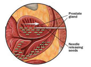
  • It is the localized radiation of the prostate, by means of trans-perineal placement of Radio-active I-125 seeds.
  • This is a nerve sparing procedure and patients have a good opportunity to maintain this.
  • The procedure takes 2-3hours excluding the anesthetic time.
  • This is an alternative to a Radical Retropubic Prostatectomy for low-intermediate risk prostate cancer. Prostates are generally smaller than 50cc.
  • Could be ideal for those patients with excessive BMI and fitting the cancer specific criteria.


How is it done?

  • Under a General Anaesthetic
  • Presence of Radiation Oncologist and Physicist
  • Lithotomy position
  • Trans-rectal placement of an ultrasound probe
  • Real-time accumulation of digital images to allow real-time placement of Radioactive Iodine seeds according to an intra-operative plan

PSA failure:

  • PSA not dropping to a nadir value, preferably 0,2ng/ml.
  • 3 consecutive PSA rises following RRP.

Complications

  • Perineal hematoma
  • Wound infections
  • Urgency frequency and weak stream
  • Limited Erectile Dysfunction, which may only surface after 18 months after treatment.
  • Lower ejaculate volume.
  • Testicular pain similar to vasectomy for 2-3 days


Post operative care:

  • A post-procedure CT scan to account all the seeds and exclude migration of seeds.
  • Normal diet
  • A salt water or Betadine Douche is required after every stool for the first week
  • A 3-month course of Flomaxtra to ease Lower Urinary Tract Symptoms
  • Wounds generally heal in 7-10 days

Catheter care

  • Your catheter will remain until you are awake.
  • Post-operative review:
  • Radio-Oncology will review a few weeks later.
  • You will review with me at 6 weeks post-procedure to check if you are doing fine.
  • Review PSA roughly 3 months after the procedure to assess PSA track
  • You should reach your Nadir (lowest PSA) at 6– 9 months after the procedure. The lower the better.
  • 6 monthly PSAs thereafter.
  • Expect a PSA Bump at approximately 9-12 months after the procedure, The PSA should drop thereafter
  • If stable with good PSA outcomes, refer back to GP for 6 monthly PSA review


Download Information Sheet

Wes Low Dose Brachytherapy

Trans Urethral Resection Prostate (TURP) – Monopolar

This is the procedure used to resect the inside (enlarged, obstructive part) of the prostate. Known generally as the “Re-Bore”.  Glycine is used as an irrigate.                                                           

Why is it done?

  • This procedure is performed when the prostate gland is enlarged to such an extent that medication cannot relieve the urinary symptoms.
  • Symptoms include
    • a weak stream,
    • nightly urination,
    • frequent urination,
    • inability to urinate,
    • kidney failure due to the obstruction,
    • bladder stones,
    • recurrent bladder infections due to retaining urine.
  • Medication such as Flomaxtra, Urorec or Minipress etc. should always be given as a first resort.
  • Step-up therapy could have been used for prostates larger than 35-50cc with either Duodart, Avodart or Proscar, where indicated and appropriate.
  • Prostate cancer first needs to be ruled out by doing a PSA, and when indicated, with a 3T MRI scan of the prostate with an abnormal PSA with a possible prostate biopsy of any suspicious lesions.

How is it done?

  • Patients will receive a general anesthesia, unless contra-indicated.
  • A cystoscopy is performed by placing a camera in the urethra with the help of a lubricant jelly and an irrigant fluid.
  • The inside of the bladder is viewed for pathology. If any suspicious lesions are seen, a biopsy will be taken.
  • A resection of the prostate is then started and should take 60-90 minutes.
  • Prophylactic antibiotics will be given to prevent any infections.


Complications

Side–effects

  • Retrograde ejaculation in more than 90% of patients. Therefore, if you have not completed your family, this procedure is not for you unless absolutely necessary.
  • Infertility as a result of the retrograde ejaculation.
  • Stress incontinence especially in the elderly and diabetic patients
  • Patients with Multiple Sclerosis, Strokes and Parkinsons have a higher risk of incontinence and risks should be discussed and accepted prior to surgery.
  • Urethral structuring in 5% of patients, requiring intermittent self-dilatation.
  • Regrowth of prostate lobes within 3-5 years requiring a second procedure.

NB! Each person is unique and for this reason, symptoms vary.


Download Information Sheet

Wes TURP Monopolar

Copyright 2019 Dr Jo Schoeman