
Flexible Cystoscopy & Urethral Dilation
A day procedure under local anaesthetic, where a flexible cystoscope is placed in the bladder via the urethra. Narrowing in the urethra is dilated.
Why is it done?
A cystoscopy is used to investigate:
- Hematuria (blood in the urine)
- Recurrent urinary tract infections
- Dilatation of Urethral narrowing/ stricture
- Abnormal cells suggestive of urothelial carcinoma, on urine cytology
Ideally a retrograde urethragram is used to diagnose this radiologically
Risk factors for strictures:
- Straddle injuries
- Catheterization or urethral instrumentation
- Infections
- Bypass cardiac surgery with long ischemic time
How is it done?
- A cystoscopy is performed by placing a camera in the urethra with the help of a lubricant jelly and saline
- Usually, you can’t move past the narrowing
- Then:
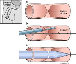
Urethral Dilatation
- If you have a urethral stricture, a guidewire will be placed and the narrowing dilated
- There may be some hemorrhaging and you may need a catheter for 3 days
- This will be removed at the hospital in 3 days or alternatively arrange for your GP to remove.
- I will review in 6 –8 weeks
Antibiotics may be given to prevent infection
Complications
What to expect after the procedure?
- You may be sent home with an indwelling catheter for 3 days
- Pain on initial passing of urine after it is removed
- Bladder infection ranging from a burning sensation to, fever, to puss (rare)
- Bloodstained urine
- Lower abdominal discomfort which will persist for a few days
- NB! Each person is unique and for this reason, symptoms vary.
Download Information Sheet
Wes Flexible Cystoscopy and Urethral Dilatation IDC
Copyright 2019 Dr. Jo Schoeman
Leave a Reply
Want to join the discussion?Feel free to contribute!