Urethral Caruncle: A Small Lump That Can Cause Big Concern
Finding a red or fleshy lump around the opening of the urethra can understandably be alarming. Fortunately, one of the most common causes, particularly in women after menopause, is a urethral caruncle.
A urethral caruncle is usually benign (non-cancerous) and may cause no problems at all. However, because several other conditions can look similar, a new, enlarging, bleeding or unusual urethral lesion deserves proper assessment.
What is a urethral caruncle?
A urethral caruncle is a small, fleshy growth arising from the lining of the urethral opening, or urethral meatus.
It typically appears as a soft red, pink or sometimes darker-red polypoid lesion at the edge of the urethral opening. It most commonly arises from the posterior or lower margin of the urethral meatus.
Caruncles occur predominantly in postmenopausal women, although they can occasionally occur in younger women.
Importantly, a urethral caruncle is not considered a precancerous condition. Its significance lies mainly in the symptoms it can produce and the fact that other urethral conditions can occasionally imitate its appearance.
What causes a urethral caruncle?
The exact cause is not completely understood, but several factors are thought to contribute.
Reduced oestrogen after menopause
This is probably the most important association.
After menopause, falling oestrogen levels cause thinning and reduced elasticity of the tissues surrounding the vagina and urethra. This is part of what is now commonly called genitourinary syndrome of menopause (GSM).
The urethral lining may become more fragile and susceptible to irritation, inflammation and prolapse.
Chronic inflammation and irritation
Repeated local irritation may contribute to inflammation around the urethral opening and subsequent formation of a caruncle.
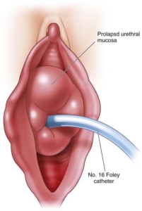
Urethral mucosal prolapse
A caruncle may represent a localised form of prolapse of the urethral lining. This helps explain why the lesion usually occurs at the edge of the urethral opening.
What does a urethral caruncle look like?
A typical caruncle is:
- Small and fleshy
- Red or pink
- Soft
- Usually located along the lower edge of the urethral opening
- Occasionally friable, meaning that it bleeds easily when touched
- Sometimes tender or inflamed
They vary considerably in size. Some are barely noticeable, while larger lesions may protrude sufficiently to be felt or seen by the patient.
Although the appearance may be quite characteristic, appearance alone should not always be relied upon when a lesion is atypical.
What symptoms can it cause?
Many urethral caruncles are discovered incidentally during a pelvic examination and cause absolutely no symptoms.
When symptoms do occur, they may include:
Bleeding
The surface of a caruncle can be delicate and vascular. Patients may notice:
- Spotting on underwear
- Blood after wiping
- Bleeding after intercourse
- Blood noticed after passing urine
One important distinction is determining whether the blood is actually coming from the urethral lesion or whether there is true blood within the urine (haematuria).
Persistent haematuria may require additional investigation.
Pain or tenderness
An inflamed caruncle can become uncomfortable, particularly with wiping, intercourse or local pressure.
Burning when passing urine
Some patients experience dysuria or a stinging sensation as urine passes across the inflamed tissue.
A visible or palpable lump
Some women first become aware of the condition because they see or feel a small lump at the urethral opening.
Urinary symptoms
Large caruncles only rarely interfere significantly with urinary flow. If there is difficulty passing urine, a weak stream or urinary retention, other causes should also be considered.
How is a urethral caruncle diagnosed?
Diagnosis usually begins with a careful examination of the urethral opening and surrounding genital tissues.
A typical small caruncle in a postmenopausal woman often has a characteristic appearance.
Your doctor may also assess for associated vaginal and urethral atrophy and ask about urinary symptoms, recurrent urinary infections, bleeding and haematuria.
Depending on the circumstances, investigations may include:
- Urinalysis
- Urine culture if infection is suspected
- Assessment for haematuria
- Cystoscopy if there are unexplained urinary symptoms or blood in the urine
- Biopsy or removal of the lesion when its appearance is atypical
Does a urethral caruncle need treatment?
Not necessarily.
A small, typical caruncle that causes no symptoms may simply be observed.
Treatment is generally considered when the lesion causes bleeding, pain, irritation or bothersome urinary symptoms, or when there is uncertainty about the diagnosis.
1. Observation
If the lesion is small, has a typical appearance and causes no symptoms, reassurance and observation may be all that is required.
This avoids treating something that is essentially harmless.
2. Topical vaginal oestrogen
In postmenopausal women, topical oestrogen therapy is frequently used, particularly when there are accompanying features of genitourinary syndrome of menopause.
Oestrogen can improve the quality and thickness of the tissues around the urethra and vagina and may allow a small caruncle to shrink considerably or resolve.
Treatment usually requires several weeks rather than several days.
The suitability of vaginal oestrogen should be discussed with your doctor, particularly if you have a history of an oestrogen-sensitive malignancy or another reason to avoid hormonal therapy.
3. Anti-inflammatory and supportive treatment
Where local inflammation is prominent, conservative measures may occasionally include:
- Avoidance of irritating soaps and products
- Treatment of associated infection if present
- Appropriate topical therapy
- Simple analgesia when required
These treatments may improve symptoms but do not necessarily remove the lesion itself.
When is surgery required?
Surgical removal may be recommended when the caruncle is:
- Persistently painful
- Repeatedly bleeding
- Large or bothersome
- Not responding to conservative treatment
- Increasing in size
- Firm, irregular, ulcerated or otherwise atypical
- Associated with uncertainty about the diagnosis
Surgery usually involves excision of the lesion at its base, often as a relatively minor procedure.
The removed tissue can then be sent to a pathologist for examination under the microscope.
Possible complications are uncommon but can include bleeding, infection, discomfort, recurrence and, rarely, scarring or narrowing of the urethral opening.
Can a urethral caruncle be cancerous?
A true urethral caruncle is benign.
The more important question is whether a lesion that looks like a caruncle could occasionally be something else.
Several conditions may resemble a caruncle, including:
- Urethral mucosal prolapse
- Urethral polyps
- Periurethral cysts
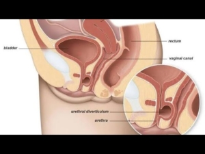
- Urethral diverticulum
- Genital warts
- Inflammatory lesions
- Melanoma
- Urethral carcinoma
- Other uncommon benign or malignant tumours
Cancer masquerading as an apparently innocent urethral lesion is uncommon, but this is precisely why an atypical lesion should not simply be labelled a caruncle and forgotten.
When should a biopsy be considered?
Biopsy or complete excision is particularly worth considering when a lesion is:
- Irregular or unusually firm
- Ulcerated
- Pigmented
- Rapidly enlarging
- Persistently bleeding
- Unusually large
- Not responding as expected to conservative treatment
- Associated with enlarged groin lymph nodes
- Clinically inconsistent with a typical caruncle
In medicine, sometimes the smallest lump deserves a second look rather than a dramatic name.
Urethral caruncle versus urethral prolapse
These conditions can look similar but are not quite the same.
A urethral caruncle generally involves only a portion of the circumference of the urethral opening, most commonly its posterior margin.
A urethral prolapse usually involves the entire circumference of the urethral lining, creating a circular or doughnut-shaped ring of tissue surrounding the opening.
The distinction can influence management.
What about blood in the urine?
This is particularly important.
A caruncle may bleed externally and make it appear that blood has come from the urine. However, visible haematuria should not automatically be attributed to a urethral caruncle.
Depending on age, symptoms and individual risk factors, haematuria may require investigation of the bladder, kidneys and urinary tract.
The presence of a convenient little red lump should therefore not distract from investigating genuine haematuria appropriately.
Will a urethral caruncle come back?
Recurrence after treatment is possible, particularly if the underlying postmenopausal tissue changes persist.
Treatment of associated genitourinary syndrome of menopause may therefore remain useful even after the immediate caruncle has improved or been removed.
When should you see a urologist?
Consider seeking medical assessment if you notice:
- A new lump around the urethral opening
- Persistent urethral pain
- Recurrent bleeding
- Blood in the urine
- A lesion that is enlarging
- Difficulty passing urine
- Recurrent urinary infections
- A lesion that does not improve with treatment
Most urethral caruncles turn out to be harmless, but examination provides reassurance and ensures that less common conditions are not overlooked.
The Bottom Line
A urethral caruncle is a common benign lesion of the urethral opening, seen particularly in postmenopausal women.
Many require no treatment at all. When symptoms occur, topical vaginal oestrogen and conservative measures are often appropriate first-line options. Persistent, troublesome or atypical lesions may require surgical excision and pathological examination.
The most important message is simple: a typical urethral caruncle is usually nothing to fear, but an unusual urethral lesion deserves proper assessment rather than assumption.
So, if you are experiencing any of the above and you are concerned, come see your local Brisbane urologist, Uro-Jo for advice on further management
Patient information disclaimer
This information is intended for general education and should not replace individual medical advice. A urethral or vulval lump, unexplained bleeding or blood in the urine should be assessed by an appropriately qualified healthcare professional.
Flexible Cystoscopy with Urethral Dilatation
A diagnostic day procedure under local anesthetic, where a flexible cystoscope is placed in the bladder via the urethra

Why is it done?
To investigate:
- Hematuria (blood in the urine)
- Recurrent urinary tract infections
- Space occupying lesions in the kidneys, ureters, bladder and urethra
- Abnormal cells suggestive of urothelial carcinoma, on urine cytology
- Possible urethral stricture
How is it done?
- A cystoscopy is performed by placing a camera in the urethra with the help of a lubricant jelly and saline
- If a narrowing is found, a guidewire will be placed and urethra dilated
- The bladder is then distended using the fluid
- The inside of the bladder is viewed for pathology.
- If any suspicious lesions are seen, a biopsy will be taken.
- Urine would have been sent for cytology prior to the procedure, to rule out the existence of cancer.
- Antibiotics may be given to prevent infection
What to expect after the procedure?
- An indwelling catheter will be placed for 3 days
- Bladder infection ranging from a burning sensation to, fever, to puss (rare)
- Blood stained urine
- Lower abdominal discomfort which will persist for a few days
- NB! Each person is unique and for this reason symptoms vary.
What next?
- This all depends on what is found during the procedure. All the options will be discussed in detail.
- With the removal of stents, the ureters have been dilated and will regain function (peristalsis) as soon as the stents are out. Thus slight pain can be expected in the first 24-48hrs.
- Urethral strictures with an IDC will require a trial of void 3 days later
- There may be some blood in the urine. This can be remedied by drinking plenty of fluids until it clears.
Urethral Dilatation
- If you have a urethral stricture, a guidewire will be placed and the narrowing dilated
- There may be some hemorrhaging and you may need a catheter for 3 days
- This will be removed at the hospital in 3 days or alternatively arrange for your GP to remove.
- I will review in 6 –8 weeks
Exision of Urethral Caruncle Prolapsed Urethral Mucosa
Why is it done?
- Prolapsed urethral mucosa causing pain and bleeding
- Occurs from childhood to old age
How is it done?
- This procedure is done under a spinal/general anesthetic, as decided by the anesthetist.
- The legs will be elevated into the lithotomy position.
- This procedure is done with cystoscopy.
- Your bladder and urethra are inspected with cystoscopy
- The prolapsed mucosa will then be excised at the external meatus.
- Dissolvable sutures will be placed for hemostasis
- A catheter will be placed until you are awake for some compression.
- Prophylactic antibiotics will be given to prevent infection.

Complications
Side–effects
- Any anesthetic has its risks, and the anesthetist will explain all such risks.
- Complications: hemorrhaging, and urine retention
- Patients’ catheter will be removed the next morning.
- If you cannot urinate after 2-3 attempts, a catheter may be inserted to empty your bladder.
- You may be required to keep the catheter for a few days if you have persistent bleeding or urinary retention.
- NB! Each person is unique and for this reason, symptoms may vary!
Download Information Sheet
Wes Urethral Caruncle – Prolapse Excision
Copyright 2019 Dr Jo Schoeman
Excision of Urethral Diverticulum – Female
Why is it done?
- Usually, an infected peri-urethral gland blocks and becomes infected
- Causes a bulge which interferes with urination
- Can mimic a vaginal prolapse
- Usually, an MRI of the urethra delineates this beautifully.
How is it done?
- This procedure is done under a general anaesthetic, as decided by the anaesthetist.
- The legs will be elevated into the lithotomy position.
- This procedure is done both cystoscopically and with an incision over the urethra.
- The urethra is evaluated. endoscopically and a catheter placed
- The vaginal mucosa will then be incised over the urethrocele.
- A Fogarty catheter will be placed inside the diverticulum and the balloon inflated to delineate the borders of the diverticulum.
- The diverticulum will be dissected out with injuring adjacent structures.
- The neck will be tied off at the level of the adjoining urethra.
- Dissolvable vaginal closure sutures will be placed for hemostasis
- A cystoscopy confirms no injury to the urethra.
- A catheter will be placed until you are awake for some compression.
- Prophylactic antibiotics will be given to prevent infection.
Complications
Side–effects
- Any anaesthetic has its risks, and the anaesthetist will explain all such risks.
- Complications: hemorrhaging, and urine retention
- Patients’ catheter will be removed the next morning
- If you cannot urinate after 2-3 attempts, a catheter may be inserted to empty your bladder.
- You may suffer temporary incontinence
- You may suffer permanent incontinence as advised by Jo, depending on the extent of the diverticulum. Make sure you have discussed this with Jo.
NB! Each person is unique and for this reason, symptoms may vary
Download Information Sheet
Wes Urethral Diverticulum Excision Female
Copyright 2019 Dr Jo Schoeman
Excision of Urethral Diverticulum – Male
Why is it done?
- Usually, an infected peri-urethral gland blocks and becomes infected, UTI symptoms
- Causes a bulge which interferes with urination
- Can mimic prostate enlargement LUTS
- Dribbling after urination
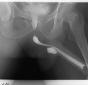
- Usually, a retrograde urethragram the urethra delineates this beautifully.

How is it done?
- This procedure is done under a general anesthetic, as decided by the anesthetist.
- The legs will be elevated into the lithotomy position.
- This procedure is done both cystoscopically and with an incision over the urethra (Bulbous Spongiosum or Perineum)
- The urethra is evaluated endoscopically and a catheter placed
- If it is a small urethral diverticulum, it can be opened into the urethra endoscopically
- If it has a narrow neck, then an external approach:
- The penis or perineum will then be incised over the urethrocele.
- A Fogarty catheter will be placed inside the diverticulum and the balloon inflated to delineate the borders of the diverticulum.
- The diverticulum will be dissected out with injuring adjacent structures.
- The neck will be tied off at the level of the adjoining urethra.
- Dissolvable closure sutures will be placed for hemostasis
- A cystoscopy confirms no injury to the urethra.
- A catheter will be placed until you are awake for some compression.
- Prophylactic antibiotics will be given to prevent infection.
Complications
Side–effects
- Any anesthetic has its risks, and the anesthetist will explain all such risks.
- Complications: hemorrhaging, and urine retention
- Catheter will be left in for 10 days
- Trial without catheter in day surgery
- If you cannot urinate after 2-3 attempts, a catheter may be inserted to empty your bladder.
- You may suffer temporary incontinence
- You may suffer permanent incontinence as advised by Jo, depending on the location of the diverticulum. Make sure you have discussed this with Jo before the surgery.
- NB! Each person is unique and for this reason, symptoms may vary!
Download Information Sheet
Wes Urethral Diverticulum Excision Male
Copyright 2019 Dr Jo Schoeman
Flexible Cystoscopy & Urethral Dilation
A day procedure under local anaesthetic, where a flexible cystoscope is placed in the bladder via the urethra. Narrowing in the urethra is dilated.
Why is it done?
A cystoscopy is used to investigate:
- Hematuria (blood in the urine)
- Recurrent urinary tract infections
- Dilatation of Urethral narrowing/ stricture
- Abnormal cells suggestive of urothelial carcinoma, on urine cytology
Ideally a retrograde urethragram is used to diagnose this radiologically
Risk factors for strictures:
- Straddle injuries
- Catheterization or urethral instrumentation
- Infections
- Bypass cardiac surgery with long ischemic time
How is it done?
- A cystoscopy is performed by placing a camera in the urethra with the help of a lubricant jelly and saline
- Usually, you can’t move past the narrowing
- Then:
Urethral Dilatation
- If you have a urethral stricture, a guidewire will be placed and the narrowing dilated
- There may be some hemorrhaging and you may need a catheter for 3 days
- This will be removed at the hospital in 3 days or alternatively arrange for your GP to remove.
- I will review in 6 –8 weeks

Antibiotics may be given to prevent infection
Complications
What to expect after the procedure?
- You may be sent home with an indwelling catheter for 3 days
- Pain on initial passing of urine after it is removed
- Bladder infection ranging from a burning sensation to, fever, to puss (rare)
- Bloodstained urine
- Lower abdominal discomfort which will persist for a few days
- NB! Each person is unique and for this reason, symptoms vary.
Download Information Sheet
Wes Flexible Cystoscopy and Urethral Dilatation IDC
Copyright 2019 Dr. Jo Schoeman
Indwelling Urethral Catheter – IDC
Non-invasive placement of a silicone tube which is secured inside the bladder and attached to a drainage bag on the outside, in order to drain an obstructed bladder (urinary retention)
Why is it done?
- This can be placed as an emergency for patients in acute urinary retention
- Prostate obstruction
- Urethral strictures
- Blood clot obstruction caused by bleeding
- Hematuria (bleeding)
- Severe urinary tract infections
- Commonly placed intra-operatively for long, non-urological surgical procedures to enable urine drainage and monitoring urine output.
- Commonly placed at the end of a Urological procedure to enable urine drainage and to enable hemostasis (stopping bleeding)
How is it done?
- This is done as a sterile procedure; therefore, the genital area will be cleaned with a non-abrasive disinfectant.
- A sterile catheter will be used
- A local anesthetic gel is placed in the urethra a few minutes prior to the placement of the catheter. This may initially sting for a few seconds until it numbs the mucosa.
- An appropriate size catheter (14-18Fr) will be inserted
- Urine should be aspirated with a syringe to confirm the correct position in the bladder.
- An anchoring balloon will be inflated with 10cc of sterile water.
- A drainage urine bag will be attached
- The catheter will be secured to your leg. (check that this is always secured)
Complications
- Urethra with resulting discomfort.
- In the presence of urethral stricture, it may be impossible to pass the catheter, and a flexible cystoscopy with dilatation of the stricture may be required prior to placement.
- If you had a large over-stretched bladder (urine retention) you may experience bleeding as the bladder empties, caused by the mucosal tears that have occurred.
- Catheters that have been placed long term, may cause irritation and possibly attract infection. Permanent catheters are usually changed every 6-8 weeks.
Download Information Sheet
Wes Catheters Indwelling Catheter
Copyright 2019 Dr Jo Schoeman