Nephrostomy Tube

What is a nephrostomy?

  • A nephrostomy tube is  a thin plastic tube inserted through the skin of your back into your kidney to drain urine when normal urine flow is blocked.
  • It is used to relieve pressure on the kidney, prevent damage, and allow urine to be collected in a bag outside the body. 

Why you might need one

  • Blocked / obstructed ureter: due to a kidney stone, tumor, or other cause
  • Injury: A hole in the ureter or bladder that causes urine to leak.
  • Preparation: To prepare for surgery or other procedures on the kidney or ureter.
  • Cancer: Cancer affecting the pelvis or treatment for cancer can block the ureter.

What to expect during the procedure

  • You will lie on your stomach on a table.
  • A local anesthetic will be injected to numb the area where the tube is inserted.
  • An interventional radiologist will use ultrasound or X-ray to guide a needle through your back into the kidney.
  • A wire is passed through the needle, and the nephrostomy tube is then threaded over the wire into the kidney.
  • The outer end of the tube is attached to a drainage bag to collect urine.
  • The procedure usually takes about 20–30 minutes.


Aftercare

  • The insertion site must be kept clean to prevent infection, with a dressing changed regularly as instructed by your doctor.
  • The tube will be secured with a stitch or dressing to prevent it from pulling.
  • You can shower with the dressing covered with plastic wrap.
  • A drainage bag will collect the urine and needs to be emptied regularly.
  • The tube may need to be flushed periodically to ensure it isn’t blocked. 

Prostate Artery Embolization – PAE

What is it?

  • Prostate artery embolization (PAE) is a minimally invasive, non-surgical procedure to treat BPH, blocking the blood supply to the prostate.
  • An interventional radiologist guides a catheter through a small incision in the wrist or groin to the prostate arteries, where tiny particles are injected to restrict blood flow, causing the gland to shrink and alleviate urinary symptoms. 
  • PAE offers a faster recovery and fewer side effects than traditional surgery.  

How it works

  • An interventional radiologist inserts a small catheter into an artery in the groin or wrist.
  • Using X-ray guidance, the catheter is maneuvered to the arteries that supply blood to the prostate.
  • Tiny particles are then injected through the catheter to block blood flow to the prostate, causing the gland to shrink. 

Benefits

  • Minimally invasive: Often performed as an outpatient or day procedure.
  • Faster recovery: Patients can often walk within hours of the procedure.
  • Relieves symptoms:
  • Fewer side effects: PAE has a lower risk of sexual dysfunction or incontinence compared to surgical options like TURP.
  • Can be repeated.

Who is it for?

  • Men with an enlarged prostate (BPH) whose symptoms are not well-controlled by medication.
  • Men who want a less invasive alternative to surgery or are unsuitable for traditional surgery.
  • Men who wish to avoid the potential risks and side effects of more invasive procedures. 

Angiomyolipoma – AML

What is it:

  • Angiomyolipoma (AML) is a benign tumor, most commonly found in the kidney.
  • Composed of fat, muscle and blood vessels.
  • It is often discovered incidentally and is usually asymptomatic,
  • Can cause pain, anemia, fever, or high blood pressure if it grows large enough.
  • Not dangerous but large or specific types, like the epithelioid variant, can carry a risk of life-threatening bleeding, which may require treatment such as surgery or embolization.  

Symptoms:

  • Most people have no symptoms.
  • If the tumor grows large, symptoms can include:
    • Pain in the flank area
    • Anemia
    • Fever
    • High blood pressure
    • Blood in the urine
    • Rarely, internal bleeding, which is a medical emergency 

Diagnosis:

  • Often found incidentally on imaging tests (like a CT scan) performed for other reasons.
  • CT scans are very effective at identifying the fat content that is characteristic of most AMLs.
  • Other imaging, such as ultrasound or MRI, can also be used.

Treatment:

  • Many small, asymptomatic AMLs may not need treatment and are simply monitored.
  • Treatment options include:
    • Embolization: Sealing off the blood vessels within the tumor to reduce the risk of bleeding. This is often suggested for tumors 4 cm or larger.
    • Surgery: To remove the tumor.
    • Medication: Drugs like everolimus can be used to block cell growth pathways, especially for large or symptomatic tumors, including those associated with tuberous sclerosis

The decision for treatment depends on the tumor’s size, symptoms, and type. 

Small renal mass ablation

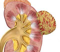
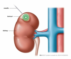
This is a minimally invasive procedure to treat a small renal mass suspicious of a renal cell carcinoma. This can be done as a day procedure with reduced effects of surgery.


Ablation techniques:

  • Microwave ablation: Commonly used here at the Wesley Hospital. Microwave energy is used to heat and destroy the tissue.
  • Radiofrequency ablation: Radiofrequency energy hits and destroy the tissue and is favored for masses smaller than 3 cm
  • Cryoablation: Utilizes a probe that creates an ice ball around the tumour freezing and destroying the cells. Often used for smaller than 4 cm masses.
  • High intensity focused ultrasound

Procedure:

  • Percutaneous technique (through the skin)
  • Done under CT guidance
  • Local anaesthetic

Indications:

  • Small renal masses less than 4 cm usually
  • Patients who are poor candidates for surgery i.e. age, multiple comorbidities, solitary kidneys and or multiple tumours

Follow-up:

  • Usually followed up with regular intervals with either CT scan or MRI scan to confirm complete destruction of the tumour.

Varicocele Embolization

How it works:

  • radiological intervention blocking off the testicular vein
  • minimally invasive procedure
  • A small puncture wound on the skin in the groin
  • An endovascular catheter that is placed through the inguinal canal into the femoral vein, with radiological imaging this coil is advanced to the testicular vein
  • A tiny coil or liquid is placed in the testicular vein to divert the blood flow.

Benefits:

  • minimally invasive
  • quicker recovery
  • success rates are comparable with surgical methods
  • reduce complications such as infection and hydrocele formation

Recovery:

  • no strenuous activity after the procedure
  • bruising can occur
  • pain relief as required