Flexible Cystoscopy & Urethral Dilation

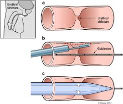
A day procedure under local anaesthetic, where a flexible cystoscope is placed in the bladder via the urethra. Narrowing in the urethra is dilated.

Why is it done?

A cystoscopy is used to investigate:

  • Hematuria (blood in the urine)
  • Recurrent urinary tract infections
  • Dilatation of Urethral narrowing/ stricture
  • Abnormal cells suggestive of urothelial carcinoma, on urine cytology

Ideally a retrograde urethragram is used to diagnose this radiologically


Risk factors for strictures:

  • Straddle injuries
  • Catheterization or urethral instrumentation
  • Infections
  • Bypass cardiac surgery with long ischemic time


How is it done?

  • A cystoscopy is performed by placing a camera in the urethra with the help of a lubricant jelly and saline
  • Usually, you can’t move past the narrowing
  • Then:

Urethral Dilatation

  • If you have a urethral stricture, a guidewire will be placed and the narrowing dilated
  • There may be some hemorrhaging and you may need a catheter for 3 days
  • This will be removed at the hospital in 3 days or alternatively arrange for your GP to remove.
  • I will review in 6 –8 weeks

Antibiotics may be given to prevent infection

Complications

What to expect after the procedure?

  • You may be sent home with an indwelling catheter for 3 days
  • Pain on initial passing of urine after it is removed
  • Bladder infection ranging from a burning sensation to, fever, to puss (rare)
  • Bloodstained urine
  • Lower abdominal discomfort which will persist for a few days
  • NB! Each person is unique and for this reason, symptoms vary.


Download Information Sheet

Wes Flexible Cystoscopy and Urethral Dilatation IDC

Copyright 2019 Dr. Jo Schoeman

Indwelling Urethral Catheter – IDC

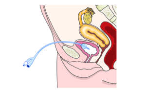
Non-invasive placement of a silicone tube which is secured inside the bladder and attached to a drainage bag on the outside, in order to drain an obstructed bladder (urinary retention)

Why is it done?

  • This can be placed as an emergency for patients in acute urinary retention
    • Prostate obstruction
    • Urethral strictures
    • Blood clot obstruction caused by bleeding
    • Hematuria (bleeding)
    • Severe urinary tract infections
  • Commonly placed intra-operatively for long, non-urological surgical procedures to enable urine drainage and monitoring urine output.
  • Commonly placed at the end of a Urological procedure to enable urine drainage and to enable hemostasis (stopping bleeding)


How is it done?

  • This is done as a sterile procedure; therefore, the genital area will be cleaned with a non-abrasive disinfectant.
  • A sterile catheter will be used
  • A local anesthetic gel is placed in the urethra a few minutes prior to the placement of the catheter. This may initially sting for a few seconds until it numbs the mucosa.
  • An appropriate size catheter (14-18Fr) will be inserted
  • Urine should be aspirated with a syringe to confirm the correct position in the bladder.
  • An anchoring balloon will be inflated with 10cc of sterile water.
  • A drainage urine bag will be attached
  • The catheter will be secured to your leg. (check that this is always secured)


Complications

  • Urethra with resulting discomfort.
  • In the presence of urethral stricture, it may be impossible to pass the catheter, and a flexible cystoscopy with dilatation of the stricture may be required prior to placement.
  • If you had a large over-stretched bladder (urine retention) you may experience bleeding as the bladder empties, caused by the mucosal tears that have occurred.
  • Catheters that have been placed long term, may cause irritation and possibly attract infection. Permanent catheters are usually changed every 6-8 weeks.


Download Information Sheet

Wes Catheters Indwelling Catheter

Copyright 2019 Dr Jo Schoeman

Meatotomy

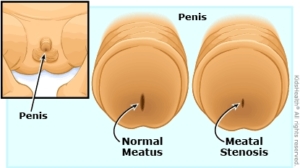
Opening of a meatal stenosis (pinhole narrowing).

Why is it done?

  • To treat a narrowing in the tip of the urethra which has formed due to previous damage/injury to the urethra.
  • Where intermittent dilatation is not desired, as discussed with the patient.
  • Causes:
    • Circumcision in early childhood where the foreskin is still attached to the glans, usually neonatal circumcision
    • After bypass surgery where a drop in blood pressure has caused an area of low blood supply to the urethra.
    • trauma to the urethra (pelvic fractures/ urethral instrumentation).
    • and sexually transmitted diseases.
  • The procedure involves surgical refashioning of your meatus.

How is it done?

  • Usually done in male patients
  • Selected Female patients can be considered.
  • Patients will receive a general anesthetic.
  • Your meatus will be refashioned, in order to leave it patent without a narrowing.
  • An indwelling catheter is left till post-operative period.
  • Prophylactic antibiotics may be given to prevent infection.

What to expect after the procedure?

  • It may be slightly uncomfortable.
  • A catheter will be inserted in the urethra and bladder. This will remain in until you are awake.
  • Catheters can be very irritating and cause some discomfort.
  • Blood stained urine will be present.
  • Painful urination may persist for a few days.
  • NB! Each person is unique and for this reason symptoms may vary!

What next?

  • Patients will be sent home after a successful attempt at voiding.
  • You may experience some discomfort with every void, that will become less over the next few voids.
  • There may be some blood in the urine. This can be remedied by drinking plenty of fluids until it clears.
  • On discharge a prescription may be issued for patients to collect.
  • Patients should schedule a follow-up appointment with Dr Schoeman within 6-8 weeks.
  • Should patients have any problems with urination, please contact the rooms for an earlier appointment.
  • Please don’t hesitate to direct any further queries to Dr Schoeman’s rooms.
  • PLEASE CONTACT THE HOSPITAL WITH ANY POST-OPERATIVE CONCERNS AND RETURN TO THE HOSPITAL IMMEDIATELY SHOULD THERE BE ANY SIGNS OF SEPSIS.

Download Information Sheet

Wes Meatotomy

One Stage Urethraplasty

Primary excision and anastomosis of a short segment urethral stricture.

Why is it done?

  • To treat urethral strictures (narrowing) caused by trauma, infection, malignancy, etc. Shorter strictures less than 2 cm in length.

How is it done?

  • This procedure is done under general anesthetic.
  • Legs are placed in a lithotomy position.
  • A single incision is made on the midline raphe on the perineum (area between scrotum and anus).
  • The corpus spongiosum identified and exposed
  • Stricture located using urethral sounds
  • The stricture is excised with a spatulated anastomosis over an indwelling catheter
  • A long-term catheter will be inserted for 10 days.
  • A dressing is then applied, which should be removed after 72 hours.
  • A local anaesthetic is injected into the wound, thus giving post-operative pain relief for the next 4-6 hours.
  • A drain may also be left for 24-48 hours to prevent the collection of serous fluids.

What to expect after the procedure?

  • Any anaesthetic has its risks and the anaesthetist will explain all such risks.
  • You will be sent home with an Indwelling catheter for 7-10 days
  • Bleeding is a common complication.
  • A haematoma (blood collection under the skin) may form and needs to be reviewed by Dr Schoeman as soon as possible. Bruising is normal.
  • An infection of the wound may occur and requires immediate attention.
  • Erectile dysfunction (15%) may occur.
  • Re-stricturing (20-30%) may occur.
  • Owing to the area of the surgery the wound should be kept clean and dry.
  • DANGER SIGNS: A scrotum that swells immediately to the size of a football, fever, or puss. Please contact Dr Schoeman or the hospital immediately as this may occur in up to 15 % of all cases.

What next?

  • The dressing should be kept dry for the initial 72 hours after surgery and then soaked in a bath until it comes off easily.
  • The dressing may sometimes adhere to the wound causing slight bleeding on removal. Don’t panic, the bleeding will stop.
  • Arrangements will be made for the removal of the catheter after 7-10 days.
  • A urinating Urethragram will be arranged with radiology within 6 weeks to determine the final result of the surgery.
  • There will be signs of bruising for at least 10 days.
  • The suture-line will be hard and indurated for at least 8-10 weeks.
  • PLEASE CONTACT THE HOSPITAL DIRECT WITH ANY POST-OPERATIVE CONCERNS AND RETURN TO THE HOSPITAL IMMEDIATELY SHOULD THERE BE ANY SIGNS OF SEPSIS.

Download Information Sheet

Wes Urethraplasty One-Stage

Optic Urethrotomy

Why is it done?

  • To treat a narrowing in the urethra which has formed due to previous damage/injury to the urethra.
  • Causes:
    • After bypass surgery where a drop in blood pressure has caused an area of low blood supply to the urethra;
    • Trauma to the urethra (pelvic fractures/ urethral instrumentation);
    • Sexually transmitted diseases.
  • The procedure entails cutting the stricture with a cold knife.

How is it done?

  • A urethroscopy is performed by placing a camera in the urethra, with the help of a lubricant jelly and an irrigate fluid, to identify the stricture.
  • A cold knife is then used to cut the stricture open.
  • The inside of the bladder is viewed for pathology.
  • If any suspicious lesions are seen, a biopsy will be taken.
  • Prophylactic antibiotics may be given to prevent infection.
  • Indwelling catheter placed.


Complications

Side–effects

  • Patients will spend the night in the hospital.
  • Patients will be sent home with a catheter for 3 days after receiving thorough catheter care Instructions.
  • Arrangements will be made to remove the catheter on day 3.
  • There may be some blood in the urine. This can be remedied by drinking plenty of fluids until it clears.
  • Review at 6 weeks.
  • There is a >50% risk of recurrence and may need further treatment
    • Re-dilatation
    • Self-dilatation
    • Urethraplasty


Download Information Sheet

Wes Optic Urethrotomy

Copyright 2019 Dr Jo Schoeman

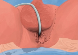
Perineostomy – Perineal Urostomy

Opening of the posterior Urethra on the Perineum

Why is it done?

  • Seldom done
  • This procedure is performed when concentric extensive scarring in the urethra (strictures) causes Urinary Retention
  • A long history of strictures.
  • This is alternative to an invasive procedure where long periods of anesthetic are contra-indicated and extensive grafts may be required
  • Usually for chronically sick patients who cannot undergo surgery yet are active enough not to want a permanent catheter.
  • Patients who don’t want to / cannot do intermittent self-dilatation of these strictures
  • Don’t want a permanent Indwelling Catheter penile/Urethral cripple


How is it done?

  • Patients will receive a General Anesthetic.
  • Flexible cystoscopy is done through your urethra or suprapubic catheter site to find normal urethra (usually posterior urethra)
  • A urethral sound is placed/ catheter
  • A perineal incision is made.
  • The urethra is divided proximal (above) the stricture
  • The opening of the urethra is brought out and attached to the skin on the perineum (the area between scrotum and anus)
  • A catheter is placed
  • Prophylactic antibiotics will be given to prevent any infections.


Complications

  • Persistent pain in penile shaft
  • Pain in Perineum when seated
  • Scarring of the opening requiring dilatation
  • Possible infection
  • NB! Each person is unique and for this reason, symptoms vary!


Download Information Sheet

Wes Perineostomy

Copyright 2019 Dr Jo Schoeman

Supra-Pubic Catheter

Invasive placement of a silicone tube in a percutaneous supra-pubic puncture site. This is secured inside the bladder (with a balloon) and attached to a drainage bag on the outside, in order to drain an obstructed bladder.

Why is it done?

  • This can be placed as an emergency for patients in acute urinary retention
  • Patients requiring long term catheterization especially spinal cord injury patients
  • Failed urethral catheterization
  • Severe prostate obstruction
  • Urethral strictures
  • Severe sepsis of the urogenital area where diverting urine away from the area is advisable
  • Urethral catheterization impossible


How is it done?

  • Usually done under general anesthesia.
  • This is done as a sterile procedure; therefore, the genital area and suprapubic area will be cleaned with a non-abrasive dis-infectant.
  • A flexible cystoscopy will be placed to inspect the bladder, allow filling with saline and visualize the puncture with a cannula from the skin (outside)
  • A 1cm incision is then made in the midline of the lower abdomen, approximately 2cm above the pubic bone
  • An appropriate size catheter (14-16Fr) will be inserted using a trocar method
  • Correct placement is confirmed with the cystoscopy (direct vision)
  • An anchoring balloon will be inflated with 10cc of sterile water.
  • A drainage urine bag will be attached
  • The catheter will be secured to your leg. (check that this is always secured)


Complications

  • Side effects from a general anesthetic.
  • Bleeding from the wound site. (Anti-coagulants should have been ceased a week prior)
  • Depending on the size of your bladder a possible bowel injury could occur, the odds of this happing will be discussed with you prior to your procedure.


Download Information Sheet

Wes Catheters Suprapubic Catheter CHANGE

Copyright 2019 Dr Jo Schoeman

Urethral Dilatation

Dilatation of urethral stricture. Dine in combination with urethroscopy and placement of IDC is advocated on daily basis at home as management of urethral stricture.

Why is it done?

  • To treat a narrowing in the urethra which has formed due to previous damage/injury to the urethra.
  • Causes: after bypass surgery where a drop in blood pressure has caused an area of low blood supply to the urethra; trauma to the urethra (pelvic fractures/ urethral instrumentation); and sexually transmitted diseases.
  • The procedure placing a dilator un your urethra on a daily basis.
  • It aids in keeping your urethra open and prevents eventual kidney damage / failure.

How is it done?

  • Patients will receive a local anesthetic.
  • A urethroscopy is performed by placing a flexible camera in the urethra, with the help of a lubricant jelly and an irrigate (fluid), to identify the stricture.
  • A guidewire is slipped through the opening of the narrowing.
  • The camera is removed.
  • The stricture is dilated using a graduated S dilators over the guide wire.
  • Prophylactic antibiotics may be given to prevent infection.

What to expect after the procedure?

  • It may be slightly uncomfortable, please don’t hesitate to tell jo and request sedation if required.
  • A catheter will be inserted in the urethra and bladder. This will remain in the bladder for 3 days.
  • Catheters can be very irritating and cause some discomfort.
  • Blood stained urine will be present.
  • Lower abdominal discomfort will persist for a few days.
  • NB! Each person is unique and for this reason symptoms may vary!


What next?

  • Patients will be sent home with a catheter for 3 days after receiving thorough catheter care instructions.
  • Arrangements will be made to remove the catheter on day 3.
  • There may be some blood in the urine. This can be remedied by drinking plenty of fluids until it clears.
  • A referral will be done to BlueCare Nurses, who will assess you to instruct you on daily self-dilatation.
  • PLEASE CONTACT THE HOSPITAL WITH ANY POST-OPERATIVE CONCERNS AND RETURN TO THE HOSPITAL IMMEDIATELY SHOULD THERE BE ANY SIGNS OF SEPSIS.

Download Information Sheet

Wes Urethroscopy & Urethral Dilatation

Urethrectomy

Removal of urethra. Usually in adjunct to a radical cystectomy. Occasionally done some time after a cystectomy where recurrences occur in urethra.


Why is it done?

  • As part of the treatment for aggressive localized Urothelial carcinoma of the bladder.
  • T1G3, > T2 disease.
  • Primary Urethral disease with:
    • Urothelial Carcinoma.
    • Squamous cell carcinoma.
    • Secondary metastatic disease to Urethra: Melanoma, Lung cancer, Breast cancer (all these are rare).
  • Advanced disease may involve a penectomy.
  • The procedure is part of the radical cystectomy, radical cysto-prostatectomy or could be and adjunct later on when recurrences are found with surveillance.
  • Part of a necrotic inflammatory condition ie: Fourniers Gangrene.

How is it done?

  • This procedure is done under general anesthetic.
  • Legs are placed in a lithotomy position.
  • A single incision is made on the midline raphe on the perineum (area between scrotum and anus). Sutures will be dissolvable.
  • The Corpora Spongiosum with the urethra inside it is mobilized off the Corpora Cavernosa.
  • Urethral meatus is removed distal and glans is closed.
  • Proximally the urethra is taken up to the sphincter and freed.
  • You will have already had a diversion of the urine with an ileostomy, or this will be done with your cystectomy part of the operation.
  • A drain will be left for 24-48 hours to prevent the collection of serous fluids.
  • A dressing is then applied, which should be removed after 72 hours.

What to expect after the procedure?

  • Any anesthetic has its risks, and the anesthetist will explain all such risks.
  • If this is the only operation you have undergone, you will be sent home as soon as the drain is removed on D2 or D3.
  • Swelling is a common complication.
  • A hematoma (blood collection under the skin) may form and needs to be reviewed by Dr Schoeman as soon as possible. Bruising is normal.
  • An infection of the wound may occur and requires immediate attention.
  • Owing to the area of the surgery the wound should be kept clean and dry.
  • DANGER SIGNS: A wound that swells immediately, fever, or puss. Please contact Dr Schoeman or the hospital immediately as this may occur in up to 15 % of all cases.

What next?

  • The dressing should be kept dry for the initial 72 hours after surgery and then soaked in a bath until it comes off easily.
  • The dressing may sometimes adhere to the wound causing slight bleeding on removal. Don’t panic, the bleeding will stop.
  • On discharge a prescription may be issued for patients to collect.
  • There will be signs of bruising for at least 10 days.
  • The suture-line will be hard and indurated for at least 8-10 weeks.
  • PLEASE CONTACT THE HOSPITAL DIRECTLY WITH ANY POST-OPERATIVE CONCERNS AND RETURN TO THE HOSPITAL IMMEDIATELY SHOULD THERE BE ANY SIGNS OF SEPSIS.

Download Information Sheet

Wes Urethrectomy

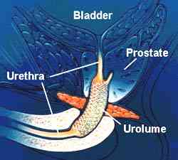
UROLUME / Memocath Urethral Stent

Minimal invasive management for the relief of lower urinary tract symptoms (LUTS) or urinary retention caused by a urethral stricture

Why is it done?

  • This procedure is performed when concentric scarring in the urethra causes LUTS and /or Urinary Retention
  • Symptoms include: a weak stream, nightly urination, frequent urination, inability to urinate, (LUTS) and Urinary Retention
  • This is alternative to an invasive procedure where long periods of anesthetic are contraindicated.
  • Usually for chronically sick patients who cannot undergo surgery yet are active enough not to want a permanent catheter.
  • Patients who don’t want to / cannot do intermittent self-dilatation of these strictures
  • Don’t want a permanent Indwelling Catheter

How is it done?

  • Patients will receive sedation with local anesthetic gel placed in the urethra.
  • A cystoscopy is performed by placing a camera in the urethra with the help of a lubricant jelly and an irrigate (fluid).
  • The measurements of the urethral are taken (length)
  • Appropriate length coil is chosen.
  • The device is placed through the cystoscopy sheath, to sit snug in the prostate urethra stretching over the length of the stricture
  • Prophylactic antibiotics will be given to prevent any infections.


Complications

Side–effects

  • Persistent pain in penile shaft
  • Pain in Perineum when seated
  • Migration of the device
  • Erosion of device
  • Possible infection
  • Long term yields the risk for encrustation and recurrent infections
  • NB! Each person is unique and for this reason, symptoms vary!


Download Information Sheet

Wes UROLUME Urethral Stent

Copyright 2019 Dr Jo Schoeman