Penile Fracture: Presentation, Investigation, Treatment and Long-Term Outlook

Penile fracture is a urological emergency. Despite its name, no bone is broken. The injury is a tear in the tunica albuginea, the tough sleeve surrounding one or both erectile cylinders (corpora cavernosa) while the penis is erect. Bleeding then occurs within and around the erectile tissues.

If you hear or feel a crack or pop during intercourse or other bending of an erect penis, followed by pain, loss of the erection, swelling or bruising, stop sexual activity and attend the nearest emergency department immediately. Do not wait to see whether it settles, and do not eat or drink until assessed in case an anaesthetic is required.

This article provides general education. It cannot diagnose an injury, replace examination, or provide individual medical advice.

How does a penile fracture happen?

During an erection, the tunica albuginea becomes much thinner and is vulnerable to sudden buckling. The most frequent mechanism is an erect penis slipping out during intercourse and striking the partner’s pubic bone or perineum. It can also occur during masturbation, intentional forceful bending, rolling onto an erect penis, or less commonly through other trauma.

Penile fracture can happen in any sexual position. It is an accident rather than evidence of wrongdoing, and embarrassment should never delay treatment.

The urethra, the tube carrying urine through the penis, may be injured at the same time. The 2026 European Association of Urology (EAU) guideline reports associated corpus spongiosum or urethral injury in approximately 10–22% of cases; rates vary among populations and mechanisms of injury.[1]

Typical presentation

The classic sequence is:

  • a sudden crack, snap or popping sensation;
  • immediate pain;
  • rapid loss of the erection (detumescence);
  • quickly developing swelling and bruising;
  • bending or deformity of the penis, often away from the injured side; and
  • sometimes a palpable defect in the tunica.

Bruising may spread into the scrotum, perineum or lower abdominal wall. The dramatic “aubergine” appearance described in medical literature is not present in every case.

Warning signs of urethral injury

Tell the treating team immediately if there is:

  • blood at the urinary opening;
  • blood in the urine;
  • pain or difficulty passing urine;
  • inability to pass urine; or
  • a weak or interrupted urinary stream after the injury.

The absence of these findings does not completely exclude urethral injury. Clinical assessment remains important.

Conditions that can look similar

Not every swollen or bruised penis after intercourse has a tunical tear. Rupture of a superficial vein or artery, injury to the suspensory ligament, or bleeding beneath the skin can produce a “false penile fracture.” Slow rather than immediate loss of erection and absence of the characteristic crack may make a true fracture less likely, but no single feature is conclusive.

Because missing a fracture can have lasting consequences, significant swelling, pain or deformity after trauma to an erect penis requires urgent medical assessment.

How is it investigated?

History and examination

When the history and examination are classic, penile fracture is primarily a clinical diagnosis. Testing should not unnecessarily delay repair. The clinician will assess the penis, scrotum and perineum and ask specifically about the mechanism, the speed of detumescence, urination and visible blood. A urine test is generally performed.

Ultrasound

Ultrasound can help locate a tear and haematoma, particularly when the diagnosis is uncertain. It is quick and widely available, but its accuracy depends on the operator, the size and position of the tear, and the amount of swelling. A negative or inconclusive ultrasound does not necessarily exclude fracture when the clinical findings are convincing.

MRI

MRI gives excellent soft-tissue detail and is generally more accurate than ultrasound for detecting a tunical tear. It may be useful in equivocal cases, but availability, cost and delay can limit its emergency use. It is not routinely required when the diagnosis is already clear.[1]

Assessment of the urethra

If urethral injury is suspected, evaluation may include retrograde urethrography (contrast imaging of the urethra) or flexible cystoscopy. Visible haematuria, blood at the meatus, difficulty voiding and bilateral corporal injury increase concern. The EAU and American Urological Association (AUA) both recommend evaluating for associated urethral injury when indicated.[1,2]

CT is not the preferred test for an isolated penile fracture, although it may be obtained when wider pelvic or abdominal trauma is suspected.

Recommended management

Prompt surgical repair

Current EAU and AUA guidance recommends prompt surgical exploration and repair for an acute penile fracture.[1,2] The operation is usually performed under general or regional anaesthesia and involves:

  1. exposing the injured erectile tissue through a circumferential degloving incision or a targeted incision over the tear;
  2. evacuating the clot and controlling bleeding;
  3. identifying and closing the tunica albuginea tear with sutures; and
  4. inspecting and repairing the urethra if it is injured.

A urinary catheter may be used, especially when the urethra has been assessed or repaired. The exact incision, catheter duration, hospital stay and follow-up depend on the injury and the surgeon’s findings.

The EAU guideline advises repair within 24 hours of presentation when feasible, but late presentation is not a reason to withhold assessment or repair.[1] A person presenting after a delay should still seek urgent urological review.

Why is conservative treatment generally avoided?

Ice, compression, pain relief, erection-suppressing medication and observation were historically used. Conservative treatment may occasionally be appropriate when imaging and specialist assessment confirm that the tunica is intact, but it is not the standard treatment for a confirmed fracture.

A meta-analysis of 58 studies involving 3,213 patients found significantly fewer overall complications and less erectile dysfunction, curvature and painful erections with surgery than with conservative management.[3] Non-operative treatment of a true fracture carries greater risks of persistent haematoma, infection or abscess, missed urethral injury, fibrosis, penile curvature and erectile dysfunction.[1,3]

Recovery after repair

Patients should follow their treating surgeon’s instructions, which may include:

  • wound and catheter care;
  • simple analgesia and other prescribed medicines;
  • avoiding strenuous activity until reviewed;
  • avoiding intercourse and masturbation, commonly for about six weeks or until healing is confirmed; and
  • follow-up to assess erections, curvature, pain and urinary function.

Night-time or spontaneous erections during healing may be uncomfortable. Medication is sometimes used selectively, but there is no universal post-operative drug regimen. Do not start or stop prescription medicines without advice.

Seek urgent review after treatment for fever, increasing redness or swelling, wound discharge, worsening pain, inability to urinate, heavy bleeding, or a catheter that stops draining.

Possible complications

Most patients do well after prompt repair, but no treatment can guarantee a complication-free outcome. Possible early or late problems include:

  • wound infection, bleeding or haematoma;
  • altered penile sensation, palpable stitches, scar or nodules;
  • painful erections;
  • penile curvature, indentation or shortening;
  • erectile dysfunction;
  • urethral narrowing (stricture), urinary spraying or reduced flow;
  • urethrocutaneous fistula after a significant urethral injury or repair;
  • need for further investigation or surgery; and
  • anxiety, avoidance of intimacy or relationship distress.

The EAU guideline summarises reported post-surgical rates of plaques or nodules, curvature and erectile dysfunction as approximately 13.9%, 2.8% and 1.9%, respectively, while noting that complications overall have been reported in up to 20% of cases.[1] These pooled figures are not a personalised prediction: published studies differ in injury severity, definitions, follow-up and measurement. Risk is higher with extensive or bilateral tears, associated urethral injury, delayed or missed diagnosis, and in some studies age over 50.[1,4]

What is the risk of erectile dysfunction?

Erectile dysfunction after a fracture may result from corporal scarring, impaired blood trapping, arterial or nerve injury, pain, anxiety, or a combination of physical and psychological factors. Prompt repair markedly reduces the risk compared with conservative management, but does not eliminate it.[1,3]

Persistent difficulty should be assessed rather than endured in silence. Evaluation can include a sexual and medical history, validated questionnaires, examination and, when indicated, penile Doppler ultrasound. Treatment is individualised and may include counselling, oral erectile-dysfunction medication when safe, vacuum therapy, injections or, rarely, reconstructive surgery or a penile prosthesis.

Does penile fracture cause Peyronie’s disease?

Healing after a penile fracture can produce a local scar, palpable nodule or post-traumatic curvature. These findings may resemble Peyronie’s disease, which is an acquired fibrotic disorder of the tunica albuginea. However, a curve after fracture should not automatically be labelled Peyronie’s disease: it may arise directly from the repaired tear, asymmetric healing or corporal fibrosis.

The distinction is made through history, examination and, if needed, photographs of the erect penis or ultrasound. Review is advisable if there is a new or worsening bend, a hard plaque, painful erections, loss of length, narrowing, instability or difficulty with intercourse. Management depends on whether the deformity is changing or stable, its severity, erectile function and how much it affects the patient. Options may include observation, penile traction in selected cases, treatment of erectile dysfunction, or reconstructive surgery once the deformity is stable. Treatment should be discussed with a urologist experienced in penile reconstruction or andrology.

The essential message

A suspected penile fracture is time-sensitive. A crack or pop followed by immediate loss of erection, swelling and bruising warrants immediate emergency assessment. Diagnosis is often clinical; ultrasound or MRI is reserved mainly for uncertainty, and the urethra must be assessed when injury is suspected. Prompt surgical repair offers the best prospect of preserving penile shape, erections and urinary function.


References

  1. European Association of Urology. EAU Guidelines on Urological Trauma. 2026 edition, sections on genital trauma and penile fracture. https://uroweb.org/guidelines/urological-trauma/chapter/urogenital-trauma-guidelines
  2. Morey AF, Brandes S, Dugi DD III, et al. Urotrauma: AUA Guideline. J Urol. 2014;192(2):327–335; amended guideline statements available from the American Urological Association. doi:10.1016/j.juro.2014.05.004. https://www.auanet.org/guidelines-and-quality/guidelines/urotrauma-guideline
  3. Amer T, Wilson R, Chlosta P, et al. Penile fracture: a meta-analysis. Urol Int. 2016;96(3):315–329. doi:10.1159/000444884.
  4. Barros R, Schulze L, Ornellas AA, Koifman L, Favorito LA. Impact of surgical treatment of penile fracture on sexual function. Urology. 2019;126:128–133. doi:10.1016/j.urology.2018.11.027.
  5. Wong NC, Dason S, Bansal RK, Davies TO, Braga LH. Can it wait? A systematic review of immediate vs delayed surgical repair of penile fractures. Can Urol Assoc J. 2017;11(1–2):53–60. doi:10.5489/cuaj.4032.
  6. Koifman L, Barros R, Júnior RAS, Cavalcanti AG, Favorito LA. Penile fracture: diagnosis, treatment and outcomes of 150 patients. Urology. 2010;76(6):1488–1492. doi:10.1016/j.urology.2010.05.043.

Website publication note

This material is general health information and was prepared from the sources listed above. It is not a substitute for emergency assessment, diagnosis, informed consent or personalised advice from a qualified practitioner. Outcomes and risks vary between individuals.

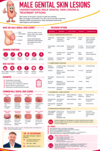
Balanitis Xerotica Obliterans and Lichen Sclerosus of the Foreskin

Balanitis xerotica obliterans—usually abbreviated to BXO, is the traditional urological name for male genital lichen sclerosus.

Lichen sclerosus is a chronic inflammatory skin condition that most commonly affects the foreskin and head of the penis. It can cause whitening, inflammation, splitting and progressive scarring of the foreskin. In some men it also narrows the urinary opening or extends into the urethra.

BXO is not simply an infection or ordinary balanitis. Early recognition and treatment can relieve symptoms and reduce permanent scarring. Long-term observation is also important because genital lichen sclerosus is associated with a small but genuine risk of penile squamous cell carcinoma.

Is BXO different from lichen sclerosus?

The names are often used interchangeably:

  • Lichen sclerosus is the preferred modern medical term.
  • Balanitis xerotica obliterans describes lichen sclerosus affecting the glans penis and foreskin, particularly when scarring has developed.

The condition may involve:

  • The inner and outer foreskin
  • The glans penis
  • The frenulum
  • The urinary opening—the meatus
  • The fossa navicularis and penile urethra

The disease is usually confined to the genital region in men, although lichen sclerosus can occasionally affect skin elsewhere.

What causes lichen sclerosus?

The exact cause remains uncertain. It is probably produced by a combination of inflammation, immune-system dysfunction, genetic susceptibility, skin injury and local environmental factors.

Possible associations include:

  • Autoimmune or inflammatory activity
  • Chronic exposure of susceptible skin to trapped urine
  • Repeated friction or minor trauma
  • A tight foreskin
  • Previous inflammation beneath the foreskin
  • Obesity and a “buried” penis
  • Diabetes or metabolic disease
  • Cigarette smoking
  • Genetic susceptibility

The moist, occluded environment beneath the foreskin may contribute to continuing inflammation in susceptible men.

Lichen sclerosus is:

  • Not caused by poor personal character or behaviour
  • Not usually a sexually transmitted infection
  • Not contagious
  • Not something that can be passed to a partner through ordinary sexual contact

Yeast or bacterial infection may occur at the same time, particularly in men with diabetes, but infection is not the underlying cause of lichen sclerosus.

Who can develop it?

Male genital lichen sclerosus can occur at any age.

It is recognised in:

  • Boys with acquired or severe scarring phimosis
  • Adolescents
  • Younger and middle-aged men
  • Older men
  • Men with a previously retractable foreskin that has gradually tightened

It is much less common in men who were circumcised early in life, although circumcision does not make the risk absolutely zero.

How does lichen sclerosus present?

The appearance varies considerably. Early disease can be subtle and may initially be mistaken for thrush, dermatitis, recurrent balanitis or ordinary phimosis.

Possible signs include:

  • White, pale or porcelain-coloured foreskin
  • A white, firm ring around the foreskin opening
  • Thickened or hardened skin
  • Shiny, crinkled or “cigarette-paper” skin
  • Red or inflamed areas
  • Small cracks or splits
  • Easy bleeding or bruising
  • Painful erosions
  • Adhesions between the foreskin and glans
  • Progressive tightening of the foreskin
  • Shortening or scarring of the frenulum
  • Whitening around the urinary opening
  • Narrowing of the meatus
  • Loss of the normal shape or definition of the glans and foreskin

Some men have significant visible disease with surprisingly little discomfort.

Symptoms patients may notice

Symptoms can include:

  • Difficulty retracting the foreskin
  • Inability to replace or retract the foreskin fully
  • Pain during erections
  • Splitting or bleeding during intercourse
  • Itching, burning or soreness
  • Recurrent “balanitis”
  • Pain or reduced enjoyment during sexual activity
  • Spraying or deflection of the urinary stream
  • A weak or narrowed stream
  • Straining to urinate
  • Dribbling after urination
  • Ballooning of the foreskin
  • Recurrent urinary infections
  • Difficulty emptying the bladder

A narrow urinary opening may be the first sign that the disease has extended beyond the foreskin.

Lichen sclerosus and phimosis

Phimosis means that the foreskin cannot be retracted comfortably behind the glans.

A non-retractile foreskin is normal in young boys and does not automatically indicate disease. However, a previously retractable foreskin that becomes tight, particularly when accompanied by a white scarred ring is suspicious for lichen sclerosus.

Forceful stretching of a scarred foreskin is not recommended. Repeated tearing may cause additional inflammation and scarring.

Paraphimosis, where a retracted tight foreskin becomes trapped behind the glans, requires urgent medical attention.

How is lichen sclerosus diagnosed?

The diagnosis is often made from the history and characteristic appearance.

Assessment should include examination of:

  • The complete foreskin
  • Glans penis
  • Frenulum
  • Urinary opening
  • Palpable tissue beneath abnormal skin
  • Urinary stream and bladder emptying when relevant
  • Groin lymph nodes if cancer is suspected

Other conditions that can resemble lichen sclerosus include:

  • Fungal or bacterial balanitis
  • Irritant or allergic dermatitis
  • Psoriasis
  • Lichen planus
  • Zoon balanitis
  • Vitiligo
  • Genital warts
  • Penile intraepithelial neoplasia: PeIN
  • Squamous cell carcinoma

Diabetes testing may be appropriate in men with recurrent inflammation or other risk factors.

Is a biopsy always necessary?

Not every typical case requires a biopsy before treatment. However, biopsy is important when:

  • The diagnosis is uncertain
  • Treatment has not produced the expected response
  • An erosion or ulcer does not heal
  • There is a persistent red or thickened area
  • A lump or hardened area can be felt
  • The skin bleeds easily without an obvious cause
  • Pigmentation changes unexpectedly
  • Penile intraepithelial neoplasia or cancer is suspected

When circumcision is performed for suspected lichen sclerosus, the foreskin should usually be sent for pathological examination.

A biopsy should be taken from any suspicious persistent lesion rather than relying only on the appearance.

Medical management

The goals of medical treatment are to:

  • Suppress inflammation
  • Relieve discomfort and itching
  • Prevent further scarring
  • Preserve foreskin and urinary function where possible
  • Identify patients who require surgery or biopsy

General skin care

Helpful measures include:

  • Washing gently with water or a non-soap cleanser
  • Avoiding perfumed soaps, antiseptics and harsh products
  • Drying the glans and foreskin gently after urination
  • Applying a bland barrier ointment or emollient
  • Avoiding forceful foreskin retraction
  • Using adequate lubrication during sexual activity
  • Stopping smoking
  • Managing diabetes carefully
  • Addressing obesity or a buried penis where relevant

Good hygiene means gentle care. Repeated scrubbing or strong antiseptics can worsen inflamed genital skin.

Potent topical corticosteroids

A prescribed potent or ultrapotent topical corticosteroid is the usual first-line medical treatment.

Clobetasol propionate 0.05% ointment is commonly used in specialist practice. A typical initial course may involve a small amount applied to the affected skin once daily for one to three months, but the exact regimen should be determined by the treating doctor.

Patients should be shown:

  • Exactly where to apply the medication
  • How much to use
  • How long to continue
  • Whether treatment should be reduced gradually
  • When follow-up is required

When used correctly for a defined course, topical corticosteroids can reduce inflammation, soreness and early tightening. They cannot always reverse established dense scar tissue.

Possible steroid-related effects include irritation, secondary infection, skin thinning or visible small blood vessels, although these are uncommon with appropriate genital use and medical supervision.

Other topical treatments

Topical calcineurin inhibitors such as tacrolimus or pimecrolimus may occasionally be considered by a dermatologist or experienced specialist when corticosteroids are unsuitable.

They are not generally preferred over potent topical corticosteroids as initial treatment. They can cause burning or irritation, and persistent suspicious lesions must be biopsied rather than repeatedly treated empirically.

Antifungal or antibiotic medication is only useful when a secondary infection is actually present. It does not treat the underlying lichen sclerosus.

When should circumcision be considered?

Circumcision is an important and often definitive treatment for foreskin lichen sclerosus.

It should be considered when there is:

  • Established scarring phimosis
  • Painful or recurrent splitting
  • Difficulty with hygiene
  • Recurrent balanitis
  • Paraphimosis
  • Failure to respond adequately to a prescribed steroid course
  • Rapid recurrence after apparently successful medical treatment
  • Significant functional or sexual difficulty
  • Concern about premalignant or malignant change
  • A need to obtain tissue for diagnosis

Complete circumcision removes the diseased foreskin and eliminates the moist, urine-exposed environment beneath it. Many men experience long-term resolution when the disease is limited to the foreskin.

Partial circumcision or simply making a slit in the scarred foreskin is generally less reliable because affected skin remains and may scar again.

Will circumcision cure lichen sclerosus?

Circumcision is often highly effective when the condition is limited to the foreskin. However, it is not an absolute guarantee of cure.

Disease may persist or recur on the:

  • Glans
  • Coronal sulcus
  • Frenulum
  • Urinary opening
  • Urethra

Ongoing symptoms or abnormal skin after circumcision should therefore be reviewed rather than assumed to be normal postoperative scarring.

Circumcision appears to reduce the risk associated with chronic inflammation and phimosis, but it does not completely eliminate the future risk of penile cancer.

Surgery for meatal narrowing

Lichen sclerosus can scar the urinary opening, producing meatal stenosis.

Symptoms may include:

  • A thin urinary stream
  • Spraying or deflection
  • Prolonged urination
  • Straining
  • Discomfort
  • Incomplete emptying

Treatment may require a meatotomy or meatoplasty, in which the opening is enlarged and reconstructed.

Simple dilatation may provide temporary improvement but often does not control active scar disease. Medical treatment of the surrounding lichen sclerosus and continued surveillance may still be required.

What if the disease involves the urethra?

In some men, scarring extends from the meatus into the penile urethra and occasionally further towards the bulbar urethra.

Assessment may include:

  • Urinary flow-rate testing
  • Post-void residual ultrasound
  • Flexible cystoscopy
  • Retrograde urethrogram
  • Voiding cystourethrogram
  • Urethral ultrasound in selected cases

Short strictures may sometimes be managed endoscopically, but repeated dilatation or urethrotomy has a significant recurrence risk when active lichen sclerosus remains.

Longer or recurrent strictures may require urethroplasty. Oral lining tissue, usually buccal mucosa from the inside of the cheek, is commonly used for reconstruction.

The European Association of Urology recommends that genital skin should not be used as a graft for lichen-sclerosus-related urethral strictures, because the disease can affect the graft and recurrence rates are poor.

Complex cases are best managed by a urologist experienced in reconstructive urethral surgery.

Other surgical options

Depending on the extent of disease, specialist procedures may include:

  • Frenuloplasty in carefully selected cases without extensive LS
  • Complete circumcision
  • Meatotomy or meatoplasty
  • Excision or biopsy of suspicious lesions
  • Glans resurfacing when premalignant disease is present
  • Oral-mucosa graft urethroplasty
  • Staged urethral reconstruction
  • Perineal urethrostomy for severe or recurrent extensive stricture disease

The operation should be tailored to the location and activity of the condition. Preserving diseased genital skin simply to avoid circumcision may produce repeated scarring and further procedures.

Lichen sclerosus and penile cancer

Male genital lichen sclerosus is associated with squamous cell carcinoma of the penis and with penile intraepithelial neoplasia, which is a precancerous change.

Published studies have reported widely differing cancer rates among men with genital lichen sclerosus, from close to zero in some groups to approximately 12.5% in highly selected specialist series. Frequently quoted estimates place the risk at roughly 2–6%, but the precise lifetime risk for an individual man remains uncertain.

This does not mean that most men with lichen sclerosus will develop penile cancer. Penile cancer remains rare, and appropriate treatment of inflammation and phimosis is expected to reduce risk.

It is also reported that lichen sclerosus is found in a substantial proportion of penile cancer specimens. This association cannot be reversed to mean that the same proportion of men with lichen sclerosus will develop cancer.

Risk is likely to be greater when there is:

  • Long-standing uncontrolled inflammation
  • Severe phimosis
  • Persistent ulceration or thickening
  • Poor visibility of the glans
  • Cigarette smoking
  • Immune suppression
  • Coexisting penile intraepithelial neoplasia
  • Failure to attend follow-up

Warning signs that require prompt review

A man with current or previous lichen sclerosus should seek medical assessment for:

  • A persistent ulcer or sore
  • A new lump or firm area
  • Skin thickening
  • An irregular red or velvety patch
  • A warty or cauliflower-like growth
  • Unexplained bleeding
  • Offensive discharge
  • Increasing pain
  • A lesion that does not respond to prescribed steroid treatment
  • A new lump in the groin

These findings do not necessarily mean cancer, but they should not be treated repeatedly as thrush or simple inflammation without further investigation.

Follow-up and self-examination

Lichen sclerosus can recur, and cancer-related changes may develop many years after initial treatment.

Patients should become familiar with the normal appearance of their penis and perform regular self-examination. When possible, the entire glans, foreskin and urinary opening should be inspected.

Medical follow-up should be tailored to disease severity. More frequent review is appropriate during active treatment, after surgery or when urethral involvement is present. Once stable, periodic or annual review may be appropriate, together with lifelong self-monitoring.

Circumcised men should still report new or persistent abnormalities.

The outlook

When recognised early, male genital lichen sclerosus can often be controlled successfully with appropriate topical corticosteroid treatment, good skin care and follow-up.

Established scarring is less likely to resolve with medication alone. Circumcision is usually highly effective for disease confined to the foreskin, while meatal and urethral disease may require more specialised reconstruction.

The important message is not to ignore a foreskin that is becoming progressively white, scarred or tight. Early assessment may prevent painful erections, urinary obstruction and more complex surgery.

The bottom line

Balanitis xerotica obliterans is the older name for male genital lichen sclerosus involving the foreskin and glans.

It commonly presents with white or scarred skin, acquired phimosis, splitting, painful erections or urinary-stream changes. Potent prescribed topical corticosteroids are the first medical treatment for suitable early disease, while circumcision is often required for established phimosis, recurrent disease or failure of medical treatment.

Meatal or urethral involvement requires urological assessment and may need reconstructive surgery.

The risk of penile squamous cell carcinoma is small but real. Persistent thickening, ulceration, bleeding, a lump or a lesion that does not respond to treatment requires prompt examination and often biopsy.

This article provides general information and does not replace personalised medical assessment. Do not use potent topical corticosteroids on genital skin without appropriate medical advice.

So guys, if you cannot retract the foreskin and has the typical appearance discussed in this article, take the time to review with your GP to have this checked out. There is effective management for this which will also then reduce your risk for penile cancer. Do not vary your head in the send and ignore this, have it seen to and let your GP refer you to your local Brisbane urologist, Dr Jo Schoeman.

References and further reading

Peyronie’s Disease: When a Curved Erection Becomes a Medical Problem

Peyronie’s disease is a condition in which scar tissue develops within the penis, causing it to bend or curve during an erection.

A slight natural curve can be completely normal. Peyronie’s disease is different because the change is usually new, may worsen over time, and can sometimes cause pain or make sexual intercourse difficult.

What causes Peyronie’s disease?

The exact cause is not always clear.

In many cases, the condition may develop after repeated minor injury or trauma to the penis. This can lead to the formation of fibrous scar tissue, known as a plaque.

The plaque does not stretch normally during an erection, causing the penis to bend toward the affected area.

Some men do not remember any specific injury.

Common symptoms

Symptoms of Peyronie’s disease can include:

  • A new bend or curve in the penis
  • A hard area or plaque beneath the skin
  • Pain during erections
  • Shortening of the penis
  • Narrowing or indentation of the shaft
  • Difficulty with sexual intercourse
  • Erectile dysfunction

The severity can vary considerably between patients.

Does Peyronie’s disease get worse?

Peyronie’s disease commonly develops in phases.

During the early or active phase, the curvature may continue to change and erections may be painful.

After a period of time, the condition often becomes more stable.

In some men the pain settles, while the curvature remains.

When should you see a urologist?

A new or worsening penile curvature should be assessed, particularly when it causes pain, affects erections or interferes with sexual activity.

A urologist can assess the degree of curvature, check for plaque formation and determine whether treatment is necessary.

Not every patient requires surgery.

Treatment options

Treatment depends on the severity of the condition, how long it has been present and whether it is affecting sexual function.

Options may include:

  • Observation
  • Medication in selected cases
  • Penile traction therapy
  • Injection treatments
  • Surgical correction

Surgery is generally considered when the condition is stable and the curvature significantly affects sexual intercourse.

Peyronie’s disease and erectile dysfunction

Some men with Peyronie’s disease also develop erectile dysfunction.

This may occur because of changes in penile structure, blood flow or anxiety related to pain and curvature.

Assessment is therefore important so that both problems can be considered together.

Specialist assessment in Brisbane

Peyronie’s disease can be uncomfortable to discuss, but it is a recognised medical condition and treatment options are available.

If you have noticed a new change in penile shape, persistent pain during erections or difficulty with sexual activity, speak with your GP or arrange an assessment with a urologist.


Penis Problems: 7 Signs You Should See a Urologist

Problems affecting the penis can be difficult or embarrassing to talk about, but they are also surprisingly common.

Pain, swelling, changes in shape, difficulty with erections or unusual lumps can have many different causes. Some are minor and temporary, while others may need proper medical assessment.

If a problem persists, becomes painful or begins affecting sexual or urinary function, speaking with your GP or a urologist in Brisbane may be the right next step.

Here are seven signs that should not simply be ignored.

1. Persistent Pain in the Penis

Penile pain can occur for several reasons.

Minor irritation or injury may settle on its own, but persistent pain should be assessed, particularly when it occurs during erections or sexual activity.

Possible causes can include injury, inflammation, infection or conditions such as Peyronie’s disease.

Sudden severe pain following an injury to an erect penis can be particularly important.

A penile fracture involves tearing of tissue within the penis and may cause sudden pain, swelling, bruising or a noticeable popping sensation.

This requires urgent medical attention.

2. A New Bend or Curve During Erections

A small natural curve in the penis is common.

However, if the penis gradually becomes noticeably more curved, particularly if erections become painful or intercourse becomes difficult, it could be a sign of Peyronie’s disease.

Peyronie’s disease occurs when scar tissue develops inside the penis.

This can cause:

  • Curvature during an erection
  • Pain
  • A palpable hard area or lump
  • Shortening or narrowing of the penis
  • Difficulty with sexual intercourse
  • Erectile dysfunction

Some men delay seeking help because they assume the problem will disappear.

If the curvature is new or worsening, a urologist can assess the condition and discuss available treatment options.

3. Difficulty Getting or Maintaining an Erection

Occasional difficulty achieving an erection can happen to many men.

Persistent erectile dysfunction is different.

Erectile dysfunction, commonly referred to as ED, can have physical, psychological or combined causes.

Possible physical factors include problems with blood flow, diabetes, cardiovascular disease, hormonal issues, prostate conditions and certain medications.

Peyronie’s disease can also contribute to erectile problems.

Because erectile dysfunction can sometimes be associated with broader health conditions, persistent ED is worth discussing with a doctor rather than simply treating the symptom yourself.

A Brisbane urologist may also be involved when the problem relates to the urinary or male reproductive system.

4. Lumps or Hard Areas in the Penis

Finding a lump or hardened area can understandably cause concern.

Not every lump is cancerous.

Some may be related to scar tissue, blocked glands or other benign conditions.

A hard plaque beneath the skin may, for example, occur with Peyronie’s disease.

However, any new or unexplained lump that persists should be properly assessed.

Changes that should be checked include:

  • A persistent lump
  • Thickening of the skin
  • A hard area beneath the skin
  • Changes in colour or texture
  • A sore that does not heal

Early assessment can determine whether further investigation is necessary.

5. Persistent Swelling, Redness or Inflammation

Temporary swelling can occur after injury or irritation.

Persistent swelling, however, may indicate inflammation, infection or another underlying problem.

The penis may become:

  • Red
  • Tender
  • Painful
  • Itchy
  • Swollen
  • Difficult to retract if the foreskin is involved

Inflammation of the head of the penis is known as balanitis and can sometimes affect the foreskin as well.

Poor hygiene, irritation and infection can all contribute.

Persistent or recurrent inflammation should be discussed with a doctor.

6. Discharge, Sores or Unusual Lesions

Discharge from the penis or the appearance of sores, ulcers or unusual lesions can sometimes be associated with a sexually transmitted infection.

Possible STIs include gonorrhoea, chlamydia, herpes and syphilis.

Not every sore or discharge is caused by an STI, but these symptoms should be assessed rather than self-diagnosed.

If you believe you may have been exposed to an STI, your GP or a sexual health clinic is usually the appropriate first point of contact.

STI testing can identify infections even when symptoms are mild or absent.

What About HIV?

HIV is a viral infection that affects the immune system and is not primarily treated by a urologist.

Importantly, HIV cannot be diagnosed by looking at the penis or by relying on symptoms alone.

Someone who believes they may have been exposed to HIV should seek medical advice and appropriate testing through a GP or sexual health service.

Modern HIV treatment is highly effective, and people receiving effective treatment can live long and healthy lives.

7. Problems With the Foreskin

Foreskin problems are another common reason men seek medical advice.

These can include:

  • Difficulty retracting the foreskin
  • Pain when retracting it
  • Recurrent infections
  • Cracking or bleeding
  • Swelling
  • Difficulty cleaning underneath the foreskin

A tight foreskin is known as phimosis.

In some cases it can cause pain, infections or difficulty urinating.

Treatment depends on the severity of the problem and may include medication or, in some circumstances, a surgical procedure.

A foreskin that has been pulled back and becomes trapped behind the head of the penis is known as paraphimosis.

This can restrict blood flow and requires urgent medical treatment.

When Should You See a Doctor?

It is worth arranging an assessment if you notice a penile problem that:

  • Persists for more than a short period
  • Is becoming worse
  • Causes significant pain
  • Affects erections or sexual activity
  • Causes difficulty urinating
  • Involves a persistent lump or sore
  • Causes recurrent swelling or inflammation
  • Is associated with discharge
  • Follows a significant injury

Some problems can be treated relatively simply once the cause has been identified.

Waiting because the issue feels embarrassing can sometimes allow the problem to become more difficult to manage.

When Is It an Emergency?

Seek urgent medical attention if you experience:

  • A significant injury to an erect penis
  • Sudden severe pain and swelling
  • Heavy bleeding
  • An erection lasting several hours that will not go away
  • Inability to urinate
  • A foreskin trapped behind the head of the penis with increasing swelling or pain

These situations should not wait for a routine appointment.

Seeing a Urologist in Brisbane

Urologists specialise in conditions affecting the urinary system and the male reproductive system.

Depending on the problem, a urologist may investigate penile pain, erectile dysfunction, penile curvature, foreskin problems, injuries and other conditions affecting male genital health.

If you have a persistent penile problem or have been referred for specialist assessment speak with your GP about whether a urology referral is appropriate.

Ignoring a problem because it feels embarrassing rarely makes it easier.

Getting the right diagnosis is the first step toward deciding whether treatment is actually needed.

Sexually Transmitted Infections in Men: What You Need to Know

More than just an uncomfortable infection

Sexually transmitted infections (STIs) are extremely common and can affect people of any age who are sexually active.

For men, an STI may present with something obvious such as penile discharge, burning when passing urine, genital ulcers or warts. However, there is an important catch:

Many sexually transmitted infections cause no symptoms at all.

Chlamydia, for example, is frequently asymptomatic. A man may therefore carry and transmit an infection without knowing it.

Most STIs can be successfully treated or controlled. The reason they should not be ignored is that untreated infection can occasionally lead to significant complications, including:

  • epididymitis or epididymo-orchitis
  • chronic testicular or pelvic discomfort
  • urethritis
  • urethral scarring and stricture disease
  • impaired fertility
  • transmission to sexual partners
  • increased susceptibility to other infections
  • and, in the case of persistent high-risk HPV infection, an increased risk of penile and other cancers.

The good news is that modern STI testing is generally straightforward, discreet and highly accurate.


What exactly is an STI?

An STI is an infection that can be transmitted during sexual contact.

Transmission does not necessarily require penetrative intercourse. Depending upon the infection, transmission can occur through:

  • vaginal intercourse
  • anal intercourse
  • oral sex
  • genital-to-genital skin contact
  • contact with infected genital lesions
  • sharing sex toys
  • exposure to infected blood.

Some infections, particularly HPV and herpes, can be transmitted by intimate skin-to-skin contact even when condoms are used correctly.


Common STIs affecting men

Chlamydia

Chlamydia trachomatis is one of the most frequently diagnosed STIs in Australia and is particularly common in younger sexually active people. Most infections produce few or no symptoms.

When symptoms occur, men may notice:

  • burning or stinging when urinating
  • clear or cloudy penile discharge
  • urethral irritation
  • testicular discomfort
  • epididymal pain or swelling.

Untreated infection can occasionally progress to epididymo-orchitis, where infection and inflammation involve structures around the testicle.

This is particularly relevant to fertility because the epididymis is part of the pathway through which sperm travel.


Gonorrhoea

Gonorrhoea is caused by Neisseria gonorrhoeae.

It classically produces:

  • significant burning during urination
  • yellow, white or green penile discharge
  • urethral discomfort
  • occasionally testicular pain.

However, gonorrhoea can also occur in the throat or rectum and may be asymptomatic.

Diagnosis is usually made with a nucleic acid amplification test (NAAT/PCR). Culture may also be obtained, particularly because monitoring antibiotic resistance is increasingly important.

Untreated gonorrhoea may lead to persistent urethritis and occasionally epididymo-orchitis.

Historically, severe gonococcal urethritis was also an important cause of urethral stricture disease.


Mycoplasma genitalium

Mycoplasma genitalium, often shortened to M. genitalium, is another cause of sexually acquired urethritis.

Men may experience:

  • burning during urination
  • penile discharge
  • urethral discomfort
  • persistent or recurrent urethritis.

It becomes particularly relevant when symptoms continue despite apparently appropriate treatment for more common infections.

Treatment needs to be carefully selected because antibiotic resistance has become an important issue.


Syphilis

Syphilis is caused by Treponema pallidum.

It has sometimes been called the “great imitator” because it can produce an extraordinary range of symptoms.

Early infection may cause a painless genital ulcer or chancre.

Later symptoms can include:

  • rash
  • swollen lymph nodes
  • fever
  • neurological symptoms
  • cardiovascular complications.

Importantly, the initial ulcer can disappear without treatment.

That does not mean the infection has gone away.

Diagnosis usually involves blood tests, although PCR/NAAT testing may sometimes be performed directly from suitable lesions.

Syphilis remains an important STI in Australia. Treatment protocols depend upon the stage of infection and individual circumstances. Australian STI guidelines should be followed because treatment recommendations and medication availability can change.


Genital herpes

Genital herpes is caused predominantly by herpes simplex virus type 1 or type 2 (HSV-1 and HSV-2).

Symptoms may include:

  • clusters of painful blisters
  • genital ulcers
  • burning
  • tingling
  • painful urination
  • swollen groin lymph nodes
  • flu-like symptoms during an initial infection.

The virus subsequently remains dormant within sensory nerves and may reactivate.

Some people experience frequent outbreaks, while others have few or no further symptoms.

Antiviral medications such as valaciclovir, aciclovir or famciclovir can reduce the duration and severity of outbreaks. Suppressive antiviral therapy can be considered for frequent or troublesome recurrences.

There is currently no treatment that completely eliminates HSV from the body.


HPV and genital warts

Human papillomavirus, or HPV, deserves particular attention in men’s urological health.

There are more than 200 recognised HPV types. Some predominantly cause benign genital warts, while persistent infection with certain high-risk HPV types can contribute to cancer.

Genital warts can appear as:

  • small raised bumps
  • flat lesions
  • clusters of lesions
  • cauliflower-like growths
  • lesions around the penile shaft, foreskin, glans, urethral opening, pubic region or anus.

Treatment options can include topical medication, cryotherapy, diathermy, laser treatment or surgical removal depending upon their location and extent.


Does HPV cause penile cancer?

This is an important question.

Certain high-risk HPV infections are associated with penile cancer.

Persistent infection with oncogenic HPV can produce precancerous cellular changes known as penile intraepithelial neoplasia (PeIN), which in some men may eventually progress to squamous cell carcinoma.

WHO recognises persistent high-risk HPV infection as being associated with cancers of the penis as well as the anus and mouth/throat.

This does not mean that having HPV means you will develop penile cancer.

HPV infection is extraordinarily common and, in most people, the immune system controls the infection without it causing cancer.

However, a penile lesion that:

  • does not heal
  • repeatedly bleeds
  • becomes ulcerated
  • changes colour
  • gradually enlarges
  • produces persistent discharge
  • or remains despite treatment

should be examined by a doctor.

Persistent penile lesions occasionally require biopsy rather than repeated creams and crossed fingers.


HPV vaccination matters for men too

HPV vaccination is not simply a cervical cancer vaccine.

Vaccination can reduce the risk of acquiring important HPV types associated with genital warts and HPV-related cancers.

Australian STI guidelines recommend considering HPV vaccination in appropriate patients who have not previously been vaccinated. Importantly, the vaccine does not treat an existing wart, but may provide protection against future acquisition of other vaccine-covered HPV types.

Your GP or sexual health clinician can advise whether vaccination is appropriate for you.


Can an STI cause infertility?

Yes, although this is not inevitable.

The male reproductive tract is rather like a carefully organised plumbing system. Sperm have to travel from the testicle through the epididymis and vas deferens before eventually joining the ejaculatory pathway.

Inflammation or infection along that pathway can occasionally interfere with sperm transport or testicular function.

Chlamydia, for example, is recognised as a potential cause of epididymo-orchitis and infertility.

Epididymo-orchitis

STIs such as chlamydia and gonorrhoea may cause inflammation of the epididymis and sometimes the testicle.

Symptoms can include:

  • unilateral testicular pain
  • scrotal swelling
  • tenderness
  • urinary symptoms
  • urethral discharge
  • occasionally fever.

Severe or recurrent inflammation can potentially damage the reproductive tract.

Bilateral disease is of greater concern for fertility.

A very important warning

Sudden severe testicular pain should never simply be assumed to be an STI.

Testicular torsion can produce similar symptoms and is a surgical emergency. A suddenly painful testicle requires urgent medical assessment.


STIs and urethral strictures

One of the less frequently discussed long-term complications of severe or recurrent urethritis is urethral stricture disease.

A urethral stricture is an area of scar tissue that narrows the urethra, the tube carrying urine from the bladder through the penis.

Repeated inflammation can injure the urethral lining. Healing may subsequently produce fibrosis and scarring.

The result can be rather like replacing a wide garden hose with a drinking straw.

Symptoms of a urethral stricture include:

  • weakening urinary stream
  • spraying or splitting of the stream
  • straining to urinate
  • prolonged urination
  • incomplete bladder emptying
  • recurrent urinary infections
  • dribbling
  • urinary retention.

In modern Australian practice, sexually transmitted infection is only one of several potential causes of urethral strictures. Previous urethral instrumentation, catheterisation, trauma, surgery and inflammatory conditions such as lichen sclerosus are also important causes.


How is a urethral stricture investigated?

Depending upon the symptoms, investigation may include:

Uroflowmetry

The patient urinates into a specialised flow meter to measure the strength and pattern of the urinary stream.

Bladder ultrasound

An ultrasound can measure how much urine remains in the bladder after urination.

Flexible cystoscopy

A small flexible telescope is passed into the urethra to identify the location and severity of narrowing.

Retrograde urethrogram

Contrast is introduced into the urethra and X-rays are obtained to demonstrate the length and position of the stricture.

Treatment depends upon the length, location and severity of the narrowing and may include dilation, endoscopic urethrotomy or reconstructive surgery (urethroplasty).


How are STIs diagnosed?

There is no single universal “STI test.”

Testing is selected according to your symptoms, sexual history and sites of sexual exposure.

Testing may include:

Urine testing

A first-pass urine sample can be tested using NAAT/PCR for infections such as:

  • chlamydia
  • gonorrhoea.

Swabs

Depending upon sexual practices and symptoms, swabs may be taken from:

  • urethra
  • throat
  • rectum
  • genital ulcers or lesions.

Testing only the urine can therefore miss an infection elsewhere.

Australian STI guidelines recommend site-specific testing according to sexual exposure.

Blood tests

Blood testing may be recommended for:

  • HIV
  • syphilis
  • hepatitis B
  • hepatitis C in appropriate circumstances.

Examination and biopsy

Genital warts are often diagnosed clinically.

Persistent, unusual, pigmented, ulcerated or suspicious penile lesions may require biopsy to exclude PeIN, penile cancer or another dermatological condition.


What happens if an STI is diagnosed?

Management depends entirely upon the infection.

Bacterial infections such as chlamydia, gonorrhoea and syphilis can usually be treated with appropriate antibiotics.

Viral infections such as HSV and HPV behave differently. Treatment may control symptoms, outbreaks or visible lesions rather than completely eliminating the virus.

Depending upon the infection, management may also involve:

1. Treating the infection

Use the recommended antibiotic or antiviral therapy and complete treatment exactly as prescribed.

2. Partner notification

Current or recent sexual partners may require testing and treatment.

This is important because treating only one person can create an unfortunate game of microbial ping-pong, with infection repeatedly passing between partners.

3. Temporarily avoiding sexual contact

Your treating clinician will advise when sexual activity can safely resume.

4. Testing for other STIs

Finding one STI may indicate exposure to others, so broader testing may be appropriate.

5. Repeat testing

Some infections require repeat testing or retesting after an appropriate interval.


What about HIV?

Modern HIV prevention and treatment have changed dramatically.

People at increased risk of HIV may benefit from pre-exposure prophylaxis (PrEP).

After a significant recent exposure, post-exposure prophylaxis (PEP) may also be appropriate and should be sought urgently because treatment needs to begin promptly.

People living with HIV who receive effective antiretroviral treatment can achieve an undetectable viral load. HIV care should be coordinated through clinicians experienced in HIV medicine.


When should I see a doctor?

Arrange medical assessment if you develop:

  • penile discharge
  • burning when urinating
  • genital ulcers
  • genital blisters
  • genital warts
  • unexplained penile lumps
  • persistent redness of the glans or foreskin
  • testicular discomfort
  • scrotal swelling
  • persistent urethral discomfort
  • weakening urinary stream
  • or concern following unprotected sexual contact.

Seek urgent medical attention for:

Sudden severe testicular pain

This may represent testicular torsion rather than infection.

Inability to pass urine

This may indicate severe obstruction.

A persistent ulcer, lump or abnormal area on the penis

Particularly if it is enlarging, bleeding or failing to heal.


“But I feel completely normal”

This is one of the most important messages about STIs:

No symptoms does not necessarily mean no infection.

Australian STI guidelines specifically recognise that STIs can exist without producing symptoms.

Testing may therefore be sensible after:

  • a new sexual partner
  • multiple partners
  • condomless sex
  • notification from a sexual partner
  • known STI exposure
  • or when recommended as part of routine sexual health screening.

The type and frequency of testing should reflect your individual circumstances rather than embarrassment, assumptions or relationship status.


Prevention

Reducing STI risk may involve several complementary strategies:

  • using condoms appropriately
  • regular STI screening when indicated
  • HPV vaccination
  • hepatitis B vaccination where appropriate
  • HIV PrEP for people at increased risk
  • prompt testing following symptoms or partner notification
  • treating infections completely
  • ensuring relevant partners are tested or treated
  • avoiding sexual contact for the recommended period following treatment.

No strategy other than abstaining from sexual contact eliminates every possible STI risk, particularly because HPV and herpes can spread through areas of skin not covered by a condom.


A urologist’s perspective

Most STIs are diagnosed and managed very effectively by GPs and sexual health clinics.

A urologist becomes particularly useful when the infection has left something behind.

This may include:

  • persistent urethral symptoms
  • recurrent epididymitis
  • chronic testicular pain
  • suspected obstruction of the reproductive tract
  • fertility concerns
  • urethral stricture
  • recurrent urinary infections
  • genital lesions requiring biopsy
  • suspected penile intraepithelial neoplasia
  • or possible penile cancer.

In these circumstances, treating the original infection may only be part of the solution. The structural or functional consequences also need assessment.


The bottom line

Sexually transmitted infections are common, and having one should be regarded as a health issue rather than a moral judgement.

The greatest problems often arise not from the initial infection but from an infection that remains undiagnosed or untreated.

For men, potentially important consequences include urethritis, epididymo-orchitis, fertility problems and urethral stricture disease. Persistent infection with certain high-risk HPV types is also associated with penile cancer.

If something looks different, burns, discharges, ulcerates, grows, hurts or simply does not seem right, getting it checked is usually far easier than spending three weeks consulting Dr Google at midnight.

Early testing → appropriate treatment → partner management → fewer complications.


Australian resources

The Australian STI Management Guidelines provide evidence-based recommendations for STI testing, diagnosis and treatment in Australia.

Australian STI Management Guidelines

Medical disclaimer

This information is intended for general patient education and does not replace individual medical advice, examination or diagnosis. STI treatment recommendations can change, particularly because of antimicrobial resistance and medication availability. Patients with symptoms or concerns about possible exposure should discuss appropriate testing and treatment with their GP, sexual health service or urologist.

So, if you have a STD and battling to get over this with the condition, come see your Brisbane Urologist, Dr Jo to discuss this further.

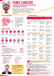
Penile Cancer: Symptoms, Diagnosis and Treatment in Australia

Penile cancer is rare, but early diagnosis matters

Penile cancer is an uncommon cancer affecting the skin and tissues of the penis. In Australia, approximately 165 men were estimated to be diagnosed with penile cancer in 2025, with the average age at diagnosis around 68 years. About 95% of penile cancers are squamous cell carcinomas (SCC).

Although the diagnosis can understandably be frightening, there is an important message:

When penile cancer is detected early, treatment is frequently curative and, in many men, much or all of the penis can be preserved.

Modern treatment has therefore moved increasingly towards penile-preserving therapy whenever this can be achieved safely.

Treatment in Australia will depend on:

  • whether the abnormality is precancerous or invasive cancer
  • the size and location of the tumour
  • how deeply it has invaded
  • the grade of the cancer
  • whether lymph nodes in the groin are involved
  • whether cancer has spread elsewhere
  • the man’s general health and personal preferences.

Because penile cancer is rare, patients with invasive or complicated disease benefit from discussion by a multidisciplinary team (MDT) involving urology, medical oncology, radiation oncology, radiology, pathology and specialist nursing.


What causes penile cancer?

There is rarely one single identifiable cause.

Important risk factors include:

  • infection with human papillomavirus (HPV)
  • smoking
  • phimosis, where the foreskin cannot be retracted
  • chronic inflammation of the penis
  • penile intraepithelial neoplasia (PeIN)
  • increasing age
  • some chronic penile skin disorders
  • immunosuppression.

HPV plays an important role in a proportion of penile cancers, although penile cancer can certainly occur without HPV infection.


What symptoms should men look for?

Penile cancer often begins as a visible or palpable abnormality, particularly involving the glans or foreskin.

Symptoms can include:

  • a red or discoloured area that does not disappear
  • persistent irritation or inflammation
  • thickening of the skin
  • a lump
  • a wart-like growth
  • an ulcer or sore that does not heal
  • bleeding
  • persistent discharge
  • an unpleasant smell from beneath the foreskin
  • increasing difficulty retracting the foreskin
  • pain or tenderness
  • swelling of the end of the penis
  • a lump beneath the foreskin
  • enlarged lymph nodes or lumps in the groin.

More advanced disease can occasionally cause difficulty passing urine, fatigue or unexplained weight loss.

A persistent penile lesion deserves examination

Most rashes, spots and lumps on the penis are not cancer. Infection, inflammation, dermatitis and benign skin conditions are much more common.

The important issue is persistence.

A penile ulcer, lump, bleeding area or unusual skin change that does not resolve should not be hidden away in the hope that it will disappear.

Embarrassment is considerably easier to treat than advanced cancer.


How is penile cancer diagnosed?

Examination

The first step is careful examination of the penis and foreskin.

The urologist will assess:

  • the site of the lesion
  • its size
  • whether it involves the glans, foreskin or shaft
  • whether deeper tissues appear involved
  • whether the foreskin can be retracted
  • whether there are enlarged lymph nodes in either groin.

Both groins are particularly important because penile cancer usually spreads first through the lymphatic system to the inguinal lymph nodes.


Biopsy

The diagnosis usually requires a biopsy.

A small piece of the abnormal tissue is removed and examined by a pathologist.

Depending upon the lesion, this may be:

  • a punch biopsy
  • an incisional biopsy, taking part of the lesion
  • an excisional biopsy, removing the whole small lesion.

A biopsy determines whether cancer is present and, importantly, identifies the type and grade of the tumour.

Histological confirmation is particularly important before treatments such as topical therapy, laser treatment or radiotherapy.


What is PeIN?

Penile intraepithelial neoplasia (PeIN) is a precancerous or very early cancerous change confined to the surface epithelium.

It has previously been described using terms such as carcinoma in situ.

PeIN is important because it can progress to invasive squamous cell carcinoma. Current European guidelines estimate progression to invasive disease despite treatment in approximately 2.6–13% of patients.

The good news is that PeIN can often be treated without removing part of the penis.


Treatment: from creams to major surgery

There is no single operation or treatment for penile cancer.

Modern management follows a ladder, beginning with the least invasive treatment capable of reliably controlling the cancer.

1. Circumcision

For abnormalities confined to the foreskin, circumcision may remove the lesion completely.

Circumcision is also frequently an important first step when PeIN involves the glans and foreskin because it allows the glans to be properly examined and treated.

For superficial disease, contemporary guidelines regard circumcision as an important primary surgical treatment.


2. Treatment with creams

Selected cases of biopsy-confirmed PeIN can be treated with medication applied directly to the penis.

The two most commonly used treatments are:

5-fluorouracil (5-FU)

5-FU is a topical chemotherapy medication.

It destroys abnormal rapidly dividing cells in the superficial layers of the penis.

Treatment usually produces inflammation of the treated area, which can include:

  • redness
  • burning
  • crusting
  • discomfort
  • erosion of the surface skin.

Imiquimod

Imiquimod is different. Rather than being conventional chemotherapy, it stimulates the local immune system to attack abnormal cells.

It can similarly cause substantial redness, swelling, ulceration or crusting during treatment.

These reactions can look alarming but often indicate that the medication is producing its intended biological effect.

Current EAU-ASCO guidance supports either 5-FU or imiquimod for appropriately selected biopsy-confirmed PeIN.

Importantly, the area must be reassessed after treatment.

If the abnormality persists, repeat biopsy or alternative treatment may be necessary. Repeated courses of topical treatment should not simply continue indefinitely when the lesion has failed to respond, because invasive cancer may be hiding beneath the surface.


3. Laser and other local treatments

Very superficial lesions can sometimes be treated using laser therapy.

Depending upon expertise and availability, other local treatments such as photodynamic therapy or cryotherapy have also been used.

These approaches can preserve the penis but require careful long-term surveillance because local recurrence is possible.


4. Penile-preserving surgery

One of the major changes in penile cancer surgery has been the move away from automatically removing a substantial portion of the penis.

Whenever oncologically safe, the aim is:

remove the cancer, obtain clear margins and preserve as much normal penis as possible.

Operations may include:

Wide local excision

The cancer and a margin of surrounding tissue are removed.

The defect may be closed directly or reconstructed with a skin graft.

Glans resurfacing

The abnormal surface layer of the glans is removed while preserving the deeper erectile tissue.

A skin graft is then placed over the glans.

This can be particularly useful for extensive superficial disease.

Glansectomy

If cancer is confined to the glans but is too extensive or invasive for simpler treatment, part or all of the glans may be removed.

Reconstruction can then be performed, often using a skin graft to create a new glans-like surface.

Current guidelines favour organ-preserving surgery for appropriately selected PeIN and T1–T2 tumours involving the glans or prepuce, provided the patient understands the need for careful follow-up.


5. Partial penectomy

More deeply invasive cancer may require removal of part of the penis.

This is called a partial penectomy.

The surgeon attempts to retain enough penile length to permit comfortable urination and, where possible, sexual function.

This may be necessary when cancer extends more deeply into the erectile tissues and cannot be reliably removed using penile-preserving techniques.


6. Total penectomy

Occasionally a cancer is too large, too proximal or too deeply invasive for part of the penis to be safely preserved.

A total penectomy may then provide the best opportunity for cure.

This understandably sounds confronting. It is generally reserved for circumstances in which less radical treatment would compromise cancer control.

Current EAU-ASCO recommendations support total penectomy with perineal urethrostomy for large invasive tumours that cannot safely be treated by partial penectomy.


What is a perineal urethrostomy?

This is sometimes mistakenly referred to as a “perineotomy”.

Following total removal of the penis, urine still needs a pathway from the bladder to the outside world.

The urethra is therefore brought to the skin of the perineum, the area between the scrotum and anus.

This opening is called a:

Perineal urethrostomy

The patient subsequently passes urine through this opening while sitting on the toilet.

A catheter is generally left temporarily while the area heals.

Once healed, most men can empty their bladder normally through the new opening and do not require a permanent catheter.

Potential problems include:

  • narrowing or stenosis of the opening
  • spraying
  • infection
  • wound complications
  • occasionally the need for further surgery.

A perineal urethrostomy does not mean that the bladder or kidneys have stopped functioning. It simply changes the final few centimetres of the urinary plumbing.


The lymph nodes are extremely important

Treating the penis itself is only one half of penile cancer management.

The other half is determining whether cancer has reached the inguinal lymph nodes in the groin.

Penile cancer generally spreads in a predictable sequence:

Penis → inguinal lymph nodes → pelvic lymph nodes → distant organs

Lymph-node involvement is the single most important prognostic factor in penile cancer.


What if there are no enlarged lymph nodes?

Unfortunately, normal-feeling groins do not completely exclude microscopic cancer.

Men with higher-risk primary tumours may therefore require further lymph-node assessment despite having no palpable lumps.

This may involve:

  • ultrasound of the groins
  • ultrasound-guided needle biopsy of abnormal nodes
  • dynamic sentinel lymph-node biopsy
  • inguinal lymph-node dissection in selected circumstances.

Current EAU-ASCO guidance recommends surgical lymph-node staging for patients at significant risk of microscopic metastatic disease, particularly T1b disease or higher.


What if a groin lymph node is enlarged?

An abnormal lymph node may be assessed using ultrasound and needle biopsy.

If metastatic penile cancer is confirmed, treatment may involve:

  • inguinal lymph-node dissection
  • pelvic lymph-node dissection in selected patients
  • chemotherapy
  • radiotherapy
  • combinations of these treatments.

Patients with clinically node-positive disease are generally staged with CT or FDG-PET/CT to assess pelvic and distant disease before definitive treatment.

Early treatment of lymph-node disease is extremely important.


Radiation therapy

Radiotherapy has an important but selective role in penile cancer.

Treatment of the primary cancer

Selected smaller T1 or T2 cancers can potentially be treated with radiation instead of surgery.

Treatment can involve:

External beam radiotherapy

Radiation is directed at the tumour from outside the body.

Brachytherapy

Radioactive sources are temporarily positioned within or very close to the tumour, allowing a concentrated radiation dose to be delivered while limiting exposure to surrounding tissues.

Radiotherapy may therefore provide another means of preserving the penis in appropriately selected patients.

Potential complications can include:

  • skin irritation
  • ulceration
  • fibrosis
  • narrowing of the urethra
  • changes in penile sensation
  • erectile dysfunction
  • tissue damage or necrosis in uncommon circumstances.

Radiotherapy is also used in selected patients for regional lymph-node disease, as part of chemoradiotherapy for advanced disease, or for palliation of symptoms from metastatic cancer.


Chemotherapy

Chemotherapy is generally reserved for more advanced penile cancer, particularly when significant lymph-node disease or metastatic disease is present.

In Australia, contemporary treatment may involve platinum-based combination chemotherapy.

One regimen used in advanced disease is:

TIP: paclitaxel + ifosfamide + cisplatin

Australia’s eviQ cancer treatment resource includes TIP as an option in neoadjuvant, adjuvant and metastatic penile cancer settings.

For bulky or fixed inguinal lymph-node disease or pelvic lymph-node involvement, chemotherapy may be given before surgery.

This is called neoadjuvant chemotherapy.

The aim is to:

  1. treat microscopic disease throughout the body,
  2. shrink the cancer in the lymph nodes,
  3. determine whether the cancer is responding,
  4. make subsequent surgery more effective or technically achievable.

Current international guidance recommends cisplatin- and taxane-based neoadjuvant chemotherapy for suitable patients with extensive inguinal or pelvic nodal disease, followed by surgery when appropriate.


Chemoradiotherapy

Radiotherapy and chemotherapy can sometimes be combined.

This may be considered for selected patients with:

  • locally advanced disease
  • unresectable cancer
  • extensive lymph-node disease
  • disease where major surgery is unsuitable
  • palliative treatment requirements.

The evidence is evolving, and these decisions should generally be made through a specialist penile cancer MDT.


What about immunotherapy?

Immunotherapy is an exciting area of cancer treatment, but its role in penile cancer is still developing.

Checkpoint inhibitors such as pembrolizumab and related drugs have demonstrated activity in some patients with advanced penile cancer, and newer combinations of chemotherapy and immunotherapy are being investigated.

However, response rates to checkpoint inhibitors alone are relatively modest and there is not yet sufficient evidence to use biomarkers such as HPV or PD-L1 routinely to determine which penile cancer patients should receive immunotherapy.

Clinical trials are particularly important for men whose cancer progresses despite standard platinum-based chemotherapy.


What happens to sexual function?

This depends enormously on treatment.

After topical treatment, circumcision, laser therapy, glans resurfacing or limited local excision, satisfactory sexual function may often be preserved.

After glansectomy or partial penectomy, intercourse may remain possible depending upon remaining penile length, erectile function and reconstruction.

After total penectomy, penetrative intercourse using the penis is no longer possible.

This does not mean that intimacy, sexual sensation or orgasm automatically disappears.

Sexual rehabilitation, psychological support and discussion with the patient and his partner can be an important component of recovery.

These conversations should ideally begin before treatment, not after it.


What is the prognosis?

Penile cancer is potentially highly curable when diagnosed before it has spread to lymph nodes.

The most important predictor of survival is not simply the size of the penile lesion but whether cancer has reached the lymph nodes.

Current EAU-ASCO data report approximate five-year cancer-specific survival according to nodal stage of:

Lymph-node stage Approximate 5-year cancer-specific survival
N0 – no lymph-node metastases 95%
N1 80%
N2 65%
N3 – advanced nodal disease 35%

These are population figures, not predictions for an individual patient.

Prognosis depends upon the tumour’s stage and grade, lymph-node involvement, response to treatment, general health and other pathological features.

The figures do, however, demonstrate why early diagnosis and appropriate assessment of the groin lymph nodes are so important.


Follow-up after treatment

Penile cancer requires ongoing surveillance.

Follow-up may include:

  • examination of the penis or reconstructed area
  • examination of both groins
  • assessment of the perineal urethrostomy where applicable
  • imaging in higher-risk patients
  • biopsy of suspicious recurrent lesions
  • monitoring urinary and sexual function
  • psychological and sexual-health support.

Penile-preserving treatment requires particularly careful surveillance because local recurrence can occur.

Importantly, a local recurrence identified early can often still be successfully treated.


Do not ignore a change in the penis

Penile cancer is uncommon, and most penile skin problems are benign.

Nevertheless, a persistent:

lump, ulcer, red patch, bleeding area, discharge, thickened foreskin or lesion that simply will not heal deserves examination.

There should be no embarrassment in asking your GP or urologist to have a look.

For early disease, treatment may be surprisingly conservative: sometimes circumcision, a cream, laser treatment or limited surgery is all that is required.

Even when invasive cancer is present, modern penile cancer surgery increasingly focuses on preserving penile tissue, urinary function and quality of life whenever this can be achieved without compromising cure.

For advanced disease, treatment becomes more complex and may involve partial or total penectomy, perineal urethrostomy, lymph-node surgery, radiotherapy and chemotherapy.

The crucial message is simple:

The earlier penile cancer is diagnosed, the greater the opportunity to cure it while preserving the penis.

Australian and international resources

Cancer Council Australia: Penile Cancer

Cancer Institute NSW: Penile Cancer

eviQ Australian Penile Cancer Chemotherapy Protocol

2026 EAU-ASCO Penile Cancer Guidelines


This information is intended for patient education and does not replace individual medical assessment. Treatment of penile cancer should be tailored to the tumour stage, pathology, lymph-node status, general health and preferences of the individual patient.

So, if you have noticed a lesion on your penis and it is growing, don’t ignore it and hope it will go away, go see your GP.

Information Sheet: Penis Cancer

Information Sheet: Penile Lesions

Information Sheet: Foreskin Problems