
Minimally Invasive Prostate Biopsy
A prostate biopsy is the definitive test used to determine whether abnormal prostate cells are cancerous. Modern biopsy techniques have changed significantly. Rather than routinely passing biopsy needles through the rectum, prostate tissue can now be sampled through the skin of the perineum, the small area between the scrotum and anus.
The TP Pivot Pro™ is a specialised needle-guidance system designed to facilitate a freehand transperineal prostate biopsy. It allows the biopsy needle to be accurately directed into different areas of the prostate using real-time ultrasound guidance, while requiring only a small number of skin puncture sites.
The transperineal approach has become increasingly attractive because it provides excellent access to the prostate while substantially reducing the risk of infection associated with passing biopsy needles through the rectum.
Why Might I Need a Prostate Biopsy?
A raised PSA does not automatically mean prostate cancer. PSA may also rise because of benign prostate enlargement, inflammation, infection, recent instrumentation or other factors.
A biopsy is generally recommended when the overall clinical picture suggests a meaningful risk of prostate cancer.
This may include:
- A persistently or progressively elevated PSA
- An abnormal prostate examination
- A suspicious lesion identified on multiparametric MRI
- An increased PSA density, which relates the PSA level to prostate volume
- A strong family history or other recognised prostate cancer risk factors
- Persistent suspicion of prostate cancer despite a previous negative biopsy
- The need to reassess a known prostate cancer as part of an active surveillance programme
Modern guidelines recommend performing prostate MRI before biopsy in most men suspected of having localised prostate cancer. MRI findings, PSA density and other individual risk factors can then be combined to determine whether biopsy is appropriate. In selected men with a negative MRI and low clinical risk, biopsy may safely be avoided in favour of PSA surveillance.
What Is a TP Pivot Pro™ Transperineal Biopsy?
TP Pivot Pro™ is a single-use needle guidance device that attaches to the transrectal ultrasound probe used to visualise the prostate.
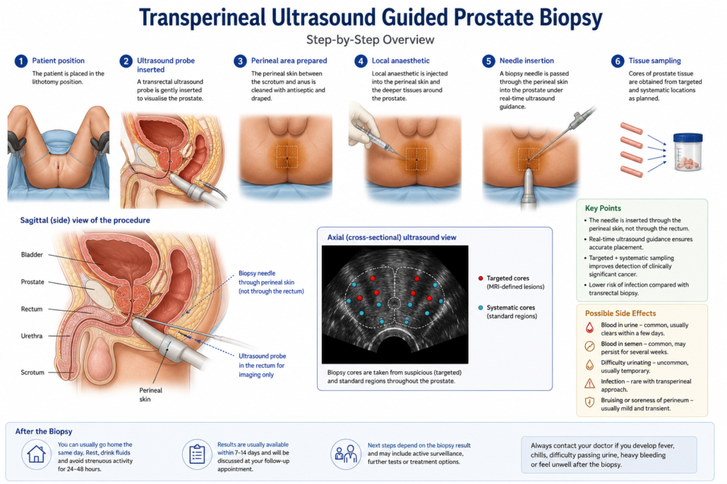
Importantly, although the ultrasound probe sits within the rectum to provide the image, the biopsy needle does not pass through the rectum.
Instead, the biopsy needle passes through the cleaned skin of the perineum.
The device incorporates an adjustable, pivoting needle guide. This allows the surgeon to change the angle and height of the biopsy needle and reach different parts of the prostate while maintaining ultrasound visualisation.
Why Use a Transperineal Approach?
Historically, many prostate biopsies were performed transrectally. During a transrectal biopsy, the needle passes from the rectum directly into the prostate.
The bowel naturally contains large numbers of bacteria. Passing multiple biopsy needles through the rectal wall therefore introduces a small but important risk of urinary infection, prostatitis and occasionally serious bloodstream infection or sepsis.
With a transperineal biopsy, the needle passes through disinfected skin instead.
A major randomised clinical trial comparing contemporary transperineal and transrectal biopsies reported no significant infections among 372 men undergoing transperineal biopsy, compared with an infection rate of 1.6% after transrectal biopsy. Cancer detection was similar between the two approaches.
The transperineal route also provides very good access to areas of the prostate that may sometimes be more difficult to sample transrectally, particularly the anterior and apical regions.
How Is the Biopsy Performed?
1. MRI and Biopsy Planning
Whenever appropriate, a multiparametric MRI of the prostate is performed before biopsy.
The MRI identifies areas that look suspicious for clinically significant prostate cancer. These are usually reported using the PI-RADS system, ranging from PI-RADS 1 to PI-RADS 5.
MRI information can then be used to guide targeted biopsy samples. Current European guidelines recommend combining targeted biopsy with appropriate regional or perilesional sampling when suspicious MRI abnormalities are present.
2. Anaesthesia
Transperineal biopsy can be performed using:
Local anaesthetic, sedation or general anaesthesia, depending on the clinical circumstances, patient preference and the technique used.
The TP Pivot Pro™ system is specifically designed to facilitate a minimally invasive freehand approach and can be used under local or general anaesthesia.
3. Ultrasound Examination
An ultrasound probe is gently inserted into the rectum.
The probe produces real-time images of the prostate, allowing the surgeon to see:
- The prostate gland
- Its boundaries
- The urethra
- The prostate apex and base
- The planned path of the biopsy needle
The ultrasound probe is used for imaging only. The biopsy needle itself enters through the perineal skin.
4. The TP Pivot Pro™ Guide Is Positioned
The TP Pivot Pro™ biopsy guide is securely attached to the ultrasound probe.
Its pivoting mechanism allows the surgeon to alter the trajectory of the introducer needle while watching its position on the ultrasound image.
This makes it possible to reach different regions of the prostate through a very small number of skin access points.
5. Biopsy Samples Are Taken
A biopsy needle is passed through the introducer and into the prostate under direct ultrasound guidance.
Each activation of the biopsy device removes a very thin cylinder of prostate tissue known as a core.
Two complementary types of biopsy may be performed.
Targeted biopsies sample abnormalities seen on MRI.
Regional or systematic biopsies sample predetermined regions of the prostate to reduce the chance of missing important cancer elsewhere in the gland.
Modern 2026 European prostate cancer guidelines favour MRI-informed targeted and regional sampling rather than simply performing the same systematic biopsy pattern in every patient.
How Long Does the Procedure Take?
The exact time varies depending on prostate size, MRI findings and the number of biopsies required.
The procedure itself is generally relatively short, although additional time is required for preparation, anaesthesia and observation afterwards.
Most patients undergoing an uncomplicated biopsy can return home the same day.
What Happens to the Biopsy Samples?
Each biopsy core is sent to a specialist anatomical pathologist.
The pathologist determines:
- Whether cancer is present
- How much cancer is present in each core
- The type of cancer
- The aggressiveness or Grade Group of the cancer
- The percentage or length of each biopsy involved
- Whether other important microscopic features are present
Prostate cancer is commonly reported using ISUP Grade Groups 1 to 5.
Broadly speaking, Grade Group 1 represents the least aggressive pattern, while Grade Group 5 represents the most aggressive.
The biopsy result is then interpreted alongside the PSA, MRI findings, prostate examination, prostate volume and the patient’s overall health.
What Are the Advantages of TP Pivot Pro™?
Potential advantages of this minimally invasive transperineal technique include:
- Very low risk of serious infection
- Avoidance of passing biopsy needles through the rectum
- Excellent access to the anterior, posterior and apical prostate
- Accurate targeting under real-time ultrasound guidance
- Ability to combine MRI-targeted and regional biopsies
- A small number of perineal skin punctures
- Ability to perform the procedure under local anaesthetic in appropriate patients
- A freehand technique that allows the needle trajectory to be adjusted during the procedure
The Pivot Pro™ does not itself diagnose cancer. It is a guidance system that assists the surgeon in accurately performing a transperineal biopsy.
Possible Complications
Transperineal prostate biopsy is generally well tolerated, but no biopsy is completely risk-free.
Blood in the Urine
A small amount of blood in the urine is common after biopsy and usually settles without treatment.
Drinking adequate fluids can help unless you have been advised to restrict your fluid intake for another medical reason.
Blood in the Semen
Blood in the semen is also common after prostate biopsy.
The semen may appear pink, red, brown or rust-coloured. This can persist intermittently for several weeks and is usually harmless.
Bruising or Minor Bleeding
Some bruising or tenderness can occur around the perineal puncture sites.
Significant bleeding is uncommon.
Difficulty Passing Urine
Temporary swelling of the prostate after biopsy can occasionally make urination difficult.
Rarely, acute urinary retention occurs and a temporary urinary catheter is required.
The risk may be greater in men with a particularly large prostate, significant pre-existing urinary symptoms or when a large number of biopsy cores are required.
In the PREVENT randomised trial, urinary retention requiring intervention occurred in approximately 0.3% of men undergoing transperineal biopsy, although rates vary between different biopsy techniques and patient populations.
Infection
Infection remains possible, but it is one of the major areas in which transperineal biopsy offers an advantage.
Because the biopsy needle does not pass through the bacteria-rich rectum, the risk of infection and sepsis is substantially lower than with the traditional transrectal route.
In the PREVENT trial, there were zero grade 2 or greater infections in the transperineal biopsy group, compared with 1.6% following transrectal biopsy.
Your surgeon will advise whether antibiotics are required according to the procedure being performed and local protocols.
When Should I Seek Medical Attention After My Biopsy?
Contact your surgeon or seek urgent medical assessment if you develop:
- Fever, chills or shaking
- Increasing difficulty passing urine
- Complete inability to urinate
- Heavy or persistent bleeding
- Large blood clots in the urine
- Increasing pain or swelling
- Feeling significantly unwell
Although serious infection is uncommon after transperineal biopsy, a fever or systemic illness following any prostate biopsy should be assessed promptly.
What Happens After the Prostate Biopsy?
The next step depends entirely on the pathology result.
No Cancer Is Found
A negative biopsy is reassuring, but it does not reduce the future risk of prostate cancer to zero.
Your urologist will consider:
PSA levels and PSA trend, MRI findings, PSA density, prostate size, family history, the adequacy of the biopsy and your overall prostate cancer risk.
Depending on these findings, management may involve routine PSA surveillance, repeat MRI or, less commonly, another biopsy if clinical suspicion remains high.
Low-Risk Prostate Cancer Is Found
Not every prostate cancer needs immediate treatment.
Men with low-risk prostate cancer, and selected men with favourable intermediate-risk disease, may be suitable for active surveillance.
Active surveillance typically involves repeated PSA testing, clinical review, MRI and repeat biopsy when appropriate.
The aim is to monitor the cancer carefully and offer treatment only if there is evidence that it is becoming more significant. Current guidelines continue to recommend active surveillance as the preferred approach for many men with low-risk prostate cancer.
Clinically Significant Prostate Cancer Is Found
If the biopsy demonstrates clinically significant cancer, the next step is to determine:
- The Grade Group
- The amount of cancer present
- Whether the cancer appears confined to the prostate
- Whether additional staging investigations are required
- Your PSA level
- Your age and general health
- Your personal treatment priorities
Some men will undergo further imaging such as PSMA PET/CT before deciding on treatment, particularly when higher-risk disease is identified.
Treatment options may include:
Active surveillance, robotic-assisted radical prostatectomy, radiotherapy, sometimes combined with hormonal therapy, or other treatments depending on the individual situation.
There is rarely a single treatment that is correct for every man. Management should be tailored to the biological behaviour of the cancer and the patient’s individual priorities.
From PSA to Diagnosis: The Modern Prostate Cancer Pathway
Abnormal PSA or clinical concern
↓
Repeat PSA and individual risk assessment
↓
Multiparametric MRI
↓
MRI and PSA-density assessment
↓
Biopsy recommended if clinically appropriate
↓
TP Pivot Pro™ transperineal targeted ± regional biopsy
↓
Pathology result
↓
No cancer → surveillance / reassessment
Low-risk cancer → consider active surveillance
Clinically significant cancer → staging and treatment discussion
Frequently Asked Questions
Is a transperineal prostate biopsy painful?
Local anaesthetic significantly reduces discomfort. Patients commonly notice pressure, vibration or the clicking of the biopsy device rather than sharp pain. Sedation or general anaesthesia can be used in appropriate circumstances.
Does the needle go through the rectum?
No. The ultrasound probe sits in the rectum to create the images, but the biopsy needle enters through the skin of the perineum.
Can the biopsy find cancers at the front of the prostate?
Yes. One of the advantages of the transperineal approach is excellent access to anterior and apical regions of the prostate.
Does everyone with a high PSA need a biopsy?
No. PSA is only one part of the assessment. Repeat PSA measurement, prostate examination, MRI, PSA density, family history and other risk factors may all influence whether biopsy is necessary.
Can an MRI replace a prostate biopsy?
MRI is extremely useful but cannot currently provide the same definitive information as examination of prostate tissue under a microscope. In selected low-risk men with a reassuring MRI, biopsy can sometimes be avoided or deferred. When clinically significant prostate cancer remains suspected, tissue biopsy remains the standard method of confirming the diagnosis.
How long will it take to receive my results?
Timing varies between pathology laboratories. Your urologist will normally arrange a follow-up appointment to discuss the pathology rather than relying on the written report alone.
A More Precise Way to Investigate Prostate Cancer
The goal of modern prostate biopsy is not simply to take more samples. It is to obtain the right samples from the right areas of the prostate while minimising unnecessary risk.
Combining high-quality prostate MRI with a minimally invasive transperineal technique such as TP Pivot Pro™ allows suspicious lesions and relevant regions of the prostate to be sampled accurately without repeatedly passing biopsy needles through the bowel.
For appropriate patients, this represents an important evolution in the diagnosis of prostate cancer: MRI-informed, targeted, transperineal and focused on reducing complications while preserving diagnostic accuracy.
This information is intended for general education and does not replace individual medical advice. The need for prostate biopsy, choice of anaesthesia, biopsy technique and subsequent treatment should be discussed with your urologist.


Leave a Reply
Want to join the discussion?Feel free to contribute!