
Superficial Urothelial Carcinoma of the Bladder
Understanding non-muscle-invasive bladder cancer, TURBT, intravesical therapy and long-term surveillance
Being told that you have a bladder tumour is understandably alarming. The reassuring part is that many bladder cancers are discovered while they are still confined to the inner layers of the bladder and have not invaded the bladder muscle.
This is called non-muscle-invasive bladder cancer (NMIBC), historically referred to as superficial bladder cancer. NMIBC includes Ta tumours, T1 tumours and carcinoma in situ (CIS). These tumours behave very differently depending on their stage, grade and other pathological features, so treatment is tailored according to the risk of the cancer coming back (recurrence) or becoming more aggressive (progression).
The good news is that most NMIBC can initially be treated through the urethra without making an incision in the abdomen. The less convenient news is that bladder cancer has a habit of returning, which is why careful surveillance becomes an important part of treatment.
What is urothelial carcinoma?
The inside of the bladder is lined by specialised cells called urothelial cells. Cancer arising from these cells is known as urothelial carcinoma.
Urothelial carcinoma can develop anywhere along the urinary tract, including the:
- renal collecting system
- ureters
- bladder
- urethra
The bladder is by far the most common site.
When a bladder tumour has not invaded the muscular wall of the bladder, it is classified as non-muscle-invasive bladder cancer.

What causes bladder cancer?
There is rarely one identifiable cause. Instead, bladder cancer develops following genetic changes within urothelial cells, often influenced by environmental exposures over many years.
Smoking
Cigarette smoking is the most important preventable risk factor for bladder cancer.
Chemicals absorbed through cigarette smoke enter the bloodstream, are filtered by the kidneys and eventually become concentrated in the urine. The bladder lining is therefore repeatedly exposed to these carcinogens.
Stopping smoking after a bladder cancer diagnosis is strongly encouraged.
Other risk factors
These include:
- increasing age
- occupational exposure to certain industrial chemicals
- previous pelvic radiotherapy
- previous treatment with cyclophosphamide
- chronic urinary tract irritation in selected circumstances
- a personal history of urothelial carcinoma
Sometimes there is no obvious risk factor at all.
How does bladder cancer present?
Blood in the urine
The classic presentation is haematuria, or blood in the urine.
This may be:
Visible haematuria
The urine may suddenly become:
- pink
- red
- burgundy
- tea-coloured
- or contain blood clots
Importantly, bleeding from a bladder tumour is frequently painless and intermittent.
The bleeding may disappear completely for days, weeks or even months. Its disappearance does not necessarily mean that the underlying problem has resolved.
Unexplained visible haematuria should always be investigated.
Microscopic haematuria
Sometimes blood is detected only on urine testing and cannot be seen with the naked eye.
Other possible symptoms
Some patients, particularly those with carcinoma in situ (CIS), may experience bladder irritation rather than obvious bleeding.
Symptoms can include:
- urinary frequency
- urgency
- burning when passing urine
- nocturia
- pelvic or bladder discomfort
- recurrent symptoms resembling a urinary tract infection
Persistent urinary symptoms with repeatedly negative urine cultures may therefore warrant further investigation.
Investigating suspected bladder cancer
The investigation usually involves a combination of urine testing, imaging and direct examination of the bladder.
Urine testing
A urine sample may be checked for:
- blood
- infection
- inflammatory cells
- abnormal malignant cells
Urine cytology
Urine cytology examines cells shed from the urinary tract under a microscope.
It is particularly useful for detecting high-grade urothelial carcinoma and CIS, but is considerably less sensitive for low-grade tumours.
A negative cytology therefore does not exclude bladder cancer.
Imaging the urinary tract
Patients with haematuria may require imaging of the kidneys, ureters and bladder.
Depending on the clinical circumstances, this may include:
- renal tract ultrasound
- CT urinary tract imaging
- CT urography
CT urography is particularly useful when investigating haematuria because urothelial carcinoma can occasionally arise within the renal collecting system or ureters as well as the bladder.
Cystoscopy
A cystoscopy allows the urologist to look directly inside the bladder.
A thin flexible telescope is passed through the urethra, usually under local anaesthetic.
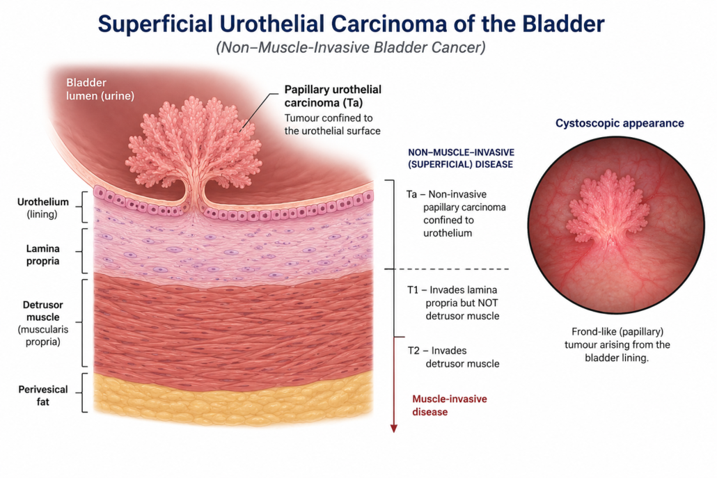
Most bladder tumours have a characteristic appearance. They may resemble delicate fronds, seaweed or a tiny underwater cauliflower attached to the bladder wall.
Flat lesions such as CIS can be much more difficult to see.
If a suspicious lesion is identified, the next step is usually a transurethral resection of bladder tumour (TURBT).
TURBT: biopsy and removal of the bladder tumour
TURBT stands for:
Transurethral Resection of Bladder Tumour
This procedure serves two important purposes:
- Treatment: removing all visible tumour where possible.
- Diagnosis and staging: providing tissue for the pathologist to determine exactly what type of tumour is present and how deeply it has invaded.
The procedure is usually performed under general or spinal anaesthesia.
A telescope called a resectoscope is passed through the urethra into the bladder. The tumour is carefully removed, usually using an electrical or bipolar resection system.
There is therefore generally no external incision or abdominal scar.
Why obtaining bladder muscle matters
An adequate TURBT should establish how deeply the tumour extends.
For many tumours, particularly T1 and high-grade lesions, the specimen should contain muscularis propria (detrusor muscle) so that the pathologist can determine whether the cancer has reached the muscle layer. The pathological report should document the presence and involvement of muscularis propria where applicable.
This distinction dramatically changes treatment.
Understanding bladder cancer staging
A simplified view of the bladder wall is:
Urine
↓
Urothelium
↓
Lamina propria
↓
Detrusor muscle
↓
Fat surrounding the bladder
The important early stages are:
Ta
The tumour is confined to the urothelial surface and has not invaded the supporting tissue underneath.
Tis: carcinoma in situ
CIS is a flat, high-grade malignant lesion confined to the urothelium.
Unlike the typical papillary bladder tumour, CIS may be difficult to identify visually.
Despite being superficial anatomically, CIS is biologically aggressive and requires appropriate treatment.
T1
The tumour has invaded into the lamina propria, but has not invaded the muscularis propria.
T1 disease, particularly high-grade T1 disease, carries a greater risk of recurrence and progression.
T2
The cancer has invaded the bladder muscle.
Once muscle invasion is identified, the disease is no longer classified as NMIBC and requires a different treatment pathway.
Stage and grade are not the same thing
This distinction frequently causes confusion.
Stage describes how deeply the cancer has travelled into the bladder wall.
Grade describes how abnormal and biologically aggressive the cancer cells appear under the microscope.
A tumour may therefore be superficial but still be high grade.
Broadly, urothelial tumours are classified as:
Low grade
These generally grow more slowly and have a relatively low risk of progressing to muscle-invasive cancer, although they may recur.
High grade
These cells look significantly abnormal and have a greater potential for:
- recurrence
- invasion
- progression
- spread beyond the bladder
Risk stratification
Following TURBT, the tumour is classified into a risk category.
Risk assessment considers factors such as:
- stage
- grade
- tumour size
- number of tumours
- previous recurrence rate
- presence of CIS
- T1 disease
- pathological features including lymphovascular invasion
- certain variant histological subtypes
This classification helps determine whether the patient requires:
TURBT alone → intravesical chemotherapy → BCG → or, in selected very-high-risk situations, consideration of radical cystectomy.
Modern guidelines emphasise risk-adapted rather than one-size-fits-all management.
Do I need another TURBT?
Sometimes.
A second-look or re-staging TURBT may be recommended when:
- the initial tumour was incompletely removed
- adequate muscle was not present in an important specimen
- high-grade disease is present in selected circumstances
- T1 disease is identified
- there is concern that the original tumour may have been understaged
- certain variant histologies are identified
The aim is to ensure that residual tumour has not been left behind and, crucially, that muscle-invasive disease has not been missed.
Intravesical treatment
Intravesical simply means that medication is placed directly into the bladder through a catheter.
This allows the treatment to come into direct contact with the bladder lining while reducing systemic exposure compared with intravenous chemotherapy.
Two important treatments are:
Mitomycin C
and
BCG
They are not interchangeable and are used for different risk groups.
Intravesical Mitomycin C
Mitomycin C is a chemotherapy drug that can be placed directly into the bladder.
Immediate Mitomycin C following TURBT
For appropriate patients with suspected low- or intermediate-risk NMIBC, a single postoperative instillation of intravesical chemotherapy may be given soon after TURBT.
Current guidelines recommend that, when used, the immediate instillation should generally occur within 24 hours of TURBT.
Its purpose is to destroy microscopic tumour cells remaining in the bladder and tumour cells released during the resection.
This reduces the risk of recurrence.
When should immediate Mitomycin C be avoided?
It should generally not be administered if there is:
- suspected bladder perforation
- significant bleeding requiring bladder irrigation
- an extensive or very deep resection where perforation is a concern
This is important because chemotherapy leaking outside the bladder can cause significant local tissue injury.
Further courses of intravesical chemotherapy
Selected patients with recurrent or intermediate-risk low-grade NMIBC may receive a course of intravesical chemotherapy rather than simply a single postoperative dose.
The exact drug, schedule and duration depend upon the tumour risk profile and local treatment protocols.
Side effects of intravesical Mitomycin C
Most patients tolerate treatment reasonably well.
Possible side effects include:
- urinary frequency
- urgency
- burning
- bladder discomfort
- haematuria
- chemical cystitis
- skin irritation if the medication contacts the genital skin
Rarely, severe bladder inflammation or tissue injury can occur.
Intravesical BCG
BCG stands for Bacillus Calmette-Guérin.
Yes, it originated as a tuberculosis vaccine. In the bladder it performs a completely different job.
BCG stimulates a powerful local immune response against urothelial cancer cells and remains one of the most effective bladder-preserving treatments for high-risk NMIBC.
Who should receive BCG?
BCG is principally considered for patients with:
- high-grade Ta tumours
- high-grade T1 tumours
- carcinoma in situ
- other appropriately selected high-risk or intermediate-risk NMIBC
For high-risk NMIBC, full-dose BCG with maintenance treatment for one to three years remains a guideline-supported treatment, while immediate radical cystectomy should also be discussed in appropriate high-risk and particularly very-high-risk disease.
How is BCG given?
BCG is inserted into the bladder through a small catheter.
A typical induction course consists of:
One treatment per week for six weeks.
The solution is retained within the bladder for a prescribed period and then passed out in the urine.
Patients who respond may subsequently receive maintenance BCG.
A commonly used maintenance approach involves three weekly treatments at defined intervals after induction. In high-risk disease, guideline schedules may continue maintenance for up to three years depending upon tumour risk, treatment tolerance and BCG availability.
When should BCG not be given?
BCG is a live attenuated organism and must be administered carefully.
Treatment should generally be postponed in patients with:
- visible haematuria
- symptomatic urinary tract infection
- traumatic catheterisation
- very recent TURBT
The EAU lists the first two weeks following TURBT, visible haematuria, traumatic catheterisation and symptomatic urinary infection as absolute contraindications to an intravesical BCG instillation.
Side effects of BCG
A degree of bladder irritation is common.
Patients may experience:
- frequency
- urgency
- burning
- bladder discomfort
- mild haematuria
- fatigue
- low-grade fever
- flu-like symptoms
These symptoms usually settle.
Rarely, BCG can cause a more significant systemic infection or inflammatory reaction.
Persistent high fever, chills, marked deterioration or severe illness following BCG requires urgent medical assessment.
What if BCG does not work?
Persistent or recurrent high-grade cancer despite adequate BCG treatment requires careful reassessment.
This situation should not simply be managed by repeatedly giving more BCG indefinitely.
Patients meeting criteria for BCG-unresponsive NMIBC should be counselled regarding further treatment, and radical cystectomy remains the oncological standard for suitable patients with BCG-unresponsive high-risk disease. Bladder-preserving alternatives may be considered for patients who are medically unsuitable for cystectomy or decline surgery, ideally within appropriate specialist or clinical-trial pathways.
When should radical cystectomy be considered?
Most patients with superficial bladder cancer will never require removal of their bladder.
However, early radical cystectomy may be discussed for very-high-risk disease, including selected patients with:
- aggressive high-grade T1 disease
- associated CIS
- variant histology
- lymphovascular invasion
- persistent high-grade disease
- BCG-unresponsive disease
- other features associated with a high risk of progression
The decision involves balancing the risks of major surgery against the danger of allowing biologically aggressive disease to progress.
Surveillance after treatment
Removing the tumour is only the first chapter.
NMIBC has a significant tendency to recur, and some high-risk tumours can progress. Consequently, regular cystoscopic surveillance is essential.
The first surveillance cystoscopy is generally performed approximately three months after TURBT.
After this, surveillance is tailored to the patient’s risk category.
A practical risk-adapted surveillance framework
| Risk group | Typical cystoscopy schedule | Cytology | Upper tract imaging |
|---|---|---|---|
| Low risk | 3 months, 12 months, then annually | Usually not routinely required | Not routinely required |
| Intermediate risk | 3 months, then approximately every 6 months for 2 years, then annually | Risk-dependent | Selected patients |
| High / very high risk | Approximately every 3 months initially, with intervals gradually extended | Usually included | Periodic upper urinary tract imaging |
The 2026 EAU guidance continues to recommend that follow-up intensity and duration be determined by the patient’s risk category, with the first cystoscopy at three months.
Individual surveillance schedules may differ depending on pathology, previous recurrences, treatment response, age, comorbidities and the treating urologist’s protocol.
Why is surveillance so important?
A recurrence does not automatically mean that treatment has failed or that the cancer has become life-threatening.
Low-grade papillary tumours may recur while remaining superficial and biologically low risk.
The purpose of surveillance is to identify recurrence early, while it can still be treated appropriately.
High-grade disease requires closer attention because the consequences of missing progression are considerably greater.
Can bladder cancer come back after years?
Yes.
This is why follow-up for intermediate- and particularly high-risk NMIBC can continue for many years.
The frequency of cystoscopy usually decreases when repeated examinations remain clear, but high-risk patients generally require prolonged surveillance.
What can I do after a diagnosis?
One of the most important modifiable factors is:
Stop smoking
Smoking cessation reduces exposure to the carcinogens responsible for many urothelial cancers and provides substantial additional cardiovascular, respiratory and general health benefits.
Patients should also:
- attend every scheduled cystoscopy
- report recurrent visible haematuria
- complete prescribed intravesical treatment
- report significant side effects from BCG or chemotherapy
- maintain appropriate hydration unless medically restricted
- discuss occupational chemical exposure where relevant
The bottom line
Non-muscle-invasive urothelial carcinoma is bladder cancer that has not invaded the muscular wall of the bladder.
The pathway typically involves:
Haematuria or urinary symptoms
↓
Urine tests + imaging
↓
Cystoscopy
↓
TURBT and pathological examination
↓
Stage + grade + risk classification
↓
Risk-adapted treatment
For some patients, TURBT followed by surveillance may be sufficient.
Others benefit from intravesical Mitomycin C or another intravesical chemotherapy to reduce recurrence.
Patients with high-grade disease, T1 cancer or CIS frequently require intravesical BCG, usually incorporating induction and maintenance treatment.
Very-high-risk or BCG-unresponsive disease may require consideration of radical cystectomy.
Most importantly, bladder cancer treatment does not finish when the initial tumour has been removed. Long-term cystoscopic surveillance is an integral part of treatment.
A final word
Bladder cancer can be a persistent visitor, but surveillance means we do not leave the front door unattended.
Early detection, complete TURBT, accurate pathological staging, appropriate intravesical therapy and structured follow-up provide the best opportunity to keep non-muscle-invasive bladder cancer under control.
This information is intended as general patient education and does not replace individual assessment or treatment advice from your urologist. Management should be tailored to the pathology, tumour risk category, general health and individual circumstances of each patient.


Leave a Reply
Want to join the discussion?Feel free to contribute!