
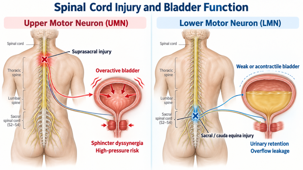
Spinal Cord Injury and the Bladder: Upper Motor Neuron versus Lower Motor Neuron Injury
Meta description: Spinal cord injury can cause urgency, leakage, urinary retention and dangerous bladder pressures. Learn how upper and lower motor neuron injuries differ, how neurogenic bladder is investigated, and which treatments may protect the kidneys and improve continence.
Best treated in an organised spinal unit.
Why can a spinal cord injury affect the bladder?
Passing urine is not simply a bladder reflex. It requires communication between the brain, the spinal cord, the sacral nerves and the urinary sphincter. During normal bladder filling, the bladder muscle, called the detrusor, remains relaxed while the outlet stays closed. When it is appropriate to urinate, the detrusor contracts and the sphincter relaxes in a coordinated fashion.
A spinal cord injury (SCI) can interrupt these pathways. The resulting problem is called neurogenic lower urinary tract dysfunction, often shortened to neurogenic bladder. A person may develop urgency, leakage, difficulty emptying, urinary retention, or a combination of these.
The most important issue is not always the symptom that is most noticeable. A bladder may feel relatively quiet yet store urine at a pressure high enough to threaten the kidneys. Conversely, severe urgency may occur without dangerous pressure. Assessment therefore aims to answer two separate questions:
- Is urine being stored and emptied at a pressure that is safe for the kidneys?
- Does the bladder routine provide acceptable continence, independence and quality of life?
The early phase: spinal shock
Immediately after an acute SCI, the bladder commonly becomes temporarily areflexic or acontractile. It fills but does not contract effectively, causing urinary retention. Bladder sensation may also be absent. Catheter drainage is usually required in this acute phase.
This phase may last days, weeks or sometimes longer. Reflex activity can then return below a suprasacral injury and the eventual bladder pattern may be very different from the early one. For this reason, the bladder should be reassessed as the neurological situation evolves.
Upper motor neuron versus lower motor neuron bladder
The traditional distinction is useful, but it is a guide—not a substitute for urodynamic testing.
| Feature | Upper motor neuron pattern | Lower motor neuron pattern |
|---|---|---|
| Typical injury site | Above the sacral micturition centre, often a suprasacral spinal cord lesion | Sacral cord, conus medullaris, cauda equina or sacral/peripheral nerve injury |
| Bladder contraction | Often involuntary detrusor contractions during filling—neurogenic detrusor overactivity | Weak or absent detrusor contraction—detrusor underactivity or acontractility |
| Outlet behaviour | The sphincter may contract instead of relaxing during a bladder contraction—detrusor-sphincter dyssynergia (DSD) | Outlet resistance may be reduced if sacral innervation is damaged, although obstruction from other causes can coexist |
| Common symptoms | Urgency, frequency, reflex voiding, urge leakage, interrupted emptying and residual urine | Reduced bladder sensation, infrequent voiding, straining, retention, overflow leakage and recurrent infection |
| Main safety concern | High storage or voiding pressure, poor compliance, reflux, hydronephrosis and renal damage | Chronic retention, over-distension, infection, stones and overflow incontinence |
| Typical emptying strategy | Often clean intermittent catheterisation, sometimes combined with bladder-relaxing treatment | Clean intermittent catheterisation is commonly required when detrusor contraction is inadequate |
Upper motor neuron injury: the overactive but poorly coordinated bladder
With an injury above the sacral spinal cord, the local sacral reflex circuit may remain intact but lose normal coordination from the brain. After spinal shock resolves, the bladder may contract unexpectedly during filling. At the same time, the external urinary sphincter may fail to relax or may tighten when the bladder contracts.
This combination of detrusor overactivity and DSD can produce:
- urgency and reflex urinary leakage;
- a stop–start urinary stream or incomplete emptying;
- high bladder pressure;
- urinary tract infection and bladder stones;
- vesicoureteric reflux, hydronephrosis and, if not controlled, renal impairment.
People with SCI at or above approximately T6 can also develop autonomic dysreflexia. Bladder distension, a blocked catheter, infection or urological procedures may trigger sudden severe hypertension, a pounding headache, sweating or flushing above the injury, anxiety, nasal congestion and a slow or irregular pulse. This is a medical emergency: sit the person upright, check the catheter and drainage system promptly, and seek urgent medical assistance if symptoms or elevated blood pressure persist.
Lower motor neuron injury: the bladder that cannot squeeze effectively
Damage involving the sacral cord, conus medullaris, cauda equina or sacral nerves can interrupt the reflex pathway to the bladder. The detrusor may contract weakly or not at all. Sensation of filling may be reduced, so the bladder can become very full without the usual warning.
Possible features include:
- difficulty starting or inability to pass urine;
- infrequent voiding and a weak stream;
- abdominal straining to empty;
- a large post-void residual;
- overflow leakage;
- recurrent infection, stones or bladder over-distension.
If outlet innervation is also impaired, stress leakage can coexist with retention. A lower motor neuron lesion is therefore not automatically a “low-pressure and harmless” bladder.
Why the neurological level does not tell the whole story
Real-life bladder dysfunction does not always fit neatly into one box. An incomplete lesion can preserve some sensation or voluntary control. Injuries around the conus may affect upper and lower motor neuron pathways together. Pre-existing prostate enlargement, urethral stricture, pelvic floor dysfunction, medications, constipation and infection can further change bladder behaviour.
Most importantly, neurological examination and lesion level do not reliably predict bladder pressure or sphincter coordination. Symptoms can also change over time. Urodynamic findings, not the label alone, should guide risk assessment and treatment.
How is bladder function investigated after SCI?
Clinical assessment
Assessment usually includes:
- the level, completeness, timing and cause of the SCI;
- bladder sensation, urgency, leakage, voiding method and catheter routine;
- recurrent infections, visible blood, stones, catheter blockage and episodes of autonomic dysreflexia;
- fluid intake, urine output and a bladder diary where practical;
- bowel function and constipation;
- mobility, hand function, cognition, carer support and personal goals;
- medicines that may affect storage or emptying;
- abdominal, genital, perineal and focused neurological examination.
Basic tests
Depending on the clinical situation, these may include:
- urinalysis and urine culture when infection is suspected;
- measurement of the post-void residual by ultrasound or catheter;
- kidney function blood tests, while recognising that serum creatinine may underestimate renal impairment in people with low muscle mass;
- renal and bladder ultrasound to look for hydronephrosis, stones, bladder wall changes and residual urine;
- other renal function assessment when clinically indicated.
A urine culture should not be used to screen for and repeatedly treat bacteria in an otherwise well catheter user. Asymptomatic bacteriuria is common and generally should not be treated, apart from recognised exceptions such as pregnancy or before selected urological procedures that breach the urinary mucosa.
Urodynamic studies
Multichannel urodynamics, sometimes combined with X-ray imaging as video-urodynamics, is central when the risk is unknown or potentially significant. It can determine:
- bladder sensation and capacity;
- detrusor overactivity;
- bladder compliance and storage pressure;
- detrusor strength during attempted voiding;
- sphincter coordination or DSD;
- leakage pressure, residual urine and, with video, reflux or outlet anatomy.
The test is not merely to explain incontinence. Its crucial role is to identify a hostile, high-pressure bladder before silent upper urinary tract damage develops. Repeat testing may be required after treatment, when symptoms change, after recurrent complications, or when renal imaging deteriorates.
Other tests
Cystoscopy is not a routine screening test for every person with SCI. It is used for a specific indication, such as visible blood in the urine, recurrent catheter blockage, suspected stones, difficult catheterisation, urethral injury or another anatomical concern. CT, MRI or nuclear renal imaging may be added when ultrasound or the clinical picture warrants it.
Management: protecting the kidneys while fitting treatment to the person
Treatment should be individualised with a urologist, rehabilitation physician, continence nurse and allied health team. The aim is low-pressure storage, reliable emptying, continence where achievable, fewer complications and a routine the person can realistically manage.
1. Reliable bladder emptying
Clean intermittent catheterisation (CIC/ISC) is often the preferred method when the bladder does not empty adequately. It avoids a continuously indwelling tube and can provide predictable, low-pressure drainage. Frequency is tailored to fluid intake, catheterised volumes, bladder pressure and the treatment plan. Hand function, access to the urethra, cognition, carer assistance, work and travel must all be considered.
If intermittent catheterisation is not feasible, an indwelling catheter may be required. When long-term indwelling drainage is necessary, a suprapubic catheter is often favoured over a urethral catheter because it avoids chronic urethral pressure and erosion, although it still carries risks of infection, blockage, leakage and stones.
Reflex voiding into a sheath system may suit selected men, but only after confirming that bladder pressures and emptying are safe. Regular reliance on abdominal straining or the Credé manoeuvre is generally discouraged because it can generate high pressure and may not empty the bladder adequately.
2. Reducing overactivity and unsafe storage pressure
Options include:
- antimuscarinic medicines, which reduce involuntary bladder contractions but can cause dry mouth, constipation, blurred vision and cognitive adverse effects;
- a beta-3 agonist, which may improve storage with a different side-effect profile but requires attention to blood pressure and other contraindications;
- carefully selected combination therapy;
- intradetrusor botulinum toxin A (Botox) when tablets are ineffective or poorly tolerated. This can markedly reduce detrusor overactivity and pressure but may increase urinary retention; the person must be willing and able to catheterise if required.
Alpha-blockers may reduce outlet resistance in selected people who void spontaneously, but they do not correct every form of DSD and may cause dizziness or low blood pressure.
3. Managing sphincter dyssynergia or outlet resistance
Where DSD prevents safe emptying, management may include intermittent catheterisation, bladder-relaxing therapy, selected alpha-blocker use, sphincter botulinum toxin, or less commonly, an outlet procedure such as sphincterotomy in carefully selected men using sheath drainage. Any procedure that lowers outlet resistance may improve emptying but can trade obstruction for incontinence.
4. Treating an acontractile lower motor neuron bladder
For a bladder that cannot contract effectively, CIC is usually the mainstay. There is no consistently effective oral medicine that restores a truly acontractile detrusor. Correcting reversible contributors: constipation, medicines, infection or mechanical obstruction remains important. Outlet surgery should only be considered when a proven obstruction is present and the likely effect on continence and catheterisation has been discussed.
5. Reconstructive surgery and urinary diversion
If conservative and minimally invasive measures cannot produce safe storage or practical drainage, options may include:
- augmentation cystoplasty to increase capacity and reduce pressure;
- creation of a continent catheterisable channel, such as a Mitrofanoff-type channel;
- selected outlet continence surgery when sphincter weakness is the main problem;
- urinary diversion, with or without bladder removal, in complex or refractory cases.
These are major procedures with long-term surveillance requirements and should be undertaken in an experienced neuro-urology service.
What about sacral neuromodulation?
Sacral neuromodulation is not a routine treatment for a complete spinal cord injury with established DSD or an acontractile bladder. It may be considered in carefully selected people with an incomplete, stable neurological lesion and suitable bladder function, but evidence in SCI is more limited than it is for non-neurogenic overactive bladder or non-obstructive retention. A test phase and specialist assessment are essential.
Follow-up is lifelong
Neurogenic bladder can change even when the spinal injury itself appears stable. Follow-up intensity is based on risk, bladder management method and previous complications. It may include review of symptoms and catheter volumes, renal function assessment, upper urinary tract imaging and repeat urodynamics in moderate- or high-risk patients.
Seek earlier review for:
- new or worsening leakage;
- difficulty catheterising or repeated catheter blockage;
- recurrent symptomatic infection or fever;
- visible blood in the urine;
- flank pain, stones or hydronephrosis;
- increasing residual urine;
- new autonomic dysreflexia;
- deterioration in kidney function.
The take-home message
An upper motor neuron injury often produces an overactive bladder that may fight against a closed sphincter. A lower motor neuron injury more often produces a poorly contracting bladder with retention. Both patterns can cause leakage, infection and kidney damage, and mixed patterns are common.
The safest plan is not based on the injury label alone. It combines symptoms, neurological assessment, kidney surveillance and where indicated, urodynamic testing. With an individualised catheter, medication, Botox or surgical plan, most people can achieve safer bladder pressures and a more predictable routine.
Medical disclaimer: This article provides general education and does not replace individual medical assessment. Sudden severe headache, sweating or flushing with high blood pressure in a person with SCI—particularly with an injury at or above T6—may represent autonomic dysreflexia and requires urgent attention.
References
- European Association of Urology. EAU Guidelines on Neuro-Urology. Current guideline edition. EAU Neuro-Urology Guidelines.
- Ginsberg DA, Boone TB, Cameron AP, et al. The AUA/SUFU Guideline on Adult Neurogenic Lower Urinary Tract Dysfunction: Diagnosis and Evaluation. J Urol. 2021;206(5):1097–1105. PubMed.
- Ginsberg DA, Boone TB, Cameron AP, et al. The AUA/SUFU Guideline on Adult Neurogenic Lower Urinary Tract Dysfunction: Treatment and Follow-up. J Urol. 2021;206(5):1106–1113. PubMed.
- American Urological Association; Society of Urodynamics, Female Pelvic Medicine & Urogenital Reconstruction. Adult Neurogenic Lower Urinary Tract Dysfunction Guideline. 2021; amended 2024. AUA/SUFU guideline.
- NSW Agency for Clinical Innovation. Management of the Neurogenic Bladder for Adults with Spinal Cord Injuries. State Spinal Cord Injury Service. NSW ACI spinal cord injury resources.
- Consortium for Spinal Cord Medicine. Bladder Management for Adults with Spinal Cord Injury: A Clinical Practice Guideline for Health-Care Providers. J Spinal Cord Med. 2006;29(5):527–573. Full text on PubMed Central.
- Pannek J, Blok B, Castro-Diaz D, et al. Guidelines on Neuro-Urology. European Association of Urology; updated annually. EAU Guidelines.
- Pannek J, Kennelly M, Kessler TM, et al. International spinal cord injury urodynamic basic data set (version 2.0). Spinal Cord Ser Cases. 2018;4:98. DOI.
- Hooton TM, Bradley SF, Cardenas DD, et al. Diagnosis, prevention, and treatment of catheter-associated urinary tract infection in adults: 2009 International Clinical Practice Guidelines from the Infectious Diseases Society of America. Clin Infect Dis. 2010;50(5):625–663. DOI.


Leave a Reply
Want to join the discussion?Feel free to contribute!