Intravesical Mitomycin C –Therapy

Why is it done?

  • Treatment for localized Superficial Urothelial Carcinoma of Bladder and Ureter


How is it done?

  • A Local anesthetic gel is administered as for a urethral catheterization procedure
  • This is done under a sterile procedure.
  • A 14-16 Fr Indwelling Catheter is placed into your bladder.
  • The pre-made-up Mitomycin solution is installed using strict administering criteria
  • Usually, 2 vials of 20mg MMC is mixed with Saline to a 50cc volume. You require 40mg.
  • The catheter is then removed
  • The MMC is required to stay in your bladder for 1 hour.
  • Body rotation is not required.
  • Can also be placed after a bladder tumour resection (TURBT)
  • WARNING: Any Fevers require urgent attention


 What next?

  • Some local discomfort may be experienced.
  • Your voiding nature will change within the next week
  • You may experience some urinary frequency
  • You could develop a fever requiring urgent attention.
  • Some patients may experience severe pain when/ if the tumour was resected very deep.

NB! Each person is unique and for this reason, symptoms may vary


Complications

Side–effects

  • This will be done every week for 6 weeks
  • 6 weeks after this a check Flexible Cystoscopy will be scheduled as part of your surveillance protocol for your Urothelial carcinoma
  • This may be repeated.
Surveillance Protocol for Superficial Urothelial Carcinoma

Low Grade: Ta, T1 (G1, G2), CIS

· Initially 3 months after the first resection

· If clear then 9months

· Then annually

High-Risk Low Grade: T1G3 with/out CIS, high volume disease

· This disease may be best treated with a radical cystectomy

· 3 monthly check cystoscopy for 12 months

· If clear, then 6 monthly for a further 12 months

· If clear then annually for 5-7 years

· My recommendation thereafter would be a 2-yearly cystoscopy with intermittent Urine Cytology

· You fall back to the beginning with any recurrences


Download Information Sheet

Wes Intravesical MMC Therapy

Copyright 2019 Dr. Jo Schoeman

Cysto-Lithotomy

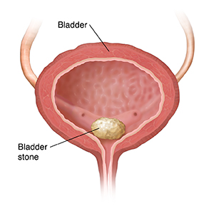
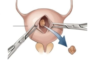
Open removal of a large bladder calculus

Why is it done?

  • To break up a large bladder calculus (stone) that cannot be done endoscopically.
  • It is done with open surgery (a cut above the pubic symphysis).

Risk factors causing this:

    • Bladder outflow obstruction
      • BPH with chronic retention.
      • Urethral stricture.
      • Neurogenic bladder.
    • Renal calculi disease.
    • Metabolic disorders.
    • Malnutrition.
    • Chronic infections.
    • Foreign objects in bladder.

How is it done?

  • A General anesthetic will be given.
  • A sterile surgical field is prepared.
  • Prophylactic antibiotics are given.
  • An indwelling catheter is inserted, and the bladder is then distended with fluid (saline).
  • A small lower abdominal incision is made, splitting the Linea alba and opening the distended bladder in the midline.
  • The stone is removed through the whole with a grasping instrument.
  • The bladder is inspected and then closed in 2 layers.
  • Skin is closed.
  • A catheter will be left for 2 weeks.

What to expect after the procedure?

  • Hematuria (blood in your urine)
  • You will have an indwelling catheter (IDC), which will remain in your bladder.
  • You may have a continuous bladder irrigation with Saline to help clear the bleeding.
  • When your urine is clear and your bowels are functioning, you will be discharged with catheter care instruction.
  • You will have this indwelling catheter for 2 weeks.
  • A cystogram will be arranged at approx. 14 days to exclude any urine leaks prior to removal of your catheter.
  • If there are any urine leaks, your catheter will remain a further 7 days, or until the leak is sealed.
  • Pain on initial passing of urine when the catheter is removed.
  • Bladder infection ranging from a burning sensation to, fever, to puss (rare).
  • Lower abdominal discomfort which will persist for a few days.
  • NB! Each person is unique and for this reason symptoms vary.

What next?

  • This all depends on what is found during the procedure. All the options will be discussed in detail.
  • There may be some blood in the urine. This can be remedied by drinking plenty of fluids until it clears.
  • Anatomical causes of the stones will be discussed and surgical options in treatment may be discussed
  • Patients should schedule a follow-up appointment within 4-6 weeks to discuss the etiology of the calculus as well as what other procedures may be involved to prevent this from occurring again.


Download Information Sheet

Wes Cysto-Lithotomy Open

Rigid Cystoscopy

Placement of a rigid cystoscope in the bladder +/- retrograde pyelogram

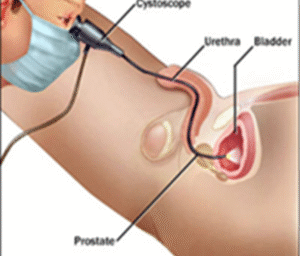
A diagnostic day procedure under general anesthetic, where a rigid cystoscope is placed in the bladder via the urethra

Why is it done?

To investigate:

  • Hematuria (blood in the urine).
  • Recurrent urinary tract infections.
  • Space occupying lesions in the kidneys, ureters and bladder.
  • Abnormal cells suggestive of urothelial carcinoma, on urine cytology.

Risk factors:

  • Strong family history of bladder cancer.
  • Smokers or passive smokers.
  • Factory workers: dyes, paints, etc.
  • Exposure to Schistosoma (Bilharzia).
  • Renal stone disease, bladder stones.

How is it done?

  • This is done under general anesthesia.
  • A cystoscopy is performed by placing a camera in the urethra with the help of a lubricant gel and saline irrigation.
  • The bladder is then distended with saline.
  • The inside of the bladder is viewed for pathology.
  • A retrograde pyelogram could be done at the same time, (placement of small silicone catheters up the kidney pipes). Through this iodine contrast is injected up into the kidney collecting systems. This facilitates the viewing of kidney pipes and kidney collecting systems on X-ray to exclude any upper tact pathology.
  • If any abnormalities are found in the kidney/ ureters, a ureteroscopy (which is the placement of a long thin camera up the ureter) will be performed.
  • If any suspicious lesions are seen, a biopsy will be taken.
  • Urine would have been sent for cytology, to rule out the existence of cancer.
  • Antibiotics may be given to prevent infection.

What to expect after the procedure?

  • Pain on initial passing of urine.
  • Bladder infection ranging from a burning sensation to, fever, to puss (rare).
  • Blood stained urine.
  • Lower abdominal discomfort which will persist for a few days.
  • NB! Each person is unique and for this reason symptoms vary.

What next?

  • This all depends on what is found during the procedure. All the options will be discussed in detail.
  • With the removal of stents, the ureters have been dilated and will regain function (peristalsis) as soon as the stents are out. Thus slight pain can be expected in the first 24-48hrs.
  • There may be some blood in the urine. This can be remedied by drinking plenty of fluids until it clears.
  • A ward prescription will be issued to patients on discharge, for own collection at any pharmacy.
  • Patients should schedule a follow-up appointment within 7 days.
  • Please don’t hesitate to direct all further queries to Jo.
  • REMEMBER: THOSE WHO SUFFER IN SILENCE, SUFFER ALONE!

Download Information Sheet

Wes Cystoscopy RIGID

Supra-Pubic Catheter

Invasive placement of a silicone tube in a percutaneous supra-pubic puncture site. This is secured inside the bladder (with a balloon) and attached to a drainage bag on the outside, in order to drain an obstructed bladder.

Why is it done?

  • This can be placed as an emergency for patients in acute urinary retention
  • Patients requiring long term catheterization especially spinal cord injury patients
  • Failed urethral catheterization
  • Severe prostate obstruction
  • Urethral strictures
  • Severe sepsis of the urogenital area where diverting urine away from the area is advisable
  • Urethral catheterization impossible


How is it done?

  • Usually done under general anesthesia.
  • This is done as a sterile procedure; therefore, the genital area and suprapubic area will be cleaned with a non-abrasive dis-infectant.
  • A flexible cystoscopy will be placed to inspect the bladder, allow filling with saline and visualize the puncture with a cannula from the skin (outside)
  • A 1cm incision is then made in the midline of the lower abdomen, approximately 2cm above the pubic bone
  • An appropriate size catheter (14-16Fr) will be inserted using a trocar method
  • Correct placement is confirmed with the cystoscopy (direct vision)
  • An anchoring balloon will be inflated with 10cc of sterile water.
  • A drainage urine bag will be attached
  • The catheter will be secured to your leg. (check that this is always secured)


Complications

  • Side effects from a general anesthetic.
  • Bleeding from the wound site. (Anti-coagulants should have been ceased a week prior)
  • Depending on the size of your bladder a possible bowel injury could occur, the odds of this happing will be discussed with you prior to your procedure.


Download Information Sheet

Wes Catheters Suprapubic Catheter CHANGE

Copyright 2019 Dr Jo Schoeman

Trans-Urethral Cauterization of Bladder Bleeders + Evacuation Clots

 Why is it done?

Primary management of:

  • Continuous or intermittent bleeding from bladder vessels
  • More prominent after radiation therapy with neo-vascularisation
  • Induced or aggravated by blood thinning and anti-platelet therapy


Risk factors:

  • Anti-coagulation therapy: Warfarin, Xaralto etc
  • Anti-platelet therapy
  • Radiation to bladder prostate or bowel
  • These need to be stopped prior to the procedure


How is it done?

  • This is done under General anaesthesia.
  • A cystoscopy is performed by placing a camera in the urethra with the help of a lubricant gel and saline irrigation.
  • The bladder is then distended with saline.
  • Clots are evacuated with an evacuator
  • A resectoscope is then placed.
  • I use Bi-polar resection, thus using Saline as irrigation.
  • The offending bleeding vessels are cauterized and sealed
  • A 3-way catheter is placed with continuous saline irrigation until your urine is clear
  • Antibiotics may be given to prevent infection.


Complications

Side–effects

  • You may have a 22 –24 French (thick) 3-way urethral catheter placed through your urethra.
  • It does have a channel for placement of constant saline irrigation and another for the drainage of the blood-stained urine.
  • The Continuous bladder irrigation will continue until your urine is clear approximately 24-48hrs.
  • This can also be remedied by drinking plenty of fluids until it clears.
  • As soon as the colour of your urine is satisfactory, your catheter will be removed.


Download Information Sheet

Wes TU cauterisation of bladder bleeders

Copyright 2019 Dr Jo Schoeman

Trans-Urethral Resection of Bladder Tumour – TURBT

Trans Urethral resection of bladder tumour with/without MMC

An endoscopic procedure where bladder tumours are excised via the urethra.

Why is it done?

Primary management of:

  • Resect a bladder lesion suspicious of bladder cancer
  • Three Types of bladder cancer:
    • Urothelial Carcinoma (85%)
    • Squamous Cell carcinoma
    • Adeno carcinoma
  • Metastatic cancer to the bladder – i.e. Breast, Cervical, Adeno carcinoma of bowel.
  • Other space occupying lesions in the bladder: infection granulomas, abscess from diverticulitis etc.
  • Abnormal cells suggestive of urothelial carcinoma, on urine cytology.

Risk factors:

  • Strong family history of bladder cancer.
  • Smokers or passive smokers.
  • Factory workers: dyes, paints, etc.
  • Exposure to Schistosoma (Bilharzia).
  • Renal stone disease, bladder stones.

How is it done?

  • This is done under General anesthesia.
  • A cystoscopy is performed by placing a camera in the urethra with the help of a lubricant gel and saline irrigation.
  • The bladder is then distended with saline.
  • A resectoscope is then placed.
  • I use Bi-polar resection, thus using saline as irrigation.
  • The tumour or tumours are resected as complete as possible.
  • Deep resection of the tumour base is done to exclude deep muscle invasive tumours.
  • In tumours where it is clearly muscle-invasive, less extensive surgery is done, as this patient may benefit from a cystectomy.
  • 40mg of Intravesical Mitomycin C is routinely placed inside your bladder for an hour after the surgery.
  • A 3-way catheter is placed with continuous saline irrigation until your urine is clear.
  • Antibiotics may be given to prevent infection.

Mitomycin C

Chemotherapeutic agent providing 40% lower incidence of Urothelial Cancer recurrence if placed within 6-8 hours inside the bladder after a TURBT.

What to expect after the procedure?

  • Blood stained urine.
  • Lower abdominal discomfort which will persist for a few days.
  • Catheter induced discomfort.
  • NB! Each person is unique and for this reason symptoms vary.
  • Small risk (<1%) of bladder perforation, causing you to have a laparoscopy with repair of bladder and wash-out of peritoneal cavity with Sterile Water.

What next?

  • You may have a 22 French (thick) 3-way urethral catheter placed through your urethra.
  • It does have a channel for placement of constant saline irrigation and another for the drainage of the blood-stained urine.
  • The continuous bladder irrigation will continue until your urine is clear approximately 24-48hrs.
  •  This can also be remedied by drinking plenty of fluids until it clears.
  • As soon as the colour of your urine is satisfactory, your catheter will be removed.
  • Staging of your cancer will be arranged to be reviewed on your review appointment.
  • A ward prescription will be issued to patients on discharge, for own collection at any pharmacy.
  • Patients should schedule a follow-up appointment within 7-14 days.
  • Please don’t hesitate to direct all further queries to Jo.
  • REMEMBER: THOSE WHO SUFFER IN SILENCE, SUFFER ALONE!

Staging

  • CT IVP.
  • CT Chest.
  • Bone scan.
  • Renal Function and Liver Function Tests.
  • MRI where an allergy to Iodine or Renal Impairment.

Download Information Sheet

Wes TURBT