
Intravesical Drugs for Non-Muscle-Invasive Bladder Cancer
Treating the tumour and reducing the risk of it coming back
Bladder cancer has an inconvenient habit: even after a visible tumour has been completely removed, new tumours can sometimes return elsewhere in the bladder.
For selected patients with non-muscle-invasive bladder cancer (NMIBC), previously often called superficial bladder cancer, Mitomycin C can be placed directly into the bladder to destroy residual cancer cells and reduce the chance of recurrence. It is one of the therapies available. It will be discussed on its own.
This treatment is called intravesical Mitomycin C.
The advantage of intravesical treatment is rather elegant: instead of sending chemotherapy around the entire body, the medication is delivered directly to the bladder lining where it is needed.
What is Mitomycin C?
Mitomycin C is an anti-cancer chemotherapy medication originally derived from Streptomyces bacteria.
When used for bladder cancer, the medication is usually introduced directly into the bladder through a urinary catheter rather than being given intravenously.
This allows a relatively high concentration of chemotherapy to come into direct contact with the urothelium while limiting systemic exposure.
Intravesical Mitomycin is used particularly following transurethral resection of bladder tumour (TURBT) and in selected patients requiring an induction course of intravesical chemotherapy.
Why is Mitomycin C used?
The first and most important treatment for most visible non-muscle-invasive bladder tumours is a TURBT.
During TURBT, the visible tumour is removed and tissue is sent to pathology to determine:
- tumour type
- tumour grade
- depth of invasion
- presence or absence of carcinoma in situ (CIS)
- whether muscle is present in the specimen
- whether cancer has invaded the bladder muscle.
The pathology results allow the tumour to be classified into a recurrence and progression risk category.
Mitomycin C may then be recommended to reduce the risk of tumour recurrence.
When is Mitomycin C used?
There are several different situations in which intravesical Mitomycin C may be considered.
1. A single dose immediately after TURBT
In appropriately selected patients, a single dose of intravesical chemotherapy can be given following TURBT.
Australian eviQ guidance describes administration within 24 hours and preferably within six hours of TURBT. AUA guidance similarly recommends considering a single postoperative instillation within 24 hours for suspected or known low- or intermediate-risk disease.
The purpose is to destroy:
- microscopic tumour cells remaining after resection
- free-floating tumour cells released during surgery
- tumour cells that might otherwise implant elsewhere on the bladder lining.
Think of TURBT as removing the weeds you can see, while the immediate intravesical treatment targets some of the microscopic seeds left behind.
Importantly, a single postoperative instillation primarily reduces recurrence. It has not been shown to provide the same benefit for progression or survival.
2. Induction Mitomycin C
Some patients with intermediate-risk NMIBC may benefit from a course of intravesical chemotherapy rather than a single treatment.
A commonly used regimen is:
Mitomycin C once weekly for six weeks.
Australian eviQ protocols include a six-week induction course for appropriate patients with Ta, T1 or CIS-containing urothelial carcinoma classified as intermediate risk, as an alternative to BCG in selected circumstances.
The exact treatment schedule should be individualised according to:
- tumour grade
- pathological stage
- tumour size
- number of tumours
- previous recurrence rate
- previous intravesical therapy
- presence of CIS
- tolerance of treatment
- overall risk of progression.
What about aggressive superficial bladder cancer?
The term “superficial bladder cancer” can be misleading.
A cancer can remain confined to the bladder lining or lamina propria and still behave aggressively.
Higher-risk features include:
- high-grade urothelial carcinoma
- T1 disease
- carcinoma in situ
- multiple tumours
- large tumours
- frequently recurrent disease
- certain adverse pathological features.
For genuinely high-risk NMIBC, particularly high-grade T1 disease and CIS, intravesical BCG immunotherapy generally has a central role when bladder preservation is appropriate.
Mitomycin C should therefore not automatically be regarded as a substitute for BCG in every patient with aggressive NMIBC.
Treatment needs to be based on the patient’s pathological risk group and previous response to therapy.
In selected circumstances, Mitomycin may nevertheless be considered, including when BCG is unsuitable, poorly tolerated, unavailable or as part of specific sequential intravesical treatment protocols.
Patients with very-high-risk disease also require discussion about whether bladder-preserving intravesical treatment remains appropriate or whether radical cystectomy should be considered.
How does Mitomycin C work?
Mitomycin C is an alkylating anti-tumour agent.
After entering a cancer cell, the drug is activated and produces reactive metabolites that bind to DNA.
It causes cross-linking of DNA strands, preventing the DNA from separating normally.
As a result, the cancer cell has difficulty:
- replicating its DNA
- dividing
- repairing damaged DNA
- continuing normal cellular function.
Ultimately, susceptible tumour cells die.
Because Mitomycin is placed directly into the bladder, it can attack residual malignant cells while generally producing much less systemic exposure than intravenous chemotherapy.
What happens during treatment?
Intravesical treatment is usually performed as an outpatient procedure.
A small urinary catheter is passed through the urethra into the bladder.
The bladder is emptied completely and the Mitomycin solution is then introduced through the catheter.
The catheter may then be removed or temporarily clamped, depending upon the treatment protocol.
The medication is usually retained within the bladder for approximately one to two hours.
Patients are generally advised to restrict fluids beforehand so that the medication is not excessively diluted. Australian eviQ protocols advise restricting fluids for approximately four to six hours before and during treatment, with appropriate adjustment of diuretic medication where relevant.
After the prescribed treatment period, the medication is passed out with the urine or drained through the catheter.
Side effects of intravesical Mitomycin C
Most patients tolerate treatment reasonably well because relatively little of the medication is normally absorbed into the bloodstream.
The commonest problems involve irritation of the bladder.
Chemical cystitis
Mitomycin can irritate the bladder lining and cause:
- burning when passing urine
- urinary frequency
- urgency
- bladder discomfort
- pelvic discomfort
- mild haematuria.
These symptoms frequently develop within hours of treatment and usually settle over the following few days.
Urinary tract infection
A urinary infection can occasionally occur following catheterisation.
Symptoms may include:
- increasing dysuria
- cloudy or offensive urine
- fever
- chills
- worsening urinary frequency
- feeling generally unwell.
A urine culture may be required if infection is suspected.
Skin irritation
Mitomycin is a cytotoxic medication and contact with the skin should be avoided.
Urine containing residual medication may irritate the skin around the urethra or genital area.
Patients should therefore follow the specific hygiene and toilet precautions provided by their treating unit.
Allergic reaction
Hypersensitivity reactions are uncommon but possible.
Symptoms can include:
- skin rash
- itching
- facial redness
- wheezing
- breathlessness
- dizziness
- fever or chills.
Severe allergic reactions are rare but require urgent medical attention.
Less common but important complications
Although uncommon, Mitomycin can occasionally produce significant complications.
Severe chemical cystitis
Repeated bladder irritation can occasionally become severe.
Persistent inflammation may lead to:
- chronic bladder pain
- reduced bladder capacity
- bladder fibrosis
- rarely, bladder contraction.
Patients who already have a small-capacity bladder require particular caution because of the potential risk of further bladder contraction.
Extravasation
One of the most important complications is Mitomycin extravasation.
This means that Mitomycin escapes through an injured or perforated bladder wall into the surrounding tissues.
Although rare, this can produce significant tissue injury.
Reported complications include:
- severe pelvic inflammation
- tissue necrosis
- abscess formation
- bladder injury
- fistula formation
- damage to surrounding genital tissues.
Symptoms can occur immediately, but importantly they may also appear weeks or even months after treatment. Persistent or unexplained pelvic or abdominal pain following intravesical Mitomycin therefore deserves investigation.
An important precaution after TURBT
Intravesical Mitomycin should not simply be administered automatically following every TURBT.
If there is any suspicion that the bladder has been perforated, the medication should not be given.
AUA guidance advises against postoperative intravesical chemotherapy following a suspected perforation or extensive resection. Australian eviQ guidance similarly excludes postoperative Mitomycin when bladder perforation has occurred.
This is important because chemotherapy escaping through a bladder perforation can cause substantial tissue injury.
When should Mitomycin treatment be postponed?
Intravesical Mitomycin is generally avoided or postponed in the presence of:
- visible haematuria
- confirmed urinary tract infection
- traumatic catheterisation
- fever or unexplained febrile illness
- known allergy to Mitomycin
- suspected or confirmed bladder perforation
- significant bladder injury following surgery.
Australian protocols specifically advise against treatment following traumatic catheterisation, with visible haematuria, active UTI or recent bladder injury.
If catheterisation is traumatic or produces bleeding on the day of treatment, the treatment is usually deferred.
Does Mitomycin cause the usual chemotherapy side effects?
Usually not.
Because the medication is placed into the bladder rather than routinely given intravenously, systemic absorption is generally very low.
Therefore classic chemotherapy problems such as:
- hair loss
- severe nausea
- widespread immunosuppression
- profound fatigue
- bone marrow suppression
are unusual with standard intravesical therapy.
Systemic absorption can occur rarely, however, particularly when the bladder lining has been significantly disrupted. Myelosuppression is considered a potential but uncommon complication, and a full blood count may be appropriate if a patient becomes unexpectedly systemically unwell.
Precautions after treatment
Mitomycin remains a cytotoxic chemotherapy drug, even when delivered directly into the bladder.
Patients should therefore follow their treating unit’s instructions carefully regarding:
- toilet hygiene
- handling urine
- washing contaminated skin
- hand washing
- fluid intake following treatment
- management of contaminated clothing.
Patients should also tell their treating team about all medications they are taking and whether they are receiving antibiotics, diuretics or other cancer treatments.
When should you seek urgent medical advice?
Contact your treating team promptly if you develop persistent or worsening urinary symptoms after treatment.
More urgent assessment is required for symptoms such as:
- temperature of 38°C or higher
- inability to pass urine
- significant or persistent haematuria
- severe pelvic or abdominal pain
- chills or shaking
- shortness of breath
- severe rash or suspected allergic reaction
- persistent symptoms that are worsening rather than settling.
Severe pelvic or abdominal pain following intravesical Mitomycin is particularly important because, although uncommon, it may indicate bladder injury or extravasation.
Mitomycin does not replace surveillance
One of the most important points about NMIBC is that successful treatment does not mean surveillance can stop.
Even after apparently complete removal and intravesical chemotherapy, urothelial tumours can recur.
Depending upon the original tumour’s risk category, ongoing surveillance may include:
- flexible cystoscopy
- urine cytology
- repeat TURBT
- upper urinary tract imaging
- further intravesical treatment.
Patients with higher-risk disease require considerably closer surveillance than those with a solitary low-grade tumour.
Mitomycin C versus BCG
These treatments are sometimes discussed together, but they work differently.
Mitomycin C is chemotherapy. It directly damages the DNA of susceptible tumour cells.
BCG is immunotherapy. It stimulates a local immune response within the bladder that attacks urothelial cancer cells.
The appropriate treatment depends upon the biological behaviour and risk category of the tumour.
For some patients, Mitomycin provides an effective and relatively well-tolerated strategy for reducing recurrence. For patients with higher-risk disease, BCG or more aggressive treatment may be preferable.
The important question is therefore not simply:
“Mitomycin or BCG?”
It is:
“What is the risk of this particular bladder cancer recurring or progressing, and which treatment provides the safest and most effective way of reducing that risk?”
The Bottom Line
Intravesical Mitomycin C is an established treatment for selected patients with non-muscle-invasive urothelial carcinoma of the bladder.
It may be used as a single treatment immediately following TURBT or as a course of intravesical chemotherapy in appropriately selected patients.
By placing chemotherapy directly into the bladder, Mitomycin can destroy residual cancer cells and reduce tumour recurrence while limiting exposure of the rest of the body.
Most side effects are related to temporary bladder irritation. Serious complications such as bladder injury, extravasation, fibrosis and tissue necrosis are uncommon but important, which is why careful patient selection and attention to precautions are essential.
And perhaps the most important message: Mitomycin reduces risk. It does not make follow-up optional.
Bladder cancer has a reputation for making return appearances, so regular cystoscopic surveillance remains an essential part of treatment.
This information is intended for general education and should not replace individual medical advice. Treatment of non-muscle-invasive bladder cancer should be tailored to the tumour’s stage, grade and recurrence/progression risk, previous treatments and the individual patient’s circumstances.


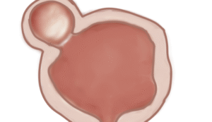
 Patients will receive a general anaesthesia, unless contra-indicated.