Flexible Cystoscopy

An atraumatic endoscopic procedure to view the bladder. Under local or sedation

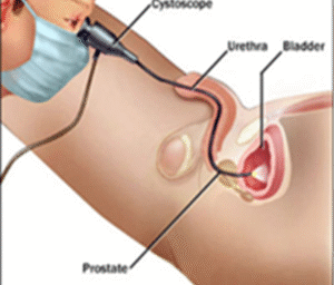
A diagnostic day procedure under local anaesthetic, where a flexible cystoscope is placed in the bladder via the urethra.
Why is it done?

To investigate:

  • Haematuria (blood in the urine).
  • Recurrent urinary tract infections.
  • Space occupying lesions in the kidneys, ureters and bladder.
  • Abnormal cells suggestive of urothelial carcinoma, on urine cytology.

Risk factors:

  • Strong family history of bladder cancer.
  • Smokers or passive smokers.
  • Factory workers: dyes, paints, etc.
  • Exposure to Schistosoma (Bilharzia).
  • Renal stone disease, bladder stones.

How is it done?

  • A cystoscopy is performed by placing a camera in the urethra with the help of a lubricant jelly and saline.
  • The bladder is then distended using the fluid.
  • The inside of the bladder is viewed for pathology.
  • If any suspicious lesions are seen, a biopsy will be taken.
  • Urine would have been sent for cytology prior to the procedure, to rule out the existence of cancer.
  • Antibiotics may be given to prevent infection.

What to expect after the procedure?

  • Pain on initial passing of urine.
  • Bladder infection ranging from a burning sensation to, fever, to puss (rare).
  • Blood stained urine.
  • Lower abdominal discomfort which will persist for a few days.
  • NB! Each person is unique and for this reason symptoms vary.

What next?

  • This all depends on what is found during the procedure. All the options will be discussed in detail.
  • With the removal of stents, the ureters have been dilated and will regain function (peristalsis) as soon as the stents are out. Thus slight pain can be expected in the first 24-48hrs.
  • There may be some blood in the urine. This can be remedied by drinking plenty of fluids until it clears.
  • A ward prescription will be issued to patients on discharge, for own collection at any pharmacy.
  • Patients should schedule a follow-up appointment within 7 days.
  • Please don’t hesitate to direct all further queries to Jo.
  • REMEMBER: THOSE WHO SUFFER IN SILENCE, SUFFER ALONE!

Download Information Sheet
Wes Flexible Cystoscopy

Wes Flexible Cystoscopy and Removal Stent

Flexible Cystoscopy

Cysto-Lithotomy

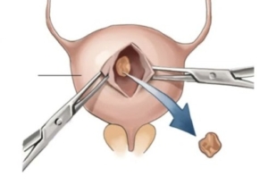
Open removal of a large bladder calculus

Why is it done?

  • To break up a large bladder calculus (stone) that cannot be done endoscopically.
  • It is done with open surgery (a cut above the pubic symphysis).

Risk factors causing this:

    • Bladder outflow obstruction
      • BPH with chronic retention.
      • Urethral stricture.
      • Neurogenic bladder.
    • Renal calculi disease.
    • Metabolic disorders.
    • Malnutrition.
    • Chronic infections.
    • Foreign objects in bladder.

How is it done?

  • A General anesthetic will be given.
  • A sterile surgical field is prepared.
  • Prophylactic antibiotics are given.
  • An indwelling catheter is inserted, and the bladder is then distended with fluid (saline).
  • A small lower abdominal incision is made, splitting the Linea alba and opening the distended bladder in the midline.
  • The stone is removed through the whole with a grasping instrument.
  • The bladder is inspected and then closed in 2 layers.
  • Skin is closed.
  • A catheter will be left for 2 weeks.

What to expect after the procedure?

  • Hematuria (blood in your urine)
  • You will have an indwelling catheter (IDC), which will remain in your bladder.
  • You may have a continuous bladder irrigation with Saline to help clear the bleeding.
  • When your urine is clear and your bowels are functioning, you will be discharged with catheter care instruction.
  • You will have this indwelling catheter for 2 weeks.
  • A cystogram will be arranged at approx. 14 days to exclude any urine leaks prior to removal of your catheter.
  • If there are any urine leaks, your catheter will remain a further 7 days, or until the leak is sealed.
  • Pain on initial passing of urine when the catheter is removed.
  • Bladder infection ranging from a burning sensation to, fever, to puss (rare).
  • Lower abdominal discomfort which will persist for a few days.
  • NB! Each person is unique and for this reason symptoms vary.

What next?

  • This all depends on what is found during the procedure. All the options will be discussed in detail.
  • There may be some blood in the urine. This can be remedied by drinking plenty of fluids until it clears.
  • Anatomical causes of the stones will be discussed and surgical options in treatment may be discussed
  • Patients should schedule a follow-up appointment within 4-6 weeks to discuss the etiology of the calculus as well as what other procedures may be involved to prevent this from occurring again.


Download Information Sheet

Wes Cysto-Lithotomy Open

Nephro / Uretero Lithotomy – Robotic Assisted

Robotic removal of stones where endoscopic procedures have failed.


Why is it done?

Open/robotic surgery for large or complicated renal and/or ureteric calculi where all other techniques have failed. Seldomly done today. Only if no other options available.

  • As a last resort to remove a large stone.
  • Robotic would be considered first
  • Where equipment is not available.
  • Same entry as for open nephrectomy.

How is it done

  • GA
  • Entry ports for the robot will be determined by where the stone is
  • A ureteric stent would already be in place, if not, this will be attempted prior to robotic access
  • Kidney:
    • Usually an area of thinned cortex
    • Opened and stone removed
    • Closed
  • Renal Pelvis:
    • Opened and stone removed
    • Closed
  • Ureter:
    • Expose the area of the ureter
    • Compression of ureter above and below the calcalculus, as to prevent migration
    • Ureter opened
    • Stone removed
    • Closed
  • Drain left

Alternatives

  • PCNL.
  • ESWL.
  • Sandwich therapy: Combination of PCNL and ESWL.
  • URSE with laser.


Download Information Sheet

Wes Nephro Uretero-lithotomy Open

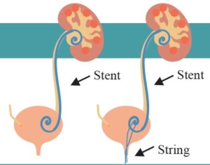
Rigid Cystoscopy, Retrograde Pyelogram, Stent Management

A therapeutic procedure under general anaesthetic, where a rigid cystoscopy is done in the bladder via the urethra, ureteric catheters are placed to enable imaging of the upper tracts with/without insertion or removal of ureteric stents

Why is it done?

To investigate:

  • Hematuria (blood in the urine)
  • Recurrent upper urinary tract infections
  • Space occupying lesions in the kidneys and ureters
  • Abnormal cells suggestive of urothelial carcinoma, on urine cytology


Risk factors:

  • Strong family history of bladder cancer
  • Smokers or passive smokers
  • Factory workers: dyes, paints, etc
  • Exposure to Schistosoma (Bilharzia)
  • Renal stone disease, bladder stones


How is it done?

  • This is done under General anaesthesia.
  • A cystoscopy is performed by placing a camera in the urethra with the help of a lubricant gel and saline irrigation.
  • The bladder is then distended with saline.
  • The inside of the bladder is viewed for pathology.
  • A retrograde pyelogram is done at the same time, (placement of small silicone catheters up the kidney pipes). Through this iodine contrast is injected up into the kidney collecting systems. This facilitates the viewing of kidney pipes and kidney collecting systems on X-ray to exclude any upper tract pathology.
  • If any abnormalities are found in the kidney/ ureters, a ureteroscopy (which is the placement of a long thin camera up the ureter) will be performed.
  • If any suspicious lesions are seen, a biopsy will be taken.
  • A ureteric stent may be placed
  • Urine would have been sent for cytology, to rule out the existence of cancer.

Antibiotics may be given to prevent infection

Complications

What to expect after the procedure?

  • Pain on initial passing of urine
  • Bladder infection ranging from a burning sensation to, fever, to puss (rare)
  • Bloodstained urine
  • Lower abdominal discomfort which will persist for a few days
  • Pain radiating from bladder to renal angle associated with urinating.
  • An infection could present with a stent being present.


Indications for a Ureteric stent

· Hematuria from upper tracts

· Dis-obstruction of the ureter caused either calculus, blood clot or tumour

· External compression of the ureter by retro-peritoneal pathology i.e.: Fibrosis, retroperitoneal lymph node compression

· Reduced renal function associated with hydronephrosis

· Sepsis associated with hydronephrosis


Download Information Sheet

Wes Cystoscopy RGP and Ureteric stents

Copyright 2019 Dr Jo Schoeman

Uretero-Renoscopic Stone Extraction with Laser (URSE)

A ureteric or renal calculus is removed with technique and may require a laser. A rigid/ flexible ureteroscope can be used.

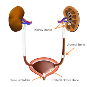
Stones in the kidney, urinary bladder and ureter. medical illustration with a cross section of the kidney and bladder. anatomy of the urinary system. Human kidney.

Why is it done?

  • Removal of renal or ureteric stones.
  • Ureteric stones can vary from 5mm to over 1 cm in size.
  • You may present with excruciating pain on the affected side. (This pain may be worse than childbirth).
  • Renal stones usually larger than 1cm obstructing the renal pelvis.
  • Or renal stones not causing any symptoms.
  • Any fevers or a single kidney is deemed an emergency!

Two Treatment Options

  • Ureteric Calculi.
    • Managed with rigid ureteroscopy.
    • Prior stenting with a ureteric stent,
    • 7-10 days after stenting the stent is removed, and the stone is addressed with laser
  • Renal Calculi.
    • Prior stenting for 7-10 days.
    • After stent removal the kidney is accessed with a flexible uretero-renoscope and the stone is fragmented with laser

How is it done?

    • Patients will receive a general anesthesia.
    • Prophylactic antibiotics is given.
    • The correct kidney is identified and marked while you are awake.
    • You would have had a cystoscopy with retrograde pyelogram 10 days prior with placement of ureteric stent to prepare your ureter.
    • A cystoscopy will be done first to remove the stent, and 2 guidewires will be placed to enable access up the ureter.
    • Depending on the position of the stone, either a rigid or flexible uretero-renoscope will be used.
    • If a stone is in the kidney a flexible uretero-renoscope will be used with access obtained with an access sheath to protect the ureter from damage.
    • Laser will be used to fragment the stone.
    • All fragments will be attempted to be cleared. Small 1-2 mm fragments may be left as “Clinically Insignificant Fragments (CISF)” and will pass spontaneously.
    • A Ureteric catheter with an indwelling catheter is left post-operatively overnight.
    • Catheters will be removed the next morning depending on the presence of blood in the urine.

What next?

  • You will spend at least one night in hospital.
  • You will have a catheter for that time.
  • On removal of your catheter, you may experience sharp colicky pain, exactly the same as your presenting renal colic. This is due to your ureter contracting back to its usual size. (The stent has dilated this to 5X its usual size).
  • You will be discharged as soon as your pain has stabilized and you can function independently.
  • Allow for a few days for stabilization of symptoms.
  • A ward prescription will be issued on your discharge, for your own collection at any pharmacy.
  • A follow-up appointment will be scheduled for 6 weeks. Stone analysis results will then be discussed in order to formulate a plan to proven recurrences
  • DON’T SUFFER IN SILENCE, OR YOU WILL SUFFER ALONE!


Types of Stones:

  • Calcium Oxalate.
  • Uric Acid.
  • Calcium Phosphate.
  • Struvite (Infection stones).
  • Cystine.


Download Information Sheet

Wes Uretero-Renoscopic Stone Extraction with Laser URSE

URSE with or without Laser