Information Sheet: Renal Cancers

Upper Tract Urothelial Carcinoma

Cancer of the Renal Pelvis and Ureter

Urothelial carcinoma is most commonly associated with the bladder, but the same specialised lining extends all the way from the bladder up the ureters and into the collecting system of each kidney.

When a urothelial cancer develops in the renal pelvis or ureter, it is called upper tract urothelial carcinoma (UTUC).

UTUC is considerably less common than bladder cancer. Importantly, its behaviour varies enormously. Some tumours are small, superficial and relatively slow growing, while others are aggressive cancers capable of invading the kidney, surrounding tissues, lymph nodes and distant organs.

The key to successful management is therefore not simply finding the tumour, but determining how aggressive it is and how likely it is to spread.


What Is the Upper Urinary Tract?

Urine produced by the kidney drains through:

Kidney → renal calyces → renal pelvis → ureter → bladder → urethra

The inner surface of the renal pelvis, ureter and bladder is covered by urothelium, previously called transitional epithelium.

Cancer arising from these cells is called urothelial carcinoma.

UTUC can therefore occur in:

  • the renal pelvis;
  • one or more renal calyces;
  • the upper, middle or lower ureter;
  • multiple areas of the same urinary tract; or
  • occasionally both upper urinary tracts.

Patients with UTUC may also develop urothelial tumours within the bladder, either at the same time or later.


What Causes Upper Tract Urothelial Cancer?

Cancer develops when genetic damage accumulates within urothelial cells, allowing them to grow and divide abnormally.

Frequently there is no single identifiable cause, but several important risk factors are recognised.

Smoking

Cigarette smoking is one of the most important preventable risk factors for urothelial carcinoma.

Carcinogenic chemicals absorbed through the lungs enter the bloodstream, are filtered by the kidneys and become concentrated in the urine. The urothelium may therefore be exposed to these substances for many years.

Stopping smoking remains important even after diagnosis because of its wider health benefits and potential relevance to future urothelial cancer risk.

Previous bladder cancer

Patients who have previously had urothelial carcinoma of the bladder have an increased risk of developing urothelial carcinoma elsewhere within the urinary tract.

Similarly, patients treated for UTUC remain at risk of subsequently developing bladder cancer.

Occupational chemical exposure

Long-term exposure to certain industrial chemicals, particularly some aromatic amines historically associated with dye, rubber, textile, leather and chemical industries, has been associated with urothelial cancer.

Lynch syndrome

A small but important proportion of UTUC occurs in people with Lynch syndrome, an inherited disorder caused by abnormalities in DNA mismatch-repair genes.

UTUC, particularly in a younger patient or someone with a strong family history of bowel, endometrial or other Lynch-associated cancers, may prompt consideration of genetic assessment.

Chronic inflammation

Long-standing urinary tract inflammation and some chronic infections may contribute to malignant change. Chronic stone disease has particularly been associated with squamous carcinoma of the renal pelvis, which is a different and much less common tumour type.

Previous analgesic exposure

Heavy historical exposure to certain analgesics, particularly phenacetin-containing preparations that are no longer routinely used in many countries, has been associated with upper tract urothelial malignancy.


What Are the Symptoms?

Blood in the urine

The most common warning sign is:

Haematuria: blood in the urine

This may be obvious, turning the urine pink, red or occasionally dark brown.

Importantly, haematuria may:

  • occur only once;
  • disappear for weeks or months;
  • be completely painless; or
  • only be detected microscopically on a urine test.

Visible blood in the urine should always be appropriately investigated, particularly in adults.

Do not assume that painless bleeding is simply due to infection, prostate enlargement or a blood-thinning medication without appropriate assessment.


Flank or loin pain

A tumour can obstruct drainage of urine from the kidney.

This may produce:

  • persistent flank discomfort;
  • loin pain;
  • renal colic;
  • hydronephrosis, where the kidney becomes dilated because urine cannot drain normally.

Blood clots passing down the ureter can occasionally produce severe colicky pain resembling a kidney stone.


Recurrent urinary symptoms

Some patients experience:

  • urinary frequency;
  • urgency;
  • burning when passing urine;
  • recurrent apparent urinary tract infections.

These symptoms are much more commonly caused by benign conditions, but persistent or unexplained symptoms may warrant further investigation.


General symptoms

More advanced disease can occasionally cause:

  • unexplained weight loss;
  • loss of appetite;
  • fatigue;
  • persistent pain;
  • anaemia.

Fortunately, many tumours are detected before these symptoms develop.


How Is UTUC Diagnosed?

Investigation usually involves several complementary tests.

1. Urine testing

Urine is assessed for:

  • microscopic blood;
  • infection;
  • renal abnormalities; and
  • sometimes malignant cells.

Urine cytology

Urine cytology examines shed urinary cells under a microscope.

It is particularly useful for detecting high-grade urothelial carcinoma, although a negative cytology result does not completely exclude cancer.

Selective urine samples may sometimes be collected directly from the affected upper urinary tract.


2. CT Urography

CT urography is one of the most important imaging investigations for suspected UTUC.

Contrast-enhanced CT imaging allows assessment of:

  • the kidneys;
  • renal collecting systems;
  • ureters;
  • bladder;
  • lymph nodes; and
  • surrounding organs.

A tumour may appear as a filling defect, thickening of the ureter or renal pelvis, or an infiltrating mass.

CT can also identify obstruction and hydronephrosis.


3. Cystoscopy

Because urothelial cancer can occur at more than one location, the bladder should generally also be examined.

A flexible cystoscope is passed through the urethra into the bladder to look for associated bladder tumours.


Ureteroscopy and Biopsy

Sometimes imaging alone cannot provide enough information about the tumour.

A ureteroscope is a very fine telescope passed through the urethra and bladder and then into the ureter and renal collecting system.

This allows the urologist to directly inspect the tumour.

During ureteroscopy it may be possible to:

  • identify the location of the tumour;
  • assess whether there is one tumour or several;
  • estimate its size;
  • obtain selective urine cytology;
  • take a biopsy; and
  • in selected low-risk tumours, treat the lesion with a laser.

Biopsy is particularly helpful in determining whether the tumour is low-grade or high-grade.

An important limitation is that ureteroscopic biopsy samples are small, so determining the exact depth of invasion before definitive surgery can sometimes be difficult.


Low-Risk Versus High-Risk Disease

Modern management increasingly divides UTUC according to its risk of invasion and progression.

Factors considered include:

  • tumour grade;
  • appearance on CT;
  • tumour size;
  • number of tumours;
  • presence of hydronephrosis;
  • urine cytology;
  • ureteroscopic appearance;
  • biopsy findings;
  • evidence of invasion; and
  • previous urothelial cancer.

This distinction is extremely important because treatment can range from relatively conservative endoscopic therapy to removal of the entire kidney and ureter.


Treatment of Low-Risk UTUC

Selected patients with low-risk disease may be suitable for kidney-sparing treatment.

The objective is to control the cancer while preserving as much functioning kidney tissue as possible.

Ureteroscopic Laser Treatment

A ureteroscope is passed to the tumour and the lesion is treated using laser energy.

Laser treatment can:

  • vaporise;
  • coagulate; or
  • fragment the tumour.

Modern flexible ureteroscopes allow access to much of the renal collecting system.

Advantages

Kidney-sparing treatment may:

  • preserve kidney function;
  • avoid major abdominal surgery;
  • reduce recovery time;
  • be particularly valuable in patients with impaired kidney function or a solitary kidney.

Disadvantages

The trade-off is the need for very careful surveillance.

UTUC has a tendency to recur, so patients may require repeated:

  • ureteroscopy;
  • urine cytology;
  • CT imaging; and
  • occasionally further laser treatments or biopsies.

Kidney preservation therefore does not mean that the tumour can simply be treated once and forgotten.


Segmental Ureterectomy

Some tumours confined to a relatively short section of ureter can be treated by removing only the affected portion.

This is known as segmental ureterectomy.

It is particularly useful for appropriately selected tumours of the distal ureter.

The affected segment is removed and the remaining ureter is either reconnected or reimplanted into the bladder.

This can preserve the kidney while providing a complete surgical specimen for pathological examination.


Treatment of High-Risk UTUC

For patients with high-risk localised disease, the standard definitive operation is generally:

Radical Nephroureterectomy

This involves removal of:

the kidney + entire ureter + a cuff of bladder surrounding the ureteric opening

Removing the complete ureter is important because leaving part of the affected ureter behind may allow cancer to recur within the remaining urothelium.

The procedure can often be performed using:

  • robotic surgery;
  • laparoscopic surgery; or
  • occasionally open surgery.

The choice depends upon tumour characteristics, anatomy, previous surgery and individual circumstances.


What About the Lymph Nodes?

For selected high-risk tumours, lymph nodes draining the affected area may also be removed.

A lymph-node dissection can provide important staging information and may be incorporated into the surgical management of appropriately selected high-risk disease.


Chemotherapy

Systemic chemotherapy plays an important role in selected patients with higher-risk UTUC.

Platinum-based chemotherapy, particularly cisplatin-containing treatment, has traditionally formed an important part of systemic treatment.

Chemotherapy may be given:

Before surgery: neoadjuvant chemotherapy

One potential advantage is that both kidneys are still present, and renal function may therefore be better able to tolerate cisplatin.

After surgery: adjuvant chemotherapy

Chemotherapy may be recommended after nephroureterectomy when the final pathology demonstrates sufficiently high-risk disease.

An important consideration is that removing a kidney can reduce renal function, potentially affecting the patient’s ability to receive certain chemotherapy drugs.

Treatment decisions are therefore ideally made by a multidisciplinary uro-oncology team.


Immunotherapy and Newer Treatments

The treatment of advanced urothelial carcinoma has changed considerably in recent years.

Depending upon the stage, previous treatment and molecular characteristics of the cancer, systemic therapy may include combinations of:

  • platinum-based chemotherapy;
  • immune checkpoint inhibitors;
  • antibody-drug conjugates; and
  • targeted treatments for selected molecular abnormalities such as FGFR alterations.

These treatments are particularly relevant to patients with locally advanced, recurrent or metastatic disease.

Treatment is increasingly individualised according to the biological characteristics of the tumour and the patient’s kidney function and overall health.


Treatment Placed Directly Into the Upper Urinary Tract

For carefully selected non-invasive disease, medication may sometimes be delivered directly into the renal pelvis and ureter.

Options can include topical chemotherapy or immunotherapy in selected circumstances.

A chemoablative gel formulation of mitomycin has also been developed for selected low-grade UTUC, allowing prolonged contact between chemotherapy and the tumour.

These treatments are specialised and are not suitable for every patient.


What Happens After Treatment?

Follow-up is particularly important because urothelial carcinoma has a characteristic ability to develop at another point along the urinary tract.

After treatment, surveillance may include:

  • cystoscopy;
  • urine cytology;
  • CT urography or other upper-tract imaging;
  • blood tests to assess kidney function;
  • ureteroscopy following kidney-sparing treatment.

The exact surveillance schedule depends upon whether the original tumour was low or high risk and what treatment was performed.


Can the Cancer Return in the Bladder?

Yes.

One of the distinctive features of UTUC is the significant risk of subsequently developing urothelial carcinoma within the bladder.

Published data have reported subsequent bladder tumours in a substantial proportion of patients following treatment of UTUC.

For this reason, regular cystoscopic surveillance remains important even when the original kidney or ureteric tumour has been completely removed.


What Is the Prognosis?

The outlook for UTUC depends predominantly upon:

  • tumour stage;
  • tumour grade;
  • lymph-node involvement;
  • metastatic spread;
  • tumour multifocality;
  • response to treatment; and
  • certain pathological and molecular characteristics.

The most important question is whether the cancer remains superficial or has invaded deeply through the wall of the renal pelvis or ureter.

Superficial, localised disease

The outlook can be excellent when a tumour is identified while still superficial and confined to the upper urinary tract. The National Cancer Institute reports that more than 90% of superficial cancers confined to the renal pelvis or ureter may be curable.

Invasive disease

Once a tumour has invaded deeply into the wall or surrounding tissues, the risk of lymph-node involvement and distant spread increases substantially.

High-grade and invasive tumours therefore usually require more aggressive treatment and closer surveillance.

Metastatic disease

When UTUC has spread to distant organs, treatment is generally systemic rather than surgical alone.

Although metastatic UTUC remains a serious disease, modern systemic treatments have expanded considerably and can provide meaningful cancer control for selected patients.


UTUC and Kidney Function

Preserving kidney function is an important part of treatment planning.

Removing one kidney is usually well tolerated when the opposite kidney is healthy, but kidney function can become particularly important in patients with:

  • pre-existing chronic kidney disease;
  • diabetes;
  • hypertension;
  • a solitary kidney;
  • bilateral upper-tract tumours; or
  • conditions likely to affect future kidney function.

The decision between kidney-sparing treatment and radical nephroureterectomy therefore involves balancing two priorities:

adequate cancer control and preservation of renal function.

Cancer safety remains the priority, but in appropriately selected low-risk disease these objectives can often coexist.


When Should You See a Urologist?

Seek medical assessment if you develop:

  • visible blood in the urine;
  • persistent microscopic haematuria;
  • unexplained flank or loin pain;
  • recurrent unexplained urinary symptoms;
  • abnormal findings on kidney imaging; or
  • haematuria with a previous history of urothelial cancer.

Blood in the urine should not automatically be attributed to prostate enlargement, infection, kidney stones or blood-thinning medication without appropriate investigation.


The Bottom Line

Upper tract urothelial carcinoma is an uncommon cancer arising from the lining of the renal pelvis or ureter.

Blood in the urine is its most important warning sign.

Investigation commonly involves CT urography, cystoscopy, urine cytology and, when required, ureteroscopy with biopsy.

Treatment is increasingly tailored to the biological risk of the tumour.

Small, low-grade tumours may sometimes be managed with kidney-sparing ureteroscopic laser treatment or segmental surgery, while high-risk invasive cancers generally require radical nephroureterectomy with removal of the bladder cuff, sometimes combined with lymph-node surgery and systemic treatment.

Long-term surveillance is essential because urothelial cancer can recur elsewhere within the urinary tract, particularly the bladder.

The reassuring part is that when UTUC is detected while still superficial and localised, the likelihood of successful treatment can be very high.


A Note for Patients

Every upper tract urothelial tumour is different. Treatment depends upon the tumour’s location, size, grade, stage, number of lesions, kidney function and your general health.

Management should therefore be individualised following discussion with your urologist and, for higher-risk disease, a multidisciplinary uro-oncology team.

This information is intended for general patient education and should not replace individual medical advice, examination or treatment recommendations from your treating specialist.

So, if you have any of the above symptoms, come see your local Brisbane Urologist, Dr Jo Schoeman at any of my 2 locations, Wesley and Caboolture Hospitals

The current 2026 EAU guideline specifically incorporates updated risk stratification, kidney-sparing management, bladder-cuff and lymph-node considerations, systemic therapy, and follow-up recommendations.

2026 EAU Upper Urinary Tract Urothelial Carcinoma Guideline
National Cancer Institute patient information on renal pelvis and ureter urothelial cancer

Small Renal Masses: When a Small Kidney Tumour Needs Watching, Treating or Removing

Finding a small mass on the kidney can be unsettling, particularly when it appears unexpectedly on an ultrasound or CT scan performed for something completely unrelated.

The reassuring news is that small does not automatically mean dangerous, and a renal mass does not automatically mean kidney cancer. Even when a small renal mass is malignant, many grow slowly and have a low risk of spreading. This means that immediate surgery is not always necessary.

Modern management has therefore moved away from a simple philosophy of “there is a lump, so it must come out”. Instead, treatment is tailored to the size and appearance of the mass, its growth rate, kidney function, the patient’s age and general health, and individual preferences.

Options range from:

  • Active surveillance
  • Percutaneous biopsy
  • Minimally invasive ablation, including cryoablation and thermal techniques
  • Robotic partial nephrectomy
  • Occasionally, radical nephrectomy

The aim is straightforward: control the cancer when treatment is necessary while preserving as much normal kidney function as possible.


What Is a Small Renal Mass?

A small renal mass (SRM) generally refers to a kidney tumour measuring 4 cm or less, corresponding to a clinical T1a renal tumour when confined to the kidney.

Many are discovered incidentally during imaging performed for abdominal pain, gallstones, bowel problems, back pain or another unrelated condition. The kidney lesion is often an unexpected passenger on the scan.

Importantly, not every small renal mass is cancer.

Benign lesions include:

  • Oncocytoma
  • Angiomyolipoma
  • Some complex renal cysts
  • Other uncommon benign tumours

Among malignant masses, renal cell carcinoma is the most important diagnosis. However, many small renal cancers demonstrate relatively indolent behaviour. Published guideline data suggest that more than 20% of small renal masses may ultimately prove benign, while many malignant lesions within this size range have relatively favourable biology.

This creates an important clinical dilemma: we want to treat a potentially dangerous cancer, but we also want to avoid unnecessary treatment of a tumour that may never have caused harm.


Step One: Properly Characterising the Mass

An incidental renal mass generally requires appropriate imaging before deciding what to do.

This usually involves a:

  • Multiphasic contrast CT scan, or
  • MRI scan

These scans help determine:

  • Exact tumour size
  • Whether the lesion is solid or cystic
  • Whether it enhances with contrast
  • Its position within the kidney
  • Relationship to blood vessels and the collecting system
  • Whether there are concerning features suggesting malignancy
  • Whether there is any evidence of disease outside the kidney

High-quality contrast CT or MRI is recommended for the characterisation of a newly discovered small renal mass.

Blood tests will usually include kidney function, particularly serum creatinine and estimated glomerular filtration rate (eGFR).


What About Complex Kidney Cysts?

Not every suspicious renal lesion is a solid tumour.

Complex renal cysts are usually classified according to the Bosniak classification, which estimates the likelihood that a cystic lesion represents malignancy.

Some Bosniak III and selected predominantly cystic lesions can also be considered for surveillance, particularly when they are small or when the risks of treatment outweigh the anticipated benefits. Larger or increasingly complex lesions are more likely to require intervention.


Do We Need a Kidney Biopsy?

Sometimes.

A renal mass biopsy involves passing a needle through the skin into the tumour, usually under ultrasound or CT guidance, and obtaining small samples for examination by a pathologist.

Biopsy can be particularly useful when:

  • The diagnosis is uncertain
  • Active surveillance is being considered
  • The biopsy result could change management
  • Ablation is planned
  • Imaging suggests a tumour that may not require surgery
  • There is a possibility of metastatic disease or another unusual diagnosis

A biopsy is not automatically necessary for every small renal mass.

Possible complications include:

  • Pain
  • Bruising
  • Bleeding around the kidney
  • Rare significant haemorrhage
  • Non-diagnostic biopsy
  • Sampling error

The important question is not simply “Can we biopsy it?” but rather:

“Will knowing the biopsy result change what we do?”


Option 1: Active Surveillance

Active surveillance is increasingly recognised as a legitimate management strategy rather than simply “doing nothing.”

There is an important difference.

Doing nothing means ignoring the tumour.

Active surveillance means deliberately monitoring it and treating it if its behaviour changes.

For appropriately selected small renal masses, particularly those less than 2 cm, active surveillance can be an excellent initial strategy. The AUA notes very favourable cancer-specific and metastasis-free outcomes in published surveillance series of these very small tumours.

The overall risk of metastatic progression among carefully selected patients undergoing surveillance for small renal masses has been reported at approximately 1–2% or less in early follow-up series, although risk varies according to tumour and patient characteristics and increases with more aggressive biological behaviour.

Who May Be Suitable for Active Surveillance?

Surveillance may be particularly attractive for:

  • Tumours less than 2 cm
  • Small tumours with favourable imaging characteristics
  • Slowly growing lesions
  • Older patients
  • Patients with significant medical problems
  • Patients with impaired kidney function
  • Patients with a solitary kidney
  • Patients at increased anaesthetic or surgical risk
  • Patients who wish to avoid or delay intervention

For renal masses measuring 2–4 cm, both surveillance and definitive treatment may be reasonable depending upon individual circumstances.


What Does an Active Surveillance Protocol Look Like?

There is no single surveillance schedule suitable for every patient.

A commonly used approach is:

Baseline

Obtain good-quality CT or MRI imaging to accurately document:

  • Tumour size
  • Tumour characteristics
  • Location
  • Complexity

A renal mass biopsy may be considered when the result would influence management.

First follow-up

Repeat cross-sectional imaging is commonly performed approximately 3–6 months after entering surveillance to determine whether there is meaningful interval growth. This approach is specifically supported by AUA guidance.

Ongoing surveillance

If the lesion remains stable, imaging may then be performed approximately:

Every 6–12 months, depending upon the patient’s age, tumour characteristics, previous growth rate and overall health.

Ultrasound can sometimes reduce the number of CT scans required, provided that the lesion can be reliably visualised and measured. CT or MRI is preferred if there is uncertainty regarding growth or tumour characteristics.

The Canadian guideline notes that commonly used protocols involve abdominal imaging every 3–6 months during the first year and every 6–12 months thereafter if stable, although there is no universally agreed schedule.


When Do We Stop Watching and Start Treating?

This is perhaps the most important aspect of active surveillance.

Treatment may be recommended if there is:

  • Significant tumour growth
  • Increasing radiological complexity
  • Growth towards or beyond 4 cm
  • A consistent growth rate greater than approximately 5 mm per year
  • Concerning biopsy findings
  • Evidence of spread
  • Development of symptoms
  • A change in the patient’s health or treatment preference

Commonly recognised triggers include tumour growth beyond 4 cm or consecutive growth exceeding approximately 0.5 cm per year, although no single measurement should be considered in isolation.

A tumour that grows by a millimetre on one scan has not necessarily sounded the alarm bell. Small differences can occur simply because of how the scan or measurement was performed.

The trend matters more than a single measurement.


Risks and Disadvantages of Active Surveillance

The main advantages are avoiding or delaying unnecessary treatment and preserving kidney function.

Potential disadvantages include:

  • Anxiety associated with living with an untreated renal mass
  • Repeated scans
  • Radiation exposure from repeated CT imaging
  • Contrast exposure
  • Possibility of tumour growth
  • Small risk of metastatic progression
  • Possibility that treatment becomes technically more difficult if the tumour grows substantially

Active surveillance therefore works best when there is a clear follow-up plan rather than a vague agreement to “scan it again sometime.”


Option 2: Minimally Invasive Tumour Ablation

For selected small renal masses, it may be possible to destroy the tumour without surgically removing it.

This is known as renal tumour ablation.

The principal techniques include:

Cryoablation

Cryoablation uses extremely low temperatures to freeze and destroy tumour cells.

Radiofrequency Ablation

Radiofrequency ablation uses thermal energy to heat and destroy tumour tissue.

Microwave Ablation

Microwave energy can similarly produce controlled heating and destruction of tumour tissue.

These treatments can often be performed percutaneously, meaning probes are inserted through the skin under CT or ultrasound guidance without requiring conventional surgery.


Who Is Suitable for Renal Ablation?

Ablation may be considered for patients with:

  • A small renal tumour
  • A tumour in a technically accessible location
  • Significant medical comorbidities
  • Increased surgical or anaesthetic risk
  • Reduced kidney function
  • A solitary kidney
  • Previous kidney surgery
  • A strong preference for a less invasive treatment

Current European guidance particularly supports tumour ablation as an alternative for patients with small cT1 renal tumours who require treatment but are unfit for surgery, while emphasising that long-term comparative evidence against partial nephrectomy remains less robust.

Renal mass biopsy is generally recommended before percutaneous ablation.


Advantages of Ablation

Potential benefits include:

  • No large surgical incision
  • Shorter hospital stay
  • Faster recovery
  • Less postoperative discomfort
  • Preservation of kidney tissue
  • Potential suitability for patients who are poor surgical candidates
  • Ability to repeat treatment in selected cases

Risks and Complications of Ablation

Possible complications include:

  • Bleeding
  • Haematoma
  • Infection
  • Pain
  • Damage to nearby organs
  • Injury to the collecting system
  • Urine leakage
  • Thermal injury to surrounding structures
  • Incomplete treatment
  • Persistent viable tumour
  • Local tumour recurrence
  • Need for repeat ablation
  • Need for subsequent surgery

Tumour size matters. European guidance advises against routinely using radiofrequency ablation for tumours larger than approximately 3 cm and cryoablation for tumours larger than approximately 4 cm, because recurrence becomes more concerning as tumour size increases.

Long-term oncological evidence for ablation is also less mature than the evidence supporting surgical excision, and local recurrence appears somewhat more frequent than following partial nephrectomy.


Option 3: Robotic Partial Nephrectomy

When definitive surgical treatment is appropriate, the modern objective is usually:

Remove the tumour, not the entire kidney.

A partial nephrectomy removes the renal tumour together with an appropriate margin while leaving the remaining healthy kidney intact.

For suitable T1a renal masses, partial nephrectomy is generally preferred over removing the whole kidney because it provides excellent cancer control while preserving more functioning renal tissue.

Increasingly, this operation is performed using robotic-assisted laparoscopic surgery.


What Happens During Robotic Partial Nephrectomy?

Several small incisions are made in the abdomen.

Using robotic instruments, the surgeon:

  1. Identifies the kidney and renal tumour.
  2. Carefully defines the tumour margins.
  3. May temporarily control blood flow to the kidney.
  4. Removes the tumour while preserving as much healthy kidney as possible.
  5. Repairs any opening into the collecting system if necessary.
  6. Controls bleeding.
  7. Reconstructs the remaining kidney.

The removed tumour is then sent to pathology, providing definitive information regarding:

  • Whether the tumour is benign or malignant
  • Type of renal tumour
  • Tumour grade
  • Exact size
  • Surgical margin status

This pathological information is one of the major advantages of surgical excision.


Who Should Consider Robotic Partial Nephrectomy?

Surgery may be preferred for:

  • Young and otherwise healthy patients
  • Enlarging tumours
  • Tumours approaching or exceeding 4 cm
  • Tumours with concerning imaging characteristics
  • Aggressive findings on biopsy
  • Patients with a long life expectancy
  • Tumours unsuitable for ablation
  • Patients who prefer definitive removal

Partial nephrectomy is particularly valuable when preserving kidney function is important, including patients with:

  • Chronic kidney disease
  • A solitary functioning kidney
  • Bilateral renal tumours
  • Conditions that may threaten future kidney function

Complications of Robotic Partial Nephrectomy

Although robotic surgery is minimally invasive, it remains a significant operation.

Potential complications include:

  • Bleeding
  • Blood transfusion
  • Infection
  • Urinary tract infection
  • Injury to surrounding organs
  • Urine leakage from the kidney
  • Damage to the renal collecting system
  • Temporary reduction in kidney function
  • Permanent loss of some kidney function
  • Blood clots
  • Anaesthetic complications
  • Hernia
  • Positive surgical margin
  • Conversion to open surgery
  • Conversion to radical nephrectomy
  • Rare loss of the kidney
  • Rare cardiovascular or other serious complications

One particular delayed complication is a renal artery pseudoaneurysm, which may cause bleeding several days or even weeks after surgery and occasionally requires radiological embolisation.

Fortunately, most patients undergoing uncomplicated robotic partial nephrectomy recover relatively quickly and retain excellent kidney function.


Does the Whole Kidney Ever Need to Be Removed?

Yes, but for a straightforward small renal mass this is increasingly avoided when partial nephrectomy is technically and oncologically appropriate.

A radical nephrectomy removes the entire kidney.

It may still be necessary when:

  • The tumour is very large
  • The tumour occupies much of the kidney
  • Tumour location makes partial nephrectomy unsafe
  • There is extensive involvement of major renal blood vessels
  • The remaining kidney tissue would provide little useful function
  • Cancer control would be compromised by attempting partial nephrectomy

The decision is therefore not simply based upon tumour diameter. Tumour anatomy matters enormously.


What Happens After Treatment?

Treating the tumour does not mean that follow-up disappears.

Continued surveillance is important following both ablation and surgery.

The intensity of follow-up depends upon:

  • Final pathology
  • Tumour stage
  • Tumour grade
  • Surgical margins
  • Treatment performed
  • Kidney function
  • Patient age and general health
  • Risk of recurrence

Follow-Up After Partial Nephrectomy

Patients will generally undergo periodic assessment including:

  • Blood pressure
  • Kidney function
  • Serum creatinine and eGFR
  • Urinalysis when indicated
  • Abdominal imaging
  • Chest imaging when clinically appropriate

CT, MRI or ultrasound may be used depending upon the patient’s recurrence risk and kidney function.

Higher-grade or higher-stage cancers generally require more intensive surveillance than a small, completely excised low-grade tumour.

Long-term follow-up is important because renal cell carcinoma can occasionally recur many years after apparently successful treatment.


Follow-Up After Ablation

Imaging is particularly important after tumour ablation because the tumour remains physically present even though the tissue has been destroyed.

Follow-up CT or MRI is therefore used to assess whether there is any persistent or recurrent contrast enhancement, which may indicate viable tumour.

Patients commonly require imaging relatively early after treatment followed by continued surveillance over subsequent years.

If residual tumour is identified, options may include:

  • Repeat ablation
  • Partial nephrectomy
  • Other surgical treatment
  • Continued surveillance in selected circumstances

And What About the Other Kidney?

Follow-up is not only about looking for recurrence.

Kidney health matters.

Patients who have undergone treatment for a renal tumour should pay attention to:

  • Blood pressure
  • Diabetes control
  • Smoking cessation
  • Healthy body weight
  • Kidney function
  • Avoidance of unnecessary kidney-toxic medications
  • Cardiovascular health

Preserving functioning kidney tissue is one reason partial nephrectomy and appropriately selected minimally invasive approaches have become so important.


The Bottom Line

A small renal mass is not automatically an emergency and not automatically a cancer requiring immediate surgery.

Modern management is increasingly personalised.

For one patient, the safest option may be:

Watch it carefully.

For another:

Biopsy it first.

For another:

Ablate it.

And for another:

Remove the tumour robotically while preserving the kidney.

The decision depends upon the tumour’s size, location, appearance, growth and biology, balanced against the patient’s age, kidney function, general health and personal preferences.

Perhaps the most important message is this:

Small renal masses deserve respect, but not necessarily panic.

Active surveillance can be an active treatment strategy in itself. Minimally invasive ablation can provide an alternative for selected patients, particularly those who are less suitable for surgery. And when definitive surgery is appropriate, robotic partial nephrectomy allows many tumours to be removed while preserving the majority of the kidney.

Whichever strategy is chosen, the story does not finish on treatment day. Continued imaging and kidney-function surveillance remain an important part of long-term care.


This information is intended for general patient education and should not replace individual assessment by a urologist. Management of a renal mass should be personalised according to imaging findings, kidney function, medical history, tumour characteristics and patient preferences.

Please note, I no longer do surgery for renal cancer but will continue with your surveillance as long as it does not require surgery. I will then refer you to a Urologist interested in doing these procedures. I can however manage this for you where focal therapy is done by the intervention radiologist.

Robotic Assisted Partial Nephrectomy

Indications:

  • Small renal cancers
  • Exophytic
  • Not involving the collecting system
  • Single kidney
  • Fit for surgery
  • Nephron-sparing

No longer offered in my practice and will be referred a Urologist who does.

Robotic Assisted Radical Nephrectomy

Indications:

  • Renal Cancer meeting the criteria for surgery

Unfortunately, this procedure is no longer done in my practice, and you will be referred to a Urologist who does.

Drainage Renal Abscess

To drain a large abscess causing low grade to high temperatures. Percutaneous or open procedure for the drainage of abscess.

Why is it done?

  • Patients presenting with low grade persistent fevers, even high fevers requiring admission to High Dependency Unit for septicemia.
  • Usually immune compromised patients: Diabetics, Corticosteroid users, Viral immune-deficiency states etc.
  • This condition requires urgent drainage.
  • The patients’ need to be resuscitated first by an emergency team with appropriate fluids and antibiotics and placed in an area where all systems can be supported (HDU).
  • As soon as the patient is stable, this abscess needs to be drained, either with open surgery or percutaneous drain placement.
  • If it is a large pyonephrosis with a non-functioning kidney, a nephrectomy should be considered.

How is it done?

  • Patients will receive a general anesthesia.
  • Appropriate resuscitation would have been started.
  • Prophylactic anti-biotics is given.
  • An indwelling catheter is placed.
  • The correct kidney is identified and marked while you are awake.
  • If it is a small abscess, an ultrasound guided needle is placed through your back or side into the fluid collection. A guidewire will be placed through the cannula and a drain fed in over the guidewire. All the pus will be drained.
  • If it is a large loculated abscess, an incision will be made over the area closest to the skin. The cavity will be opened, drained and rinsed, after which a drain will be placed.
  • If you have a non-functioning kidney associated with this, your kidney may be removed at the same time.
  • A drain is left post-operatively.

What next?

  • You will spend up to 7 or more nights in hospital.
  • You may be on life support depending on the degree of sepsis.
  • You will have intravenous fluids, antibiotics and circulatory supporting drugs being administered. Either a central venous line for monitoring, an arterial line and a peripheral infusion line.
  • You will have a catheter for that time.
  • A drain for 2-3 days.
  • Your drain will be removed with minimal drainage present.
  • You will a trial without the catheter as soon as you are back in the ward.
  • You will be discharged as soon as your renal function has stabilized, and you have opened your bowels.
  • Allow for 6 weeks for stabilization of symptoms.
  • A follow-up appointment will be scheduled for 6 weeks.
  • DON’T SUFFER IN SILENCE, OR YOU WILL SUFFER ALONE!

Risks

  • This a potentially dangerous condition, which could result in death. It requires urgent management!
  • May lose your kidney in serious cases.
  • May risk dialysis when in septic shock.
  • Wound Infection.
  • Prolonged stay in HDU.
  • Post-operative hernia formations especially associated in the elderly with atrophic abdominal muscles.
  • NB! Each person is unique and for this reason symptoms vary


Download Information Sheet

Wes Drainage renal abcess

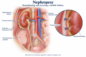
Nephropexy – Robotic Assisted

Why is it done?

  • Nephroptosis causing abdominal pain
  • Confirmed on standing Urogram with hydronephrosis caused by kinking of the ureter as the kidney falls down due to loss of supporting structures
  • Done with robotic assistance

Very Important!!

The correct side for surgery should be checked and confirmed with you,

Mark correct side,

CT scan present

How is it done?

  • GA
  • Prophylactic anti-biotics is given.
  • An indwelling catheter is placed.
  • The correct kidney is identified and marked while you are awake
  • Depending on the affected side of 5 incisions will be made.
  • Porst placed and attached to the Da Vinci robotic system
  • The colon is reflected to reveal the retro-peritoneal space
  • The ureter is identified and cleared up to the hilum
  • The upper pole of the kidney is mobilized with its surrounding fat.
  • A proximal spot on the psoas muscle is cleared
  • 2 non-dissolvable sutures are used to fix the upper pole of the kidney to the Psoas Muscle
  • A drain is placed


Complications

Side–effects

  • Minimal Blood loss
  • Wound Infection.
  • Post-operative hernia formations especially associated with the elderly with atrophic abdominal muscles
  • NB! Each person is unique and for this reason, symptoms vary

Repeat CT in 6 weeks


Download Information Sheet

Wes RA Nephropexy

Copyright 2019 Dr Jo Schoeman

Pelvi-Ureteric Junction Repair (PUJ) – Robotic Assisted

A congenital or acquired narrowing in the ureteric pelvis junction. This narrowing is excised with a reconnection. There are several techniques described in repairing this: I prefer the Dismembered Pyeloplasty

Why is it done?

  • High grade obstruction.
  • Causing deterioration of renal function.
  • Thinning of renal cortex.
  • Chronic pain.
  • Chronic infection.
  • Recurrent renal calculi.

Causes

  • Congenital lack of muscle, or neuro transmission in this area, causing a non-functioning part leading to obstruction.
  • Vesico-ureteric reflux, longstanding can also cause this.
  • Usually diagnosed in kids.
  • Crossing vessel.

How is it done?

Robotic assisted pyeloplasty.

  • Types
      • Dismembered.
      • Foley’s Y-V Pyeloplasty.
      • Culp-Dewierd.
      • Pelvi-calyceal pyeloplasty.
      • Endopyelotomy with laser.
    • Patients will receive a general anaesthesia.
    • Prophylactic antibiotics is given.
    • The correct ureteric system is identified and marked while you are awake.
    • This will be mostly a robotic / laparoscopic procedure.
    • The endoscopic procedure is reserved as a second line in my practice.
    • Laparoscopic ports are placed
    • The affected ureter is exposed, the defect cut out with a re-anastomosis of a spatulated ureter to a trimmed renal pelvis over a ureteric stent.
    • An indwelling catheter is placed. A drain is placed.

What next?

  • You may be in hospital for 3 days
  • Your drain will be removed when there is no urine draining.
  • Your catheter will be removed the following day.
  • As soon as you are comfortable with no signs of pain and emptying your bladder sufficiently, you will be discharged
  • A ward prescription may be issued on your discharge, for your own collection at any pharmacy.
  • A follow-up appointment will be scheduled for 6 weeks to remove your ureteric stent under local anaesthesia with a Flexible Cystoscope.
  • A review with a CT IVP will be scheduled 6 weeks after this to check on the end result of the ureter.
  • Any pain or signs of fever require an urgent review.
  • DON’T SUFFER IN SILENCE, OR YOU WILL SUFFER ALONE!

Possible Complications

  • Re-stenosis with recurrent obstruction.
  • Second procedure.
  • With further deterioration of renal function, you may require a nephrectomy where affected kidney contributes < 15-20% of total renal function.
  • Urine leak, Urinoma, requiring drainage.
  • Infection possible sepsis requiring long-term antibiotics.


Download Information Sheet

Wes RA reair

Rigid Cystoscopy, Retrograde Pyelogram, Stent Management

A therapeutic procedure under general anaesthetic, where a rigid cystoscopy is done in the bladder via the urethra, ureteric catheters are placed to enable imaging of the upper tracts with/without insertion or removal of ureteric stents

Why is it done?

To investigate:

  • Hematuria (blood in the urine)
  • Recurrent upper urinary tract infections
  • Space occupying lesions in the kidneys and ureters
  • Abnormal cells suggestive of urothelial carcinoma, on urine cytology


Risk factors:

  • Strong family history of bladder cancer
  • Smokers or passive smokers
  • Factory workers: dyes, paints, etc
  • Exposure to Schistosoma (Bilharzia)
  • Renal stone disease, bladder stones


How is it done?

  • This is done under General anaesthesia.
  • A cystoscopy is performed by placing a camera in the urethra with the help of a lubricant gel and saline irrigation.
  • The bladder is then distended with saline.
  • The inside of the bladder is viewed for pathology.
  • A retrograde pyelogram is done at the same time, (placement of small silicone catheters up the kidney pipes). Through this iodine contrast is injected up into the kidney collecting systems. This facilitates the viewing of kidney pipes and kidney collecting systems on X-ray to exclude any upper tract pathology.
  • If any abnormalities are found in the kidney/ ureters, a ureteroscopy (which is the placement of a long thin camera up the ureter) will be performed.
  • If any suspicious lesions are seen, a biopsy will be taken.
  • A ureteric stent may be placed
  • Urine would have been sent for cytology, to rule out the existence of cancer.

Antibiotics may be given to prevent infection

Complications

What to expect after the procedure?

  • Pain on initial passing of urine
  • Bladder infection ranging from a burning sensation to, fever, to puss (rare)
  • Bloodstained urine
  • Lower abdominal discomfort which will persist for a few days
  • Pain radiating from bladder to renal angle associated with urinating.
  • An infection could present with a stent being present.


Indications for a Ureteric stent

· Hematuria from upper tracts

· Dis-obstruction of the ureter caused either calculus, blood clot or tumour

· External compression of the ureter by retro-peritoneal pathology i.e.: Fibrosis, retroperitoneal lymph node compression

· Reduced renal function associated with hydronephrosis

· Sepsis associated with hydronephrosis


Download Information Sheet

Wes Cystoscopy RGP and Ureteric stents

Copyright 2019 Dr Jo Schoeman

Uretero-Renoscopic Stone Extraction with Laser (URSE)

A ureteric or renal calculus is removed with technique and may require a laser. A rigid/ flexible ureteroscope can be used.

Stones in the kidney, urinary bladder and ureter. medical illustration with a cross section of the kidney and bladder. anatomy of the urinary system. Human kidney.

Why is it done?

  • Removal of renal or ureteric stones.
  • Ureteric stones can vary from 5mm to over 1 cm in size.
  • You may present with excruciating pain on the affected side. (This pain may be worse than childbirth).
  • Renal stones usually larger than 1cm obstructing the renal pelvis.
  • Or renal stones not causing any symptoms.
  • Any fevers or a single kidney is deemed an emergency!

Two Treatment Options

  • Ureteric Calculi.
    • Managed with rigid ureteroscopy.
    • Prior stenting with a ureteric stent,
    • 7-10 days after stenting the stent is removed, and the stone is addressed with laser
  • Renal Calculi.
    • Prior stenting for 7-10 days.
    • After stent removal the kidney is accessed with a flexible uretero-renoscope and the stone is fragmented with laser

How is it done?

    • Patients will receive a general anesthesia.
    • Prophylactic antibiotics is given.
    • The correct kidney is identified and marked while you are awake.
    • You would have had a cystoscopy with retrograde pyelogram 10 days prior with placement of ureteric stent to prepare your ureter.
    • A cystoscopy will be done first to remove the stent, and 2 guidewires will be placed to enable access up the ureter.
    • Depending on the position of the stone, either a rigid or flexible uretero-renoscope will be used.
    • If a stone is in the kidney a flexible uretero-renoscope will be used with access obtained with an access sheath to protect the ureter from damage.
    • Laser will be used to fragment the stone.
    • All fragments will be attempted to be cleared. Small 1-2 mm fragments may be left as “Clinically Insignificant Fragments (CISF)” and will pass spontaneously.
    • A Ureteric catheter with an indwelling catheter is left post-operatively overnight.
    • Catheters will be removed the next morning depending on the presence of blood in the urine.

What next?

  • You will spend at least one night in hospital.
  • You will have a catheter for that time.
  • On removal of your catheter, you may experience sharp colicky pain, exactly the same as your presenting renal colic. This is due to your ureter contracting back to its usual size. (The stent has dilated this to 5X its usual size).
  • You will be discharged as soon as your pain has stabilized and you can function independently.
  • Allow for a few days for stabilization of symptoms.
  • A ward prescription will be issued on your discharge, for your own collection at any pharmacy.
  • A follow-up appointment will be scheduled for 6 weeks. Stone analysis results will then be discussed in order to formulate a plan to proven recurrences
  • DON’T SUFFER IN SILENCE, OR YOU WILL SUFFER ALONE!


Types of Stones:

  • Calcium Oxalate.
  • Uric Acid.
  • Calcium Phosphate.
  • Struvite (Infection stones).
  • Cystine.


Download Information Sheet

Wes Uretero-Renoscopic Stone Extraction with Laser URSE

URSE with or without Laser