
Renal angiomyolipoma (AML): when to watch, when to treat and when to seek urgent help
An angiomyolipoma, usually shortened to AML, is a growth in the kidney made up of blood vessels, muscle-like tissue and fat. Most are found by chance on a scan and never cause trouble. The main concern is that some AMLs can bleed, occasionally quite severely. The right plan depends on the diagnosis, the growth’s features and your circumstances, not its diameter alone. [1,2]
In this article, “AML” means angiomyolipoma, not acute myeloid leukaemia.
Is an AML cancer?
A classic renal AML is generally a benign tumour. Most occur on their own (sporadic AML). Some occur with tuberous sclerosis complex (TSC) or, less commonly, lymphangioleiomyomatosis (LAM); these can be multiple, affect both kidneys and require a different surveillance and treatment approach. A rare epithelioid AML can behave aggressively and needs specialist assessment. Not every solid renal mass presumed to be an AML can be confidently identified on a scan, particularly when little visible fat is present. [1,3,4]
What symptoms can it cause?
Many AMLs have no symptoms. Possible symptoms include aching or pain in the side, visible blood in the urine, or, less commonly, a palpable swelling. A sudden bleed into or around the kidney may cause abrupt, severe flank or abdominal pain, sometimes with dizziness, fainting, weakness or a racing heart. Blood in the urine and flank pain can also have other causes, including stones or cancer, so they should be assessed rather than attributed automatically to a known AML. [1–3]
Emergency warning: If you develop sudden severe side or abdominal pain, especially with faintness, collapse, a racing heart or visible bleeding, call 000 in Australia or attend an emergency department immediately. A bleeding AML can cause major blood loss even when blood is not visible in the urine. Do not wait for a routine appointment. [1,3]
How is it diagnosed?
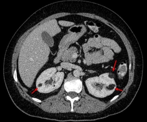
An AML may first appear on an ultrasound performed for another reason. CT or MRI helps confirm whether it contains fat and assess its size, location, blood vessels and any evidence of bleeding. Contrast-enhanced imaging may be needed for an indeterminate lesion or procedural planning, with the choice adapted to kidney function, pregnancy and other factors. A classic fat-rich AML is often diagnosed on imaging alone. A fat-poor AML may resemble kidney cancer; specialist review, further imaging or a biopsy may be appropriate if the result would change management. Biopsy has limitations and is chosen case by case. [1,3,4]
The assessment may also include blood pressure, kidney function and a blood count if bleeding is suspected. Multiple or bilateral AMLs, especially in a younger person, should prompt consideration of TSC or LAM and appropriate specialist input. [3,5]
Is there a critical size that must be treated?
There is no single diameter at which every AML must be removed. The traditional rule to treat an AML once it reaches 4 cm is no longer considered an automatic trigger. The European Association of Urology (EAU) states that the relationship between size and bleeding risk is uncertain and that no universal intervention threshold exists. Some larger AMLs can still be watched safely; a smaller one may need treatment if it bleeds or causes persistent symptoms. [1,2]
Size remains one part of the discussion. The team may also consider growth, vascular features or aneurysms seen on imaging, prior bleeding, pain, kidney function, pregnancy plans, blood-thinning medicines and access to urgent care. An aneurysm measurement such as 5 mm has been proposed as a risk marker, but recent evidence questions how reliably it predicts bleeding on its own. These findings guide an individual discussion; they do not supply a stand-alone rule. [1,3,6]
Management options
Active surveillance
For a confidently diagnosed, symptom-free AML, monitoring is often appropriate. It avoids exposing people with stable lesions to procedure risks. A surveillance plan records the starting size and imaging characteristics, checks for new symptoms and uses ultrasound, MRI or CT when appropriate to look for change. There is no single imaging interval suitable for all sporadic AMLs. A small, stable lesion may need little or no ongoing imaging after specialist review; a larger or changing lesion usually needs closer follow-up. Agree on the next scan, who will review it and what changes should prompt earlier contact. [1–3]
Selective arterial embolisation
An interventional radiologist passes a catheter into the blood vessels feeding the AML and blocks selected branches. Embolisation can control active bleeding and can be considered in advance for selected higher-risk or symptomatic tumours, while preserving kidney tissue. Pain, fever and nausea can occur afterwards; infection, non-target embolisation and repeat treatment are possible. In the EAU systematic review, around 30% of embolised cases required a further intervention, although individual outcomes vary. [1,2]
Kidney-sparing surgery
A partial nephrectomy removes the AML while aiming to preserve the rest of the kidney. Open, laparoscopic or robotic approaches may be considered depending on anatomy and surgical expertise. Surgery may be useful when imaging is uncertain, symptoms persist, embolisation is unsuitable or a definitive removal is preferred. It carries anaesthetic, bleeding, urine leak and kidney-function risks. Removal of the whole kidney is generally reserved for situations where kidney-sparing care is not feasible, especially in an emergency. [1–3]
Ablation and medicines
Thermal ablation is used less often for AML, and the evidence is more limited than for surveillance, embolisation and surgery. For TSC-associated AML, specialist teams may use an mTOR inhibitor such as everolimus for an asymptomatic but growing lesion over 3 cm when treatment is appropriate. This TSC recommendation should not be applied automatically to an isolated sporadic AML. Medicines require monitoring for adverse effects and interactions. [1,2,5]
What happens if an AML bleeds?
In hospital, the immediate priorities are resuscitation, blood tests and imaging to locate the bleed. Treatment may include intravenous fluids, blood transfusion and urgent selective arterial embolisation to stop the bleeding. Surgery is considered if embolisation is unavailable, unsuccessful or otherwise unsuitable. After recovery, follow-up checks the AML, remaining kidney function and whether further treatment is needed. [1,3]
How is surveillance different with tuberous sclerosis?
TSC-related AMLs require a coordinated long-term plan because lesions may occur in both kidneys and kidney function needs protection over a lifetime. The 2021 international TSC recommendations advise abdominal MRI every 1–3 years, with blood pressure, kidney function and urine protein checked at least yearly; the interval is tailored to individual risk. Growing AMLs greater than 3 cm may merit first-line mTOR treatment when intervention is indicated, while acute bleeding is generally managed with embolisation. A TSC specialist team can help balance treatment against the need to preserve kidney tissue. [5]
Questions to bring to your appointment
- Is the imaging diagnosis of a classic AML secure, or is more investigation needed?
- What are its size, growth pattern and vascular features?
- What is my personal risk of bleeding, and how does kidney function affect my options?
- If we monitor it, when is my next scan and what result would change the plan?
- If treatment is recommended, why is embolisation, surgery or another option best for me?
This article provides general information and does not replace individual assessment or advice.
References
- European Association of Urology. EAU Guidelines on Renal Cell Carcinoma, section on other renal tumours and AML. Accessed September 2026.
- Fernández-Pello S, et al. Management of sporadic renal angiomyolipomas: a systematic review to guide EAU recommendations. European Urology Oncology. 2020;3:57–72. doi:10.1016/j.euo.2019.04.005.
- Guo Y, et al. Canadian Urological Association best practice report: Diagnosis and management of sporadic angiomyolipomas. Canadian Urological Association Journal. 2020;14–E536. doi:10.5489/cuaj.6942.
- Wilson MP, et al. Diagnostic performance of MRI in detection of renal lipid-poor angiomyolipomas: systematic review and meta-analysis. Radiology. 2020;296:511–520.
- Northrup H, et al. Updated international TSC diagnostic criteria and surveillance and management recommendations. Pediatric Neurology. 2021;123:50–66. See also Tuberous Sclerosis Australia: Kidneys.
- Swärd J, et al. Renal angiomyolipoma: investigating radiological signs indicative of bleeding risk. Insights into Imaging. 2025.