
Ureteroceles: symptoms, investigations and treatment
A ureterocoele (also spelt ureterocele) is a balloon-like swelling at the lower end of a ureter: the tube carrying urine from the kidney to the bladder. The swelling usually projects into the bladder. Its opening may be narrow, slowing urine drainage and sometimes causing infection, stones or pressure on the kidney.
Some ureterocoeles cause no problems and are found incidentally. Others need treatment to improve drainage and protect kidney function. [1,2]
Congenital or acquired?
Most true ureteroceles are congenital, meaning they develop before birth. They may be detected on pregnancy ultrasound, during childhood or much later in adult life. A diagnosis in adulthood does not necessarily mean the ureterocele developed recently.
A ureterocoele can occur with a single ureter or a duplex collecting system, in which a kidney has two drainage systems. In a duplex kidney, the ureterocoele commonly involves the ureter draining the upper part of the kidney.
An intravesical or orthotopic ureterocoele lies within the bladder. An ectopic ureterocoele extends into the bladder neck or urethra and may interfere with bladder emptying. Adults more often have an intravesical ureterocoele associated with a single drainage system. [1,3]
The term “acquired ureterocoele” needs care. An acquired swelling of the lower ureter that resembles a ureterocoele is often called a pseudoureterocoele. A stone or a lesion involving the bladder near the ureteric opening can cause this appearance. An irregular or unusual swelling should therefore be investigated rather than assumed to be a harmless congenital abnormality. [4]
Presentation and symptoms
A ureterocoele may be discovered during investigations for an unrelated problem. When symptoms occur, they can include:
- Recurrent urinary tract infections.
- Pain in the side, back or lower abdomen.
- Burning, urgency or frequent urination, particularly with infection.
- Blood in the urine.
- Stones trapped within the ureterocoele.
- Difficulty emptying the bladder or, occasionally, urinary retention.
Children may present with unexplained fever, feeding difficulties or poor growth associated with infection. Rarely, a large ureterocoele can prolapse through the urethra.
Symptoms depend on whether the swelling obstructs drainage, contains a stone or affects the bladder outlet. Prolonged obstruction and repeated kidney infections can damage kidney function. [2,5]
What investigations are needed?
Investigations establish the anatomy, assess drainage and kidney function, and look for infection or reflux. Not everyone needs every test.
| Investigation | What it helps assess |
|---|---|
| Urine testing and culture | Infection and the appropriate antibiotic |
| Blood tests | Kidney function; inflammatory markers when infection is suspected |
| Kidney and bladder ultrasound | The ureterocoele, stones and swelling of the kidney or ureter |
| CT, sometimes CT urography | Stones and detailed urinary tract anatomy, particularly in adults |
| MR urography | Complex anatomy, including duplicated drainage systems |
| MAG3 or DMSA kidney scan | Drainage and relative function, or functioning kidney tissue and scarring |
| Micturating cystourethrogram (MCUG/VCUG) | Urine flowing backwards from the bladder into a ureter |
| Cystoscopy | Direct inspection of the bladder and ureteric openings |
CT involves radiation; contrast use depends on kidney function and the clinical question. Reflux testing is particularly relevant in children or when infection, complex anatomy or postoperative concerns warrant it. Cystoscopy can clarify an uncertain diagnosis and allow treatment during the same procedure. [1,3,5]
Does every ureterocoele need surgery?
No. Observation with planned follow-up may be appropriate when there are no troublesome symptoms, significant obstruction or recurrent infections, and kidney function is satisfactory.
Monitoring may include ultrasound and review of symptoms. Antibiotics treat infection but do not correct the swelling. Preventive antibiotics are considered selectively, particularly in children at increased infection risk. [6]
An infected, obstructed kidney requires urgent assessment and drainage, alongside antibiotics. Drainage may involve endoscopic opening of the ureterocoele or a nephrostomy, a tube placed through the skin into the kidney. Antibiotics alone may be insufficient when infected urine cannot drain. [1]
Endoscopic incision or deroofing
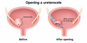
Treatment can often be performed through a telescope passed along the urethra into the bladder, usually under general anaesthesia.
Incision or puncture creates an opening in the wall of the ureterocoele. Deroofing removes part of its wall to create a broader opening. Both aim to let urine drain freely and reduce the balloon-like swelling.
The choice depends on anatomy, obstruction and associated stones. Instruments using electrical energy or a laser may be used. A stone within the sac can sometimes be fragmented and removed at the same operation.
A bladder catheter may be needed temporarily. Endoscopic treatment can be sufficient for a straightforward adult intravesical ureterocoele, while complex duplex or ectopic cases may need further surgery. [5,7,8]
Where does a ureteric stent fit in?
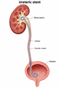
A double-J stent is a soft internal tube extending from the kidney to the bladder. It may be used selectively to support drainage after treatment, during associated stone surgery or while swelling settles. It is not required in every case and does not necessarily provide a permanent solution to the ureterocoele. [8]
Stents can cause frequency, urgency, bladder discomfort, blood in the urine and flank pain, sometimes after urination. Infection, displacement and mineral encrustation are other possible problems.
Every temporary stent needs a clear removal or exchange plan. The duration depends on the procedure and clinical circumstances. Contact the treating team if the removal date is unclear or overdue. [9]
Possible complications, including vesicoureteric reflux
Possible complications of endoscopic treatment include bleeding, infection, discomfort, injury to nearby urinary tract structures, persistent obstruction or narrowing as the opening heals. Repeat treatment or reconstructive surgery may be needed. [3,5,7]
Vesicoureteric reflux (VUR) means urine travels backwards from the bladder towards the kidney. It may already be present before treatment, particularly in a duplex system, or develop after opening the ureterocoele alters the natural anti-reflux mechanism.
Reflux does not automatically mean kidney damage or another operation. Its significance depends on its severity, infections, kidney function and bladder function. Monitoring may be appropriate; persistent significant reflux with recurrent infections can require further treatment, including ureteric reimplantation.
Risk varies with anatomy and technique. Ectopic ureterocoeles and duplex systems have higher rates of postoperative reflux and additional surgery than simpler intravesical cases. [1,7]
In one small adult study, reflux was detected in two of 23 patients followed after endoscopic incision, persisting in one at six months. These results illustrate the possibility of reflux; they do not predict an individual patient’s risk. Adult evidence largely comes from small studies. [7]
Further surgery and follow-up
Reconstructive options include reimplanting the ureter into the bladder or joining it to another healthy ureter in a duplex system. Occasionally, a severely damaged, poorly functioning kidney segment causing problems may require removal. Specialist assessment determines whether reconstruction, observation or removal is appropriate. [1]
Follow-up checks that drainage has improved and looks for recurrent infection, obstruction or reflux. Ultrasound is commonly used; further imaging is selected according to the findings. Symptom improvement alone does not confirm that drainage is normal. [5,7]
Seek urgent medical care for fever or shaking chills with flank pain, inability to pass urine, severe worsening pain, persistent vomiting or feeling seriously unwell. These symptoms may indicate infection or obstruction, including after treatment or with a stent in place. [1,9]
This article provides general education. Treatment should be tailored to the patient’s anatomy, symptoms and kidney function in consultation with their urologist.
References
- European Association of Urology. EAU Guidelines on Paediatric Urology: Ureterocele and ectopic ureter. Current online guideline, accessed 5 October 2026. https://uroweb.org/guidelines/paediatric-urology/chapter/ureterocele-and-ectopic-ureter
- European Association of Urology. Patient information: Ureterocele. https://patients.uroweb.org/condition/duplex-congenital-malformations-of-the-urinary-tract/ureterocele
- The Outcome of Transurethral Electro-Endoscopic Resection of Orthotopic Single System Ureterocoeles in Adults Nigerians. https://pmc.ncbi.nlm.nih.gov/articles/PMC11073836/
- Thornbury JR, Silver TM, Vinson RK. Ureteroceles vs. pseudoureteroceles in adults. Urographic diagnosis. Radiology. 1977;122(1):81–84. doi:10.1148/122.1.81. https://pubmed.ncbi.nlm.nih.gov/830358/
- An adult ureterocele complicated by a large stone: A case report. International Journal of Surgery Case Reports. 2018. https://pmc.ncbi.nlm.nih.gov/articles/PMC5927814/
- European Association of Urology. Watchful waiting for ureterocele. https://patients.uroweb.org/condition/duplex-congenital-malformations-of-the-urinary-tract/ureterocele/wait-and-see-ureterocele
- Vijay MK, et al. The safety and efficacy of endoscopic incision of orthotopic ureterocele in adult. Saudi Journal of Kidney Diseases and Transplantation. 2011;22(6):1169–1174. https://pubmed.ncbi.nlm.nih.gov/22089776/
- A Large Stone Within a Ureteroceles: A Diagnostic Pitfall and the Utility of Holmium Laser Deroofing as a Viable Surgical Option. https://pubmed.ncbi.nlm.nih.gov/29468199/
- British Association of Urological Surgeons. Information about your ureteric stent. https://www.baus.org.uk/_userfiles/pages/files/Patients/Leaflets/Stent%20advice.pdf