Trans Perineal Ultrasound Guided Prostate Biopsies – Minimally Invasive

You would have had a 3T MRI study prior to pinpoint any suspicious high-grade lesions. PET PSMA is another means of determining if you have any suspicious areas. Usually, targeted biopsies are taken of the prostate via the skin between scrotum and anus (perineal area).

The 3 possibilities of an elevated PSA is:

  • Benign Prostate Hyperplasia,
  • Prostatitis or
  • Prostate Cancer.
  • (Cyclist can have a higher PSA -saddle irritation)

Why is it done?

  • This is a diagnostic procedure used to make a diagnosis for an elevated PSA.
  • It is done as a day surgery procedure.
  • You are required to remain starved 6-8 hours prior to the procedure.


How is it done?

  • This procedure: is done under GA as a day procedure and takes approximately 30min (Incl anesthetic time)
  • Lithotomy position
  • Sterile field preparation
  • Prophylactic antibiotics are essential and is provided with your anaesthetic.
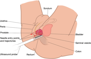
  • Minimally invasive technique with 2 skin puncture sites using: PIVOT-PRO or PRECISION POINT technique
  • This is a transcutaneous access – through the skin between your scrotum and anus.

Complications

Side–effects

  • Hematuria (blood in urine) 2-3days
  • Hematospermia (blood in ejaculate) will become less the more often you ejaculate.
  • Bacteraemia (infection) with low-grade fever and feeling unwell < 0.5%


ANY FEVERS REQUIRES URGENT ATTENTION


Download Information Sheet

Wes TPUS Biopsy Prostate PIVOT PRO


Copyright 2019 Dr Jo Schoeman

Trans Urethral Bladder Neck Resection

For those guys with normal sized prostates, but with a prominent bladder neck causing all the irritating symptoms of an enlarged prostate (LUTS).

Why is it done?

  • This procedure is performed when the bladder neck has become stenotic (narrow) and tight, usually after a TURP, can also be found in the young man, with an overactive bladder neck.
  • Usually seen in the younger more anxious men who keeps his finger on the pulse of everything.
  • Symptoms include:
    • a weak stream,
    • nightly urination,
    • frequent urination,
    • inability to urinate,
    • kidney failure due to the weak urination (obstruction),
    • bladder stones,
    • recurrent bladder infections.
  • Medication such as Flomaxtra, Urorec and Minipress etc. should always be given as a trial first.
  • Prostate cancer first needs to be ruled out by doing a PSA and when indicated a 3T MRI scan is required prior to targeted prostate biopsies to exclude a malignancy.

How is it done?

  • Patients will receive a general anesthesia unless otherwise indicated.
  • A cystoscopy is performed by placing a camera in the urethra with the help of a lubricant jelly and an irrigate (fluid).
  • The inside of the bladder is viewed for pathology. If any suspicious lesions are seen, a biopsy will be taken.
  • And incision of the bladder neck is made until a largely patent bladder neck is present, using bipolar current.
  • Laser can also be utilized and is probably preferred due to lack of bleeding.
  • Prophylactic antibiotics will be given to prevent any infections.

What can go wrong?

  • Any anesthesia has its risks, and the anesthetist will explain this to you.
  • You may in extreme cases experience blood loss, which may require a blood transfusion.
  • You will wake up with a catheter in your urethra and bladder. This will remain in the bladder for 1-3 days depending on the technique used and incidence of post-operative bleeding.
  • You may have a continuous bladder irrigate running in and out of your bladder to prevent clot formation.
  • Lower abdominal discomfort for a few days.
  • NB! Each person is unique and for this reason symptoms vary!

What next?

  • You will spend 1-3 days in hospital.
  • You will a trial without catheter as soon as your urine is clear.
  • You will be discharged as soon as you can completely empty your bladder.
  • You may initially suffer from urge incontinence and will improve within the next 6 weeks.
  • Allow for 6 weeks for stabilization of symptoms.
  • There may be some blood in your urine. You can remedy this by drinking plenty of fluids until it clears.
  • A ward prescription will be issued on your discharge, for your own collection at any pharmacy.
  • A follow-up appointment will be scheduled for 6 weeks. Should your pathology be worrisome, you will be contacted for an earlier appointment.
  • Don’t hesitate to ask me if you have any queries.
  • DON’T SUFFER IN SILENCE, OR YOU WILL SUFFER ALONE!

Side-effects

  • Retrograde ejaculation in more than 90% of patients. Therefore, if you have not completed your family, this procedure is not for you unless absolutely necessary.
  • Infertility as a result of the retrograde ejaculation.
  • Stress incontinence especially in the elderly and the diabetic patients.
  • Patients with Multiple Sclerosis, Strokes and Parkinsons have a higher risk of incontinence and risks should be discussed and accepted prior to surgery.
  • Urethral structuring in 2-3% of patients, requiring intermittent self-dilatation.
  • Scarring of the bladder neck can occur within 3-5 years requiring a second procedure.
  • NB! Each person is unique and for this reason symptoms vary!

Download Information Sheet

Wes Bladder Neck Incision

Trans-Urethral Cauterization of Bladder Bleeders + Evacuation Clots

 Why is it done?

Primary management of:

  • Continuous or intermittent bleeding from bladder vessels
  • More prominent after radiation therapy with neo-vascularisation
  • Induced or aggravated by blood thinning and anti-platelet therapy


Risk factors:

  • Anti-coagulation therapy: Warfarin, Xaralto etc
  • Anti-platelet therapy
  • Radiation to bladder prostate or bowel
  • These need to be stopped prior to the procedure


How is it done?

  • This is done under General anaesthesia.
  • A cystoscopy is performed by placing a camera in the urethra with the help of a lubricant gel and saline irrigation.
  • The bladder is then distended with saline.
  • Clots are evacuated with an evacuator
  • A resectoscope is then placed.
  • I use Bi-polar resection, thus using Saline as irrigation.
  • The offending bleeding vessels are cauterized and sealed
  • A 3-way catheter is placed with continuous saline irrigation until your urine is clear
  • Antibiotics may be given to prevent infection.


Complications

Side–effects

  • You may have a 22 –24 French (thick) 3-way urethral catheter placed through your urethra.
  • It does have a channel for placement of constant saline irrigation and another for the drainage of the blood-stained urine.
  • The Continuous bladder irrigation will continue until your urine is clear approximately 24-48hrs.
  • This can also be remedied by drinking plenty of fluids until it clears.
  • As soon as the colour of your urine is satisfactory, your catheter will be removed.


Download Information Sheet

Wes TU cauterisation of bladder bleeders

Copyright 2019 Dr Jo Schoeman

Trans Urethral De-Roofing of Prostate Abscess

Non invasive opening of an abscess found in the prostate, Very similar to a TURP with the exception that the bladder neck is preserved

Why is it done?

  • This procedure is performed when you have been diagnosed with a prostatic abscess, usually after an MRI investigation.
  • You would have had a history of swinging low to high grade fevers, a history of catheterization or instrumentation.
  • Not improving on antibiotics.
  • You may possibly be very sick with High Dependancy Unit (HDU) admission on intravenous antibiotics.
  • This procedure is done where the abscess or fluid collection in the parenchyma of the prostate is in the transitional zone of the prostate and easily accessible via endoscopic technique.
  • It is done under a general anaesthetic with prophylactic antibiotics in place, or appropriate Antibiotics as per your serum cultures.

How is it done?

  • Patients will receive a general Anaesthesia unless otherwise indicated.
  • A cystoscopy is performed by placing a camera in the urethra with the help of lignocaine gel.
  • The inside of the prostatic urethra and bladder is viewed for pathology and especially signs of the abcess. If any suspicious lesions are seen, a biopsy will be taken.
  • The area of the prostatic urethra above the abcess is resected until the abcess is opened and drained.
  • The bladder neck and urethral sphincter is preserved.
  • Laser can also be utilized and is probably preferred due to lack of bleeding.
  • Prophylactic antibiotics will be given to prevent any infections.

What can go wrong?

  • Any anaesthesia has its risks and the anaethiatist will explain this to you.
  • You will wake up with a catheter in your urethra and bladder. This will remain in the bladder for 1-3 days depending on the technique used and incidence of post-operatve bleeding. And until signs of sepsis have cleared.
  • You may have a continuous bladder irrigate running in and out of your bladder to prevent clot formation until the urine clears.
  • Lower abdominal discomfort for a few days.
  • NB! Each person is unique and for this reason symptoms vary!

What next?

  • You will be hospitalized until all signs of sepsis have cleared.
  • You will a trial without catheter as soon as your urine is clear.
  • You will be discharged as soon as you can completely empty your bladder.
  • You may initially suffer from urge incontinence and will improve within the next 6 weeks.
  • Allow for 6 weeks for stabilization of symptoms.
  • You may have a change in ejaculate volume and could even suffer retrograde ejaculation.
  • There may be some blood in your urine. You can remedy this by drinking plenty of fluids until it clears.
  • A follow-up appointment will be scheduled for 6 weeks. Should your pathology be worrisome, you will be contacted for an earlier appointment.
  • DON’T SUFFER IN SILENCE, OR YOU WILL SUFFER ALONE!


Download Information Sheet

Wes TUR Deroofing of Prostate Abscess

Trans-Urethral Resection of Bladder Tumour – TURBT

Trans Urethral resection of bladder tumour with/without MMC

An endoscopic procedure where bladder tumours are excised via the urethra.

Why is it done?

Primary management of:

  • Resect a bladder lesion suspicious of bladder cancer
  • Three Types of bladder cancer:
    • Urothelial Carcinoma (85%)
    • Squamous Cell carcinoma
    • Adeno carcinoma
  • Metastatic cancer to the bladder – i.e. Breast, Cervical, Adeno carcinoma of bowel.
  • Other space occupying lesions in the bladder: infection granulomas, abscess from diverticulitis etc.
  • Abnormal cells suggestive of urothelial carcinoma, on urine cytology.

Risk factors:

  • Strong family history of bladder cancer.
  • Smokers or passive smokers.
  • Factory workers: dyes, paints, etc.
  • Exposure to Schistosoma (Bilharzia).
  • Renal stone disease, bladder stones.

How is it done?

  • This is done under General anesthesia.
  • A cystoscopy is performed by placing a camera in the urethra with the help of a lubricant gel and saline irrigation.
  • The bladder is then distended with saline.
  • A resectoscope is then placed.
  • I use Bi-polar resection, thus using saline as irrigation.
  • The tumour or tumours are resected as complete as possible.
  • Deep resection of the tumour base is done to exclude deep muscle invasive tumours.
  • In tumours where it is clearly muscle-invasive, less extensive surgery is done, as this patient may benefit from a cystectomy.
  • 40mg of Intravesical Mitomycin C is routinely placed inside your bladder for an hour after the surgery.
  • A 3-way catheter is placed with continuous saline irrigation until your urine is clear.
  • Antibiotics may be given to prevent infection.

Mitomycin C

Chemotherapeutic agent providing 40% lower incidence of Urothelial Cancer recurrence if placed within 6-8 hours inside the bladder after a TURBT.

What to expect after the procedure?

  • Blood stained urine.
  • Lower abdominal discomfort which will persist for a few days.
  • Catheter induced discomfort.
  • NB! Each person is unique and for this reason symptoms vary.
  • Small risk (<1%) of bladder perforation, causing you to have a laparoscopy with repair of bladder and wash-out of peritoneal cavity with Sterile Water.

What next?

  • You may have a 22 French (thick) 3-way urethral catheter placed through your urethra.
  • It does have a channel for placement of constant saline irrigation and another for the drainage of the blood-stained urine.
  • The continuous bladder irrigation will continue until your urine is clear approximately 24-48hrs.
  •  This can also be remedied by drinking plenty of fluids until it clears.
  • As soon as the colour of your urine is satisfactory, your catheter will be removed.
  • Staging of your cancer will be arranged to be reviewed on your review appointment.
  • A ward prescription will be issued to patients on discharge, for own collection at any pharmacy.
  • Patients should schedule a follow-up appointment within 7-14 days.
  • Please don’t hesitate to direct all further queries to Jo.
  • REMEMBER: THOSE WHO SUFFER IN SILENCE, SUFFER ALONE!

Staging

  • CT IVP.
  • CT Chest.
  • Bone scan.
  • Renal Function and Liver Function Tests.
  • MRI where an allergy to Iodine or Renal Impairment.

Download Information Sheet

Wes TURBT

Uretero-Renoscopic Stone Extraction with Laser (URSE)

A ureteric or renal calculus is removed with technique and may require a laser. A rigid/ flexible ureteroscope can be used.

Stones in the kidney, urinary bladder and ureter. medical illustration with a cross section of the kidney and bladder. anatomy of the urinary system. Human kidney.

Why is it done?

  • Removal of renal or ureteric stones.
  • Ureteric stones can vary from 5mm to over 1 cm in size.
  • You may present with excruciating pain on the affected side. (This pain may be worse than childbirth).
  • Renal stones usually larger than 1cm obstructing the renal pelvis.
  • Or renal stones not causing any symptoms.
  • Any fevers or a single kidney is deemed an emergency!

Two Treatment Options

  • Ureteric Calculi.
    • Managed with rigid ureteroscopy.
    • Prior stenting with a ureteric stent,
    • 7-10 days after stenting the stent is removed, and the stone is addressed with laser
  • Renal Calculi.
    • Prior stenting for 7-10 days.
    • After stent removal the kidney is accessed with a flexible uretero-renoscope and the stone is fragmented with laser

How is it done?

    • Patients will receive a general anesthesia.
    • Prophylactic antibiotics is given.
    • The correct kidney is identified and marked while you are awake.
    • You would have had a cystoscopy with retrograde pyelogram 10 days prior with placement of ureteric stent to prepare your ureter.
    • A cystoscopy will be done first to remove the stent, and 2 guidewires will be placed to enable access up the ureter.
    • Depending on the position of the stone, either a rigid or flexible uretero-renoscope will be used.
    • If a stone is in the kidney a flexible uretero-renoscope will be used with access obtained with an access sheath to protect the ureter from damage.
    • Laser will be used to fragment the stone.
    • All fragments will be attempted to be cleared. Small 1-2 mm fragments may be left as “Clinically Insignificant Fragments (CISF)” and will pass spontaneously.
    • A Ureteric catheter with an indwelling catheter is left post-operatively overnight.
    • Catheters will be removed the next morning depending on the presence of blood in the urine.

What next?

  • You will spend at least one night in hospital.
  • You will have a catheter for that time.
  • On removal of your catheter, you may experience sharp colicky pain, exactly the same as your presenting renal colic. This is due to your ureter contracting back to its usual size. (The stent has dilated this to 5X its usual size).
  • You will be discharged as soon as your pain has stabilized and you can function independently.
  • Allow for a few days for stabilization of symptoms.
  • A ward prescription will be issued on your discharge, for your own collection at any pharmacy.
  • A follow-up appointment will be scheduled for 6 weeks. Stone analysis results will then be discussed in order to formulate a plan to proven recurrences
  • DON’T SUFFER IN SILENCE, OR YOU WILL SUFFER ALONE!


Types of Stones:

  • Calcium Oxalate.
  • Uric Acid.
  • Calcium Phosphate.
  • Struvite (Infection stones).
  • Cystine.


Download Information Sheet

Wes Uretero-Renoscopic Stone Extraction with Laser URSE

Ureteric Injury

Infrequently ureteric injuries can occur with other abdominal surgery i.e.:

  • Hysterectomy.
  • Ovarian mass resection.
  • Bowel resections.
  • Sacro Colpopexy.

If the injury is below the pelvic brim, then a re-implantation with is recommended with Boari-flap, otherwise a primary end-to-end anastomosis

Why is it done?

  • Stricturing or narrowing of the ureter causing significant hydronephrosis.
  • Injury to ureter with Urological surgery can cause this: Ureteroscopy.
  • Injury to the ureter from other surgeries: Colorectal, Gynecological.
  • This injury if not noted may lead to chronic infection, peritonitis, sepsis etc.
  • End-result is loss of renal function and an ICU stay.
  • An end-to-end anastomosis can be considered in the mid ureter.
  • A Reimplantation into the bladder with lower ureteric strictures.
  • A pelvi-ureteric junction repair in higher ureteric strictures.
  • A trans uretero-ureteric anastomosis joining one ureter to the other where long defects are present.
  • Renal Auto Transplantation where ureter is completely damaged or an ileal ureteric substitution where the whole ureter is damaged.

How is it done?

Robotic technique.

  • Patients will receive a general anesthesia.
  • Prophylactic antibiotics is given.
  • The correct ureteric system is identified and marked while you are awake.
  • Robotic ports are placed,
  • The affected ureter is exposed, the defect cut out with a re-anastomosis of a spatulated proximal ureter to the distal ureter over a ureteric stent.
  • An indwelling catheter is placed.
  • A drain is placed.

What next?

  • You may be in hospital for a few days
  • A drain will be left overnight and removed the next day if not draining any fluid.
  • Your catheter will be removed the following day. Or as soon as your urine is clear.
  • As soon as you are comfortable with no signs of pain and emptying your bladder sufficiently, you will be discharged.
  • A ward prescription may be issued on your discharge, for your own collection at any pharmacy.
  • A follow-up appointment will be scheduled for 6 weeks to remove your ureteric stent under local anesthesia with a Flexible Cystoscope.
  • A review with a CT IVP will be scheduled 6 weeks after this to check on the end result of the ureter.
  • DON’T SUFFER IN SILENCE, OR YOU WILL SUFFER ALONE!


Wes Ureteric Injury

Ureteric Reimplantation

  • Surgical repair of VUJ obstruction, robotic or open
  • Mainly a procedure for pediatric urology to correct grade 4-5 vesicoureteral reflux
  • Distal ureteric injuries.

Why is it done?

  • Grade 4-5 Vesical-Ureteric Reflux where conservative management has failed with a progressive deterioration in renal function.
  • Distal ureterectomy due to stricture disease.
  • Iatrogenic injury to lower ureter during surgical procedure: hysterectomy, colectomy, sacrocolpopexy etc.
  • Ureteric involvement in pelvic oncological (cancer) conditions, i.e.: colon cancer, rectal cancer, ovarian cancer, etc.
  • Ureteric involvement in pelvic inflammatory conditions: Diverticular abscesses, Pelvic Inflammatory disease etc.

How is it done?

  • Patients will receive a general anesthesia.
  • Prophylactic antibiotics is given.
  • The correct kidney is identified and marked while you are awake.
  • This will be a combined robotic and endoscopic procedure.
  • A cystoscopy will be done with placement of ureteric catheter or stent, if not already done.
  • Patients with complete closure of the ureter may have a nephrostomy tube into their kidney via the back.
  • An indwelling catheter is placed.
  • Robotic ports are placed, and the pelvic cavity is entered.
  • The ureter is identified, and the affected area of the lower ureter is identified and cut off above the injury / diseased area.
  • The bladder is opened, bi-valved and the ureter is re-implanted either as refluxing or non-refluxing.
  • A Psoas-hitch procedure will be performed where the bladder is fixed onto the affected side’s Psoas muscle as to take off tension from the anastomosis / reimplantation.
  • A Boari-flap may be considered with considerable length of defect.
  • In the case of VUR, the ureter is not cut, rather loosened in the bladder and re-tunneled in a non-refluxing technique under the mucosa of the bladder. Several techniques have been described.
  • A ureteric stent is placed for 6 weeks and an indwelling catheter for 10 days.
  • A drain is also placed for post-operative drainage for a couple of days.

What next?

  • You may be in hospital for at least 3-5 days.
  • You may have continuous intravenous antibiotics on board.
  • You will have a drain and an indwelling catheter.
  • The drain will be removed on D2-3 as soon as the drainage is less than 20-30cc per 24 hours.
  • The indwelling catheter will remain for 10-14 days until a cystogram reveals no leaks.
  • Your stent will be removed on a separate occasion in 6 weeks after all the fibrosis has settled.
  • A follow-up appointment will be scheduled for 6 weeks to remove the stent.
  • A further follow-up is arranged with a CT IVP to check on the end result of the ureter.
  • DON’T SUFFER IN SILENCE, OR YOU WILL SUFFER ALONE!

Possible complications:

  • Vesical Ureteric Reflux.
  • Stricturing / Narrowing of the implanted ureter.
  • Persistent Reflux.
  • Re-implantation.
  • VUR.
  • Stenosis and narrowing with persistent hydronephrosis.


Download Information Sheet

Wes Ureteric Reimplantation VUR

Urethral Dilatation

Dilatation of urethral stricture. Dine in combination with urethroscopy and placement of IDC is advocated on daily basis at home as management of urethral stricture.

Why is it done?

  • To treat a narrowing in the urethra which has formed due to previous damage/injury to the urethra.
  • Causes: after bypass surgery where a drop in blood pressure has caused an area of low blood supply to the urethra; trauma to the urethra (pelvic fractures/ urethral instrumentation); and sexually transmitted diseases.
  • The procedure placing a dilator un your urethra on a daily basis.
  • It aids in keeping your urethra open and prevents eventual kidney damage / failure.

How is it done?

  • Patients will receive a local anesthetic.
  • A urethroscopy is performed by placing a flexible camera in the urethra, with the help of a lubricant jelly and an irrigate (fluid), to identify the stricture.
  • A guidewire is slipped through the opening of the narrowing.
  • The camera is removed.
  • The stricture is dilated using a graduated S dilators over the guide wire.
  • Prophylactic antibiotics may be given to prevent infection.

What to expect after the procedure?

  • It may be slightly uncomfortable, please don’t hesitate to tell jo and request sedation if required.
  • A catheter will be inserted in the urethra and bladder. This will remain in the bladder for 3 days.
  • Catheters can be very irritating and cause some discomfort.
  • Blood stained urine will be present.
  • Lower abdominal discomfort will persist for a few days.
  • NB! Each person is unique and for this reason symptoms may vary!


What next?

  • Patients will be sent home with a catheter for 3 days after receiving thorough catheter care instructions.
  • Arrangements will be made to remove the catheter on day 3.
  • There may be some blood in the urine. This can be remedied by drinking plenty of fluids until it clears.
  • A referral will be done to BlueCare Nurses, who will assess you to instruct you on daily self-dilatation.
  • PLEASE CONTACT THE HOSPITAL WITH ANY POST-OPERATIVE CONCERNS AND RETURN TO THE HOSPITAL IMMEDIATELY SHOULD THERE BE ANY SIGNS OF SEPSIS.

Download Information Sheet

Wes Urethroscopy & Urethral Dilatation

Urethrectomy

Removal of urethra. Usually in adjunct to a radical cystectomy. Occasionally done some time after a cystectomy where recurrences occur in urethra.


Why is it done?

  • As part of the treatment for aggressive localized Urothelial carcinoma of the bladder.
  • T1G3, > T2 disease.
  • Primary Urethral disease with:
    • Urothelial Carcinoma.
    • Squamous cell carcinoma.
    • Secondary metastatic disease to Urethra: Melanoma, Lung cancer, Breast cancer (all these are rare).
  • Advanced disease may involve a penectomy.
  • The procedure is part of the radical cystectomy, radical cysto-prostatectomy or could be and adjunct later on when recurrences are found with surveillance.
  • Part of a necrotic inflammatory condition ie: Fourniers Gangrene.

How is it done?

  • This procedure is done under general anesthetic.
  • Legs are placed in a lithotomy position.
  • A single incision is made on the midline raphe on the perineum (area between scrotum and anus). Sutures will be dissolvable.
  • The Corpora Spongiosum with the urethra inside it is mobilized off the Corpora Cavernosa.
  • Urethral meatus is removed distal and glans is closed.
  • Proximally the urethra is taken up to the sphincter and freed.
  • You will have already had a diversion of the urine with an ileostomy, or this will be done with your cystectomy part of the operation.
  • A drain will be left for 24-48 hours to prevent the collection of serous fluids.
  • A dressing is then applied, which should be removed after 72 hours.

What to expect after the procedure?

  • Any anesthetic has its risks, and the anesthetist will explain all such risks.
  • If this is the only operation you have undergone, you will be sent home as soon as the drain is removed on D2 or D3.
  • Swelling is a common complication.
  • A hematoma (blood collection under the skin) may form and needs to be reviewed by Dr Schoeman as soon as possible. Bruising is normal.
  • An infection of the wound may occur and requires immediate attention.
  • Owing to the area of the surgery the wound should be kept clean and dry.
  • DANGER SIGNS: A wound that swells immediately, fever, or puss. Please contact Dr Schoeman or the hospital immediately as this may occur in up to 15 % of all cases.

What next?

  • The dressing should be kept dry for the initial 72 hours after surgery and then soaked in a bath until it comes off easily.
  • The dressing may sometimes adhere to the wound causing slight bleeding on removal. Don’t panic, the bleeding will stop.
  • On discharge a prescription may be issued for patients to collect.
  • There will be signs of bruising for at least 10 days.
  • The suture-line will be hard and indurated for at least 8-10 weeks.
  • PLEASE CONTACT THE HOSPITAL DIRECTLY WITH ANY POST-OPERATIVE CONCERNS AND RETURN TO THE HOSPITAL IMMEDIATELY SHOULD THERE BE ANY SIGNS OF SEPSIS.

Download Information Sheet

Wes Urethrectomy