
Superficial Bladder Cancer: Diagnosis, Treatment and the Risk of Progression
“Superficial bladder cancer” is an older term for cancer confined to the bladder lining or the tissue immediately beneath it. The preferred modern term is non–muscle-invasive bladder cancer, usually abbreviated to NMIBC.
Although these cancers have not invaded the bladder muscle, they do not all behave in the same way. Some are small, low-grade tumours with a relatively low risk of causing serious harm. Others, particularly high-grade T1 cancer and carcinoma in situ, can recur frequently and may progress into the bladder muscle.
Accurate staging and risk classification are therefore essential when deciding between surveillance, intravesical treatment and removal of the bladder.
What is non-muscle-invasive bladder cancer?
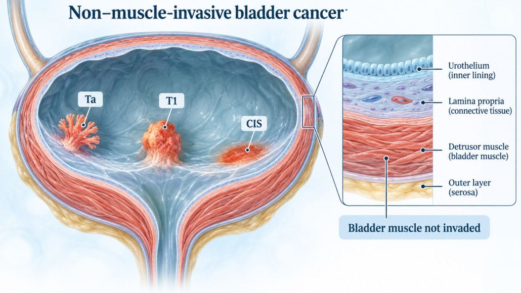
The bladder wall consists of several layers. Most bladder cancers begin in the urothelium, the specialised lining on the inside of the bladder.
NMIBC includes three main stages:
- Ta: a papillary tumour growing from the bladder lining without invading the supporting tissue
- T1: cancer that has invaded the connective tissue beneath the lining but has not reached the bladder muscle
- Carcinoma in situ/CIS or Tis: a flat, usually high-grade cancer confined to the bladder lining
CIS can be difficult to see because it may look like a red or inflamed area rather than a typical bladder tumour. Despite being confined to the surface, CIS is biologically aggressive and requires active treatment.
Once cancer enters the bladder muscle, it becomes muscle-invasive bladder cancer—stage T2 or higher. This usually requires a different and more intensive treatment approach.
How common is superficial bladder cancer?
Approximately 70–75% of bladder cancers are non–muscle-invasive when first diagnosed. The remaining patients generally have muscle-invasive or metastatic disease at presentation.
Bladder cancer is considerably more common in men than women and occurs most frequently in people over 60. Women sometimes experience delays in diagnosis when blood in the urine is initially attributed to urinary infection.
Most bladder cancers are urothelial carcinomas. Less common types include squamous cell carcinoma, adenocarcinoma and small-cell or neuroendocrine carcinoma.
What symptoms can bladder cancer cause?
The most common presentation is visible blood in the urine, haematuria.
The urine may appear:
- Pink
- Red
- Rust-coloured
- Tea-coloured
- Normal between episodes
The bleeding is often painless and may disappear for days or weeks. Its disappearance does not mean the underlying problem has resolved.
Other possible symptoms include:
- Microscopic blood detected on a urine test
- Urinary frequency
- A sudden need to urinate
- Burning or discomfort when passing urine
- Recurrent symptoms resembling a urinary tract infection
- Difficulty emptying the bladder
- Pelvic discomfort
CIS may cause urinary urgency, frequency and burning without producing a large visible tumour.
Blood in the urine should always be investigated, particularly in an older adult or someone with a history of smoking. Infection, stones and benign prostate enlargement are common alternative explanations, but bladder and upper urinary tract cancers must be excluded.
What causes bladder cancer?
Bladder cancer develops when genetic damage causes cells in the bladder lining to grow abnormally. In many patients there is no single identifiable cause.
Cigarette smoking
Smoking is the most important preventable risk factor. Carcinogens from tobacco enter the bloodstream, are filtered by the kidneys and remain in contact with the bladder lining in the urine.
The risk increases with the amount and duration of smoking. Stopping smoking remains valuable even after diagnosis because continued smoking may increase the risk of recurrence and progression.
Occupational chemical exposure
Long-term exposure to certain aromatic amines and industrial chemicals can increase risk. Historically, higher-risk industries have included:
- Dye and pigment manufacturing
- Rubber and leather production
- Painting
- Printing
- Metal processing
- Petroleum and chemical industries
Modern workplace protections have reduced—but not eliminated—these exposures.
Other risk factors
Additional risk factors include:
- Increasing age
- Male sex
- Previous pelvic radiotherapy
- Previous cyclophosphamide chemotherapy
- Chronic bladder irritation or inflammation
- Long-term urinary catheterisation
- Certain inherited cancer syndromes, particularly Lynch syndrome
- A personal history of cancer elsewhere in the urinary tract
Bladder cancer is not generally considered hereditary, although familial and genetic risks exist in a minority of patients.
How is bladder cancer investigated?
Medical history and urine testing
Assessment begins with a history of the bleeding, urinary symptoms, smoking and occupational exposure. Urine testing may identify blood, infection or abnormal cells.
A negative urine test after an episode of visible haematuria does not remove the need for investigation.
Urine cytology
Urine cytology examines shed urinary cells under a microscope.
It is most useful for detecting:
- High-grade urothelial cancer
- Carcinoma in situ
- Cancer elsewhere in the urinary tract
Cytology is less sensitive for low-grade tumours, so a negative result does not exclude bladder cancer.
Urinary molecular-marker tests may occasionally provide additional information, but they do not usually replace cystoscopy.
Imaging of the urinary tract
A CT urogram is commonly used to assess:
- Kidneys
- Renal pelvises
- Ureters
- Bladder
- Enlarged lymph nodes or other abnormalities
An ultrasound may be appropriate for selected patients, particularly when CT contrast or radiation should be avoided. However, ultrasound cannot reliably exclude small bladder tumours or CIS.
Flexible cystoscopy
A flexible cystoscope is passed through the urethra under local anaesthetic to inspect the bladder directly.
If a suspicious lesion is found, the next step is generally a formal resection under anaesthesia.
Transurethral resection of bladder tumour: TURBT
TURBT is the central procedure for diagnosing and treating NMIBC.
A rigid telescope is passed through the urethra, and the visible tumour is removed using an electrical loop, bipolar instrument or other resection technique. Tissue is sent to a pathologist to determine:
- Cancer type
- Tumour grade
- Depth of invasion
- Whether bladder muscle is present in the specimen
- Whether muscle invasion has occurred
- Whether variant histology or lymphovascular invasion is present
A complete TURBT should remove all visible tumour where safely possible and include adequate sampling of the underlying bladder muscle.
Enhanced cystoscopy using blue-light fluorescence or narrow-band imaging may help identify small tumours or CIS in selected patients.
When is a second TURBT required?
A repeat resection, usually within approximately two to six weeks, may be recommended when:
- The first resection was incomplete
- No bladder muscle was present in the specimen, apart from selected clearly low-risk Ta tumours
- The tumour is high-grade T1
- There is uncertainty about staging
- Residual tumour is suspected
Repeat TURBT may find residual cancer and occasionally identifies previously unrecognised muscle invasion. It can therefore materially change treatment.
Understanding low-, intermediate-, high- and very-high-risk disease
Treatment is based on more than the word “superficial.” Important risk factors include:
- Ta, T1 or CIS stage
- Low-grade or high-grade pathology
- Number of tumours
- Tumour size
- First occurrence or recurrence
- Frequency of previous recurrences
- Presence of CIS
- Depth and extent of T1 invasion
- Variant histology
- Lymphovascular invasion
- Involvement of the prostatic urethra
- Response to previous BCG treatment
Low-risk NMIBC
This usually involves a first, solitary, small, low-grade Ta tumour without CIS.
These cancers commonly recur but have a very low risk of progressing to muscle-invasive disease.
Intermediate-risk NMIBC
This is a broad group between low and high risk. It may include recurrent, multiple or larger low-grade tumours and selected other tumours without high-risk features.
The pattern and frequency of recurrence help determine treatment intensity.
High-risk NMIBC
High-risk disease includes most:
- High-grade T1 tumours
- Carcinoma in situ
- High-grade Ta tumours with adverse features
- Tumours with other aggressive pathological findings
These cancers have a meaningful risk of entering the bladder muscle and require more intensive treatment and surveillance.
Very-high-risk NMIBC
Very-high-risk disease may include combinations such as extensive high-grade T1 cancer with CIS, lymphovascular invasion, certain aggressive variant histologies or involvement of the prostatic urethra.
For these patients, early radical cystectomy may provide the best chance of cure.
Initial treatment after TURBT
Surveillance for selected low-risk disease
For a completely removed low-risk tumour, treatment may consist of:
- TURBT
- A single immediate dose of intravesical chemotherapy when safe
- Follow-up cystoscopy
Small, recurrent low-grade tumours may sometimes be treated with office fulguration or carefully selected surveillance, depending on the patient and tumour history.
Intravesical treatment
“Intravesical” means that a medication is placed directly into the bladder through a catheter. The medicine is retained for a prescribed time and then drained or passed in the urine.
Because the treatment remains mainly inside the bladder, it generally causes fewer whole-body effects than intravenous chemotherapy.
The role of intravesical mitomycin C
Mitomycin C is a chemotherapy medicine that damages the DNA of rapidly dividing cancer cells.
A single immediate postoperative dose
A single dose may be placed into the bladder shortly after TURBT—preferably within 24 hours—when the procedure has been uncomplicated.
Its purpose is to destroy floating tumour cells and reduce the chance that they implant elsewhere in the bladder. It also treats microscopic tumour cells remaining at the resection site.
This treatment is particularly useful for low-risk tumours and selected intermediate-risk tumours.
Mitomycin should not be administered immediately when there is:
- Suspected bladder perforation
- A very deep or extensive resection
- Significant ongoing bleeding
- A need for continuous bladder irrigation
- Concern that the drug could leak outside the bladder
A course of mitomycin
Patients with intermediate-risk disease may receive weekly mitomycin treatments followed by a variable maintenance schedule. The exact schedule depends on tumour characteristics, previous recurrence pattern and local protocol.
Side effects of mitomycin
Possible side effects include:
- Burning when urinating
- Urinary frequency and urgency
- Bladder discomfort
- Blood in the urine
- Chemical cystitis
- Skin irritation or a rash involving the hands or genital region
- Reduced bladder capacity after repeated severe inflammation
- Infection
- Rare injury if the medication leaks outside the bladder
Patients should follow the treatment unit’s instructions regarding fluid intake, urine handling and washing after treatment.
The role of intravesical BCG
BCG, Bacillus Calmette–Guérin, is a live, weakened form of Mycobacterium bovis. It was originally developed as a tuberculosis vaccine but also stimulates a powerful immune response against bladder cancer cells.
BCG is generally the preferred bladder-preserving treatment for:
- Carcinoma in situ
- High-risk high-grade Ta cancer
- High-grade T1 cancer after adequate resection
- Selected recurrent or aggressive intermediate-risk tumours
How is BCG given?
The usual initial course consists of one bladder instillation each week for six weeks. This is called induction BCG.
Patients who respond may then receive maintenance BCG. For high-risk disease, treatment may continue intermittently for one to three years, depending on tolerance, availability and individual risk.
Maintenance therapy is important because induction BCG alone provides less durable protection against recurrence and progression.
Side effects of BCG
Common short-term effects include:
- Burning when urinating
- Frequency and urgency
- Mild blood in the urine
- Bladder discomfort
- Fatigue
- Low-grade fever
- Flu-like symptoms
These effects usually settle within one or two days.
Less common but potentially serious complications include:
- Severe bacterial urinary infection
- Prostatitis
- Epididymo-orchitis
- Granulomatous inflammation
- Joint inflammation
- Hepatitis or pneumonitis
- Systemic BCG infection or sepsis
A high or persistent fever, shaking chills, breathing difficulty, confusion or severe illness after BCG requires urgent medical assessment.
When should BCG be postponed or avoided?
BCG should not be given:
- Within the early healing period after TURBT, generally the first two weeks
- After traumatic catheterisation
- When visible haematuria is present
- During a symptomatic urinary tract infection
- When bladder perforation is suspected
- In some patients with significant immune suppression
- When previous BCG caused a severe systemic reaction
BCG is handled differently from routine chemotherapy because it contains live bacteria. Patients must follow the treatment centre’s hygiene and urine-disposal instructions.
Mitomycin or BCG: which is better?
Neither treatment is best for every patient.
- Low-risk disease: a single immediate chemotherapy instillation is usually sufficient after complete TURBT.
- Intermediate-risk disease: a course of chemotherapy or one year of BCG may be considered according to recurrence and progression risk.
- High-risk disease: induction and maintenance BCG is generally preferred when bladder preservation is appropriate.
- Very-high-risk disease: early radical cystectomy should be discussed, although BCG may remain an option in carefully selected patients who understand the risk.
BCG is more effective than chemotherapy for preventing recurrence and progression in appropriately selected high-risk disease, particularly when maintenance BCG is completed. It also tends to cause more local and systemic side effects.
What is the chance of developing muscle-invasive cancer?
There is no single percentage that applies to every NMIBC patient.
Across all NMIBC categories, approximately 10–20% of patients may eventually develop muscle-invasive disease, but this average hides enormous differences between low- and high-risk tumours.
Using contemporary EAU risk categories, estimated five-year progression risks can range approximately from:
- Around 1% or less for low-risk disease
- Several per cent for intermediate-risk disease
- Around 10% or higher for high-risk disease
- Approximately 40% or more for very-high-risk disease
At ten years, the estimated risk in very-high-risk patients may exceed 50% without effective additional treatment. These figures are estimates from risk models and do not precisely predict an individual patient’s outcome. BCG, repeat resection, early cystectomy and other treatments can substantially change the risk.
Progression risk is particularly concerning with:
- Persistent or recurrent high-grade T1 cancer
- T1 cancer associated with CIS
- Extensive or multifocal CIS
- Deep invasion into the lamina propria
- Lymphovascular invasion
- Aggressive variant histology
- Prostatic urethral involvement
- Failure to respond to adequate BCG
- Early high-grade recurrence following BCG
Recurrence and progression are different. A small low-grade Ta tumour may recur several times without becoming muscle invasive, while a high-grade T1 tumour may progress after relatively few visible recurrences.
What is BCG-unresponsive bladder cancer?
BCG-unresponsive disease is a specific high-risk situation in which high-grade cancer persists or returns despite an adequate course of BCG within a defined period.
Continuing the same BCG treatment in genuinely BCG-unresponsive disease is unlikely to provide meaningful benefit and could delay curative surgery.
For a patient fit enough for major surgery, radical cystectomy is generally the preferred oncological treatment for BCG-unresponsive high-risk NMIBC.
Alternative bladder-preserving treatments or clinical trials may be considered when a patient:
- Is medically unfit for cystectomy
- Declines cystectomy after informed discussion
- Has a strong preference for bladder preservation and accepts the additional risk
However, the possibility of losing the optimal window for curative surgery must be discussed clearly.
When should removal of the bladder be considered?
Radical cystectomy means removing the bladder, nearby lymph nodes and certain adjacent organs, followed by creating a new way for urine to leave the body.
It may be considered for NMIBC when there is:
- Very-high-risk NMIBC at initial diagnosis
- Persistent high-grade T1 cancer after repeat TURBT
- High-grade T1 cancer with CIS
- Lymphovascular invasion
- Aggressive variant histology, such as micropapillary, plasmacytoid or selected sarcomatoid differentiation
- Extensive CIS that does not respond adequately to BCG
- High-grade recurrence following adequate BCG
- BCG-unresponsive disease
- Tumour involvement of the prostatic urethra or ducts
- Disease that cannot be completely controlled endoscopically
- Frequent, extensive high-grade recurrences
- Progression to muscle-invasive bladder cancer
Cystectomy may sound excessive for a cancer described as “superficial,” but high-grade T1 disease can already possess the biological ability to spread. Delaying surgery until muscle invasion or metastasis develops can reduce the chance of cure.
What does radical cystectomy involve?
In men, surgery commonly removes the:
- Bladder
- Prostate
- Seminal vesicles
- Pelvic lymph nodes
In women, surgery is tailored individually and may involve removal of the bladder, pelvic lymph nodes and selected reproductive organs. Organ-preserving approaches may be possible in carefully selected patients.
Urinary reconstruction options include:
- Ileal conduit: urine drains through a short segment of bowel to a stoma and external bag
- Orthotopic neobladder: bowel is used to create an internal reservoir connected to the urethra
- Continent catheterisable reservoir: an internal pouch is emptied using a catheter through a small abdominal opening
The most appropriate option depends on cancer location, kidney function, bowel health, manual dexterity, general fitness and patient preference.
Radical cystectomy is major surgery. Potential effects on urinary, sexual and bowel function must be balanced against the danger of progression.
Why lifelong surveillance is important
NMIBC has a strong tendency to recur, even after apparently complete treatment. Follow-up commonly includes:
- Regular cystoscopy
- Urine cytology in higher-risk patients
- Periodic upper urinary tract imaging
- Biopsy or repeat TURBT when abnormalities are found
- Monitoring for late treatment complications
Low-risk patients generally require less intensive surveillance. High-risk patients need frequent cystoscopy and cytology, particularly during the first two years, followed by long-term or lifelong monitoring.
The exact schedule should be tailored to the patient’s EAU risk group, pathology, treatment response and general health.
Can recurrence be prevented?
Not every recurrence can be prevented, but patients can improve their general and bladder health by:
- Stopping smoking
- Avoiding occupational carcinogen exposure
- Completing recommended intravesical treatment
- Attending every surveillance cystoscopy
- Reporting recurrent blood in the urine promptly
- Treating urinary infections appropriately
- Maintaining good hydration unless medically restricted
Smoking cessation remains the most important modifiable step.
The bottom line
Most bladder cancers are diagnosed before they enter the bladder muscle, but the term “superficial” should not be mistaken for harmless.
Low-grade Ta tumours frequently recur but rarely progress. High-grade T1 cancer and CIS behave much more aggressively and require complete TURBT, appropriate intravesical therapy and close surveillance.
Mitomycin C is particularly useful for reducing recurrence after TURBT and treating selected low- or intermediate-risk disease. BCG is the main bladder-preserving treatment for high-risk NMIBC and CIS.
Radical cystectomy should be discussed early—not only after muscle invasion—in patients with very-high-risk features, persistent high-grade T1 cancer or BCG-unresponsive disease. For these patients, timely surgery may offer the best chance of cure.
This article provides general information and does not replace individual medical advice. Treatment should be based on formal pathology review, complete staging, medical fitness and multidisciplinary discussion.
References and further reading
- European Association of Urology—Non–Muscle-Invasive Bladder Cancer Guidelines
- American Urological Association—Non–Muscle-Invasive Bladder Cancer Guideline
- Healthdirect Australia—Bladder cancer
- Cancer Council Australia—Bladder cancer
- Cancer Australia—Bladder cancer
So, if you are experiencing blood in your urine and have been identified by your GP as having a possible bladder cancer, come see your Urologist in Brisbane, Dr Jo Schoeman to discuss options with you.





Leave a Reply
Want to join the discussion?Feel free to contribute!