
Small Renal Masses: When a Small Kidney Tumour Needs Watching, Treating or Removing
Finding a small mass on the kidney can be unsettling, particularly when it appears unexpectedly on an ultrasound or CT scan performed for something completely unrelated.
The reassuring news is that small does not automatically mean dangerous, and a renal mass does not automatically mean kidney cancer. Even when a small renal mass is malignant, many grow slowly and have a low risk of spreading. This means that immediate surgery is not always necessary.
Modern management has therefore moved away from a simple philosophy of “there is a lump, so it must come out”. Instead, treatment is tailored to the size and appearance of the mass, its growth rate, kidney function, the patient’s age and general health, and individual preferences.
Options range from:
- Active surveillance
- Percutaneous biopsy
- Minimally invasive ablation, including cryoablation and thermal techniques
- Robotic partial nephrectomy
- Occasionally, radical nephrectomy
The aim is straightforward: control the cancer when treatment is necessary while preserving as much normal kidney function as possible.
What Is a Small Renal Mass?
A small renal mass (SRM) generally refers to a kidney tumour measuring 4 cm or less, corresponding to a clinical T1a renal tumour when confined to the kidney.
Many are discovered incidentally during imaging performed for abdominal pain, gallstones, bowel problems, back pain or another unrelated condition. The kidney lesion is often an unexpected passenger on the scan.
Importantly, not every small renal mass is cancer.
Benign lesions include:
- Oncocytoma
- Angiomyolipoma
- Some complex renal cysts
- Other uncommon benign tumours
Among malignant masses, renal cell carcinoma is the most important diagnosis. However, many small renal cancers demonstrate relatively indolent behaviour. Published guideline data suggest that more than 20% of small renal masses may ultimately prove benign, while many malignant lesions within this size range have relatively favourable biology.
This creates an important clinical dilemma: we want to treat a potentially dangerous cancer, but we also want to avoid unnecessary treatment of a tumour that may never have caused harm.
Step One: Properly Characterising the Mass
An incidental renal mass generally requires appropriate imaging before deciding what to do.
This usually involves a:
- Multiphasic contrast CT scan, or
- MRI scan
These scans help determine:
- Exact tumour size
- Whether the lesion is solid or cystic
- Whether it enhances with contrast
- Its position within the kidney
- Relationship to blood vessels and the collecting system
- Whether there are concerning features suggesting malignancy
- Whether there is any evidence of disease outside the kidney
High-quality contrast CT or MRI is recommended for the characterisation of a newly discovered small renal mass.
Blood tests will usually include kidney function, particularly serum creatinine and estimated glomerular filtration rate (eGFR).
What About Complex Kidney Cysts?
Not every suspicious renal lesion is a solid tumour.
Complex renal cysts are usually classified according to the Bosniak classification, which estimates the likelihood that a cystic lesion represents malignancy.
Some Bosniak III and selected predominantly cystic lesions can also be considered for surveillance, particularly when they are small or when the risks of treatment outweigh the anticipated benefits. Larger or increasingly complex lesions are more likely to require intervention.
Do We Need a Kidney Biopsy?
Sometimes.
A renal mass biopsy involves passing a needle through the skin into the tumour, usually under ultrasound or CT guidance, and obtaining small samples for examination by a pathologist.
Biopsy can be particularly useful when:
- The diagnosis is uncertain
- Active surveillance is being considered
- The biopsy result could change management
- Ablation is planned
- Imaging suggests a tumour that may not require surgery
- There is a possibility of metastatic disease or another unusual diagnosis
A biopsy is not automatically necessary for every small renal mass.
Possible complications include:
- Pain
- Bruising
- Bleeding around the kidney
- Rare significant haemorrhage
- Non-diagnostic biopsy
- Sampling error
The important question is not simply “Can we biopsy it?” but rather:
“Will knowing the biopsy result change what we do?”
Option 1: Active Surveillance
Active surveillance is increasingly recognised as a legitimate management strategy rather than simply “doing nothing.”
There is an important difference.
Doing nothing means ignoring the tumour.
Active surveillance means deliberately monitoring it and treating it if its behaviour changes.
For appropriately selected small renal masses, particularly those less than 2 cm, active surveillance can be an excellent initial strategy. The AUA notes very favourable cancer-specific and metastasis-free outcomes in published surveillance series of these very small tumours.
The overall risk of metastatic progression among carefully selected patients undergoing surveillance for small renal masses has been reported at approximately 1–2% or less in early follow-up series, although risk varies according to tumour and patient characteristics and increases with more aggressive biological behaviour.
Who May Be Suitable for Active Surveillance?
Surveillance may be particularly attractive for:
- Tumours less than 2 cm
- Small tumours with favourable imaging characteristics
- Slowly growing lesions
- Older patients
- Patients with significant medical problems
- Patients with impaired kidney function
- Patients with a solitary kidney
- Patients at increased anaesthetic or surgical risk
- Patients who wish to avoid or delay intervention
For renal masses measuring 2–4 cm, both surveillance and definitive treatment may be reasonable depending upon individual circumstances.
What Does an Active Surveillance Protocol Look Like?
There is no single surveillance schedule suitable for every patient.
A commonly used approach is:
Baseline
Obtain good-quality CT or MRI imaging to accurately document:
- Tumour size
- Tumour characteristics
- Location
- Complexity
A renal mass biopsy may be considered when the result would influence management.
First follow-up
Repeat cross-sectional imaging is commonly performed approximately 3–6 months after entering surveillance to determine whether there is meaningful interval growth. This approach is specifically supported by AUA guidance.
Ongoing surveillance
If the lesion remains stable, imaging may then be performed approximately:
Every 6–12 months, depending upon the patient’s age, tumour characteristics, previous growth rate and overall health.
Ultrasound can sometimes reduce the number of CT scans required, provided that the lesion can be reliably visualised and measured. CT or MRI is preferred if there is uncertainty regarding growth or tumour characteristics.
The Canadian guideline notes that commonly used protocols involve abdominal imaging every 3–6 months during the first year and every 6–12 months thereafter if stable, although there is no universally agreed schedule.
When Do We Stop Watching and Start Treating?
This is perhaps the most important aspect of active surveillance.
Treatment may be recommended if there is:
- Significant tumour growth
- Increasing radiological complexity
- Growth towards or beyond 4 cm
- A consistent growth rate greater than approximately 5 mm per year
- Concerning biopsy findings
- Evidence of spread
- Development of symptoms
- A change in the patient’s health or treatment preference
Commonly recognised triggers include tumour growth beyond 4 cm or consecutive growth exceeding approximately 0.5 cm per year, although no single measurement should be considered in isolation.
A tumour that grows by a millimetre on one scan has not necessarily sounded the alarm bell. Small differences can occur simply because of how the scan or measurement was performed.
The trend matters more than a single measurement.
Risks and Disadvantages of Active Surveillance
The main advantages are avoiding or delaying unnecessary treatment and preserving kidney function.
Potential disadvantages include:
- Anxiety associated with living with an untreated renal mass
- Repeated scans
- Radiation exposure from repeated CT imaging
- Contrast exposure
- Possibility of tumour growth
- Small risk of metastatic progression
- Possibility that treatment becomes technically more difficult if the tumour grows substantially
Active surveillance therefore works best when there is a clear follow-up plan rather than a vague agreement to “scan it again sometime.”
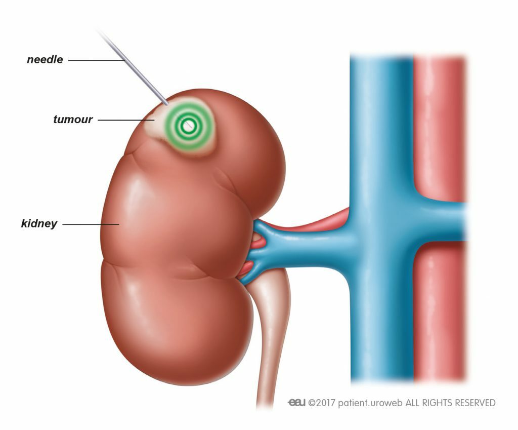
Option 2: Minimally Invasive Tumour Ablation
For selected small renal masses, it may be possible to destroy the tumour without surgically removing it.
This is known as renal tumour ablation.
The principal techniques include:
Cryoablation
Cryoablation uses extremely low temperatures to freeze and destroy tumour cells.
Radiofrequency Ablation
Radiofrequency ablation uses thermal energy to heat and destroy tumour tissue.
Microwave Ablation
Microwave energy can similarly produce controlled heating and destruction of tumour tissue.
These treatments can often be performed percutaneously, meaning probes are inserted through the skin under CT or ultrasound guidance without requiring conventional surgery.
Who Is Suitable for Renal Ablation?
Ablation may be considered for patients with:
- A small renal tumour
- A tumour in a technically accessible location
- Significant medical comorbidities
- Increased surgical or anaesthetic risk
- Reduced kidney function
- A solitary kidney
- Previous kidney surgery
- A strong preference for a less invasive treatment
Current European guidance particularly supports tumour ablation as an alternative for patients with small cT1 renal tumours who require treatment but are unfit for surgery, while emphasising that long-term comparative evidence against partial nephrectomy remains less robust.
Renal mass biopsy is generally recommended before percutaneous ablation.
Advantages of Ablation
Potential benefits include:
- No large surgical incision
- Shorter hospital stay
- Faster recovery
- Less postoperative discomfort
- Preservation of kidney tissue
- Potential suitability for patients who are poor surgical candidates
- Ability to repeat treatment in selected cases
Risks and Complications of Ablation
Possible complications include:
- Bleeding
- Haematoma
- Infection
- Pain
- Damage to nearby organs
- Injury to the collecting system
- Urine leakage
- Thermal injury to surrounding structures
- Incomplete treatment
- Persistent viable tumour
- Local tumour recurrence
- Need for repeat ablation
- Need for subsequent surgery
Tumour size matters. European guidance advises against routinely using radiofrequency ablation for tumours larger than approximately 3 cm and cryoablation for tumours larger than approximately 4 cm, because recurrence becomes more concerning as tumour size increases.
Long-term oncological evidence for ablation is also less mature than the evidence supporting surgical excision, and local recurrence appears somewhat more frequent than following partial nephrectomy.
Option 3: Robotic Partial Nephrectomy
When definitive surgical treatment is appropriate, the modern objective is usually:
Remove the tumour, not the entire kidney.
A partial nephrectomy removes the renal tumour together with an appropriate margin while leaving the remaining healthy kidney intact.
For suitable T1a renal masses, partial nephrectomy is generally preferred over removing the whole kidney because it provides excellent cancer control while preserving more functioning renal tissue.
Increasingly, this operation is performed using robotic-assisted laparoscopic surgery.
What Happens During Robotic Partial Nephrectomy?
Several small incisions are made in the abdomen.
Using robotic instruments, the surgeon:
- Identifies the kidney and renal tumour.
- Carefully defines the tumour margins.
- May temporarily control blood flow to the kidney.
- Removes the tumour while preserving as much healthy kidney as possible.
- Repairs any opening into the collecting system if necessary.
- Controls bleeding.
- Reconstructs the remaining kidney.
The removed tumour is then sent to pathology, providing definitive information regarding:
- Whether the tumour is benign or malignant
- Type of renal tumour
- Tumour grade
- Exact size
- Surgical margin status
This pathological information is one of the major advantages of surgical excision.
Who Should Consider Robotic Partial Nephrectomy?
Surgery may be preferred for:
- Young and otherwise healthy patients
- Enlarging tumours
- Tumours approaching or exceeding 4 cm
- Tumours with concerning imaging characteristics
- Aggressive findings on biopsy
- Patients with a long life expectancy
- Tumours unsuitable for ablation
- Patients who prefer definitive removal
Partial nephrectomy is particularly valuable when preserving kidney function is important, including patients with:
- Chronic kidney disease
- A solitary functioning kidney
- Bilateral renal tumours
- Conditions that may threaten future kidney function
Complications of Robotic Partial Nephrectomy
Although robotic surgery is minimally invasive, it remains a significant operation.
Potential complications include:
- Bleeding
- Blood transfusion
- Infection
- Urinary tract infection
- Injury to surrounding organs
- Urine leakage from the kidney
- Damage to the renal collecting system
- Temporary reduction in kidney function
- Permanent loss of some kidney function
- Blood clots
- Anaesthetic complications
- Hernia
- Positive surgical margin
- Conversion to open surgery
- Conversion to radical nephrectomy
- Rare loss of the kidney
- Rare cardiovascular or other serious complications
One particular delayed complication is a renal artery pseudoaneurysm, which may cause bleeding several days or even weeks after surgery and occasionally requires radiological embolisation.
Fortunately, most patients undergoing uncomplicated robotic partial nephrectomy recover relatively quickly and retain excellent kidney function.
Does the Whole Kidney Ever Need to Be Removed?
Yes, but for a straightforward small renal mass this is increasingly avoided when partial nephrectomy is technically and oncologically appropriate.
A radical nephrectomy removes the entire kidney.
It may still be necessary when:
- The tumour is very large
- The tumour occupies much of the kidney
- Tumour location makes partial nephrectomy unsafe
- There is extensive involvement of major renal blood vessels
- The remaining kidney tissue would provide little useful function
- Cancer control would be compromised by attempting partial nephrectomy
The decision is therefore not simply based upon tumour diameter. Tumour anatomy matters enormously.
What Happens After Treatment?
Treating the tumour does not mean that follow-up disappears.
Continued surveillance is important following both ablation and surgery.
The intensity of follow-up depends upon:
- Final pathology
- Tumour stage
- Tumour grade
- Surgical margins
- Treatment performed
- Kidney function
- Patient age and general health
- Risk of recurrence
Follow-Up After Partial Nephrectomy
Patients will generally undergo periodic assessment including:
- Blood pressure
- Kidney function
- Serum creatinine and eGFR
- Urinalysis when indicated
- Abdominal imaging
- Chest imaging when clinically appropriate
CT, MRI or ultrasound may be used depending upon the patient’s recurrence risk and kidney function.
Higher-grade or higher-stage cancers generally require more intensive surveillance than a small, completely excised low-grade tumour.
Long-term follow-up is important because renal cell carcinoma can occasionally recur many years after apparently successful treatment.
Follow-Up After Ablation
Imaging is particularly important after tumour ablation because the tumour remains physically present even though the tissue has been destroyed.
Follow-up CT or MRI is therefore used to assess whether there is any persistent or recurrent contrast enhancement, which may indicate viable tumour.
Patients commonly require imaging relatively early after treatment followed by continued surveillance over subsequent years.
If residual tumour is identified, options may include:
- Repeat ablation
- Partial nephrectomy
- Other surgical treatment
- Continued surveillance in selected circumstances
And What About the Other Kidney?
Follow-up is not only about looking for recurrence.
Kidney health matters.
Patients who have undergone treatment for a renal tumour should pay attention to:
- Blood pressure
- Diabetes control
- Smoking cessation
- Healthy body weight
- Kidney function
- Avoidance of unnecessary kidney-toxic medications
- Cardiovascular health
Preserving functioning kidney tissue is one reason partial nephrectomy and appropriately selected minimally invasive approaches have become so important.
The Bottom Line
A small renal mass is not automatically an emergency and not automatically a cancer requiring immediate surgery.
Modern management is increasingly personalised.
For one patient, the safest option may be:
Watch it carefully.
For another:
Biopsy it first.
For another:
Ablate it.
And for another:
Remove the tumour robotically while preserving the kidney.
The decision depends upon the tumour’s size, location, appearance, growth and biology, balanced against the patient’s age, kidney function, general health and personal preferences.
Perhaps the most important message is this:
Small renal masses deserve respect, but not necessarily panic.
Active surveillance can be an active treatment strategy in itself. Minimally invasive ablation can provide an alternative for selected patients, particularly those who are less suitable for surgery. And when definitive surgery is appropriate, robotic partial nephrectomy allows many tumours to be removed while preserving the majority of the kidney.
Whichever strategy is chosen, the story does not finish on treatment day. Continued imaging and kidney-function surveillance remain an important part of long-term care.
This information is intended for general patient education and should not replace individual assessment by a urologist. Management of a renal mass should be personalised according to imaging findings, kidney function, medical history, tumour characteristics and patient preferences.


Leave a Reply
Want to join the discussion?Feel free to contribute!