
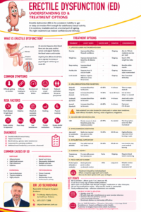
The Long Foreskin: When a Little Extra Skin Is Just… Extra
Foreskins come in all shapes and sizes. Some are short, some sit neatly over the tip of the penis, and others extend well beyond the glans, occasionally giving the penis the appearance that it has ordered the extra-long sleeve option.
A long foreskin, sometimes described medically as a redundant or elongated prepuce, is usually simply a normal anatomical variation. Length alone is not a disease and does not automatically require treatment.
The important question is not “How long is it?” but rather:
Does it cause any problems?
What is a long or redundant foreskin?
The foreskin, or prepuce, is the fold of skin covering the glans (head) of the penis. Its length varies considerably between men.
In some men, the foreskin extends only slightly beyond the glans when the penis is flaccid. In others, there may be a considerable amount of skin extending beyond the tip.
The appearance can also change substantially between the flaccid and erect states. A foreskin that looks impressively generous when flaccid may retract quite normally during an erection.
There is therefore no particular measurement at which a foreskin suddenly becomes “too long.”
If it retracts comfortably, causes no symptoms and can be kept clean, there is generally no medical reason to shorten it.
Can a very long foreskin cause problems?
Most men with a long foreskin have no problems whatsoever.
Occasionally, however, additional foreskin can contribute to several issues.
Hygiene
A longer foreskin may make cleaning beneath the foreskin slightly more important.
Smegma, dead skin cells and moisture can accumulate underneath the foreskin if it is not regularly retracted and washed. This may produce irritation or an unpleasant smell.
Fortunately, the solution is usually wonderfully low-tech: water, gentle washing and regular hygiene.
Aggressive scrubbing, strong soaps, antiseptics and fragranced products can actually irritate this rather sensitive real estate.
Recurrent balanitis
Balanitis is inflammation of the glans. When both the glans and foreskin are inflamed, the condition is called balanoposthitis.
Symptoms may include:
- Redness
- Swelling
- Itching or burning
- Discomfort
- Discharge
- Unpleasant odour
- Difficulty retracting the foreskin
Recurrent inflammation deserves assessment because there may be an underlying cause such as infection, dermatitis, diabetes or a chronic inflammatory skin condition.
Difficulty retracting the foreskin
A long foreskin and a tight foreskin are not the same thing.
A very long foreskin that retracts easily is generally harmless. If the opening is too narrow to retract comfortably over the glans, however, this is phimosis.
Phimosis can cause discomfort, recurrent infections, painful erections or difficulty with sexual activity.
Paraphimosis
Occasionally a tight foreskin is pulled behind the glans and becomes trapped there. The foreskin swells and cannot be returned to its normal position.
This is called paraphimosis.
Unlike having a long foreskin, paraphimosis is not merely an interesting anatomical conversation starter. It can compromise blood flow to the glans and requires urgent medical attention.
Problems during sex
Some men with considerable redundant foreskin notice excessive movement or bunching of the skin during intercourse.
Others experience:
- Irritation
- Small tears
- Discomfort
- Difficulty using condoms
- Foreskin becoming trapped behind the glans
- Problems associated with an accompanying short frenulum
Importantly, many men with long foreskins have completely normal and comfortable sexual function.
Urination
A long foreskin may occasionally balloon during urination or cause the urinary stream to spray.
A little post-urination dribbling from urine retained beneath a long foreskin can also occur.
If the foreskin opening is normal and there are no other symptoms, this is often more inconvenient than medically significant. Persistent ballooning associated with a very narrow opening, pain or difficulty passing urine should be assessed.
Does a long foreskin increase the risk of cancer?
Having a long foreskin by itself does not mean that a man will develop penile cancer.
Penile cancer is uncommon, particularly in Australia. Important risk factors include persistent HPV infection, smoking, chronic inflammation and certain foreskin disorders such as lichen sclerosus.
Any persistent penile ulcer, lump, thickened area, bleeding lesion, unusual discharge or change in the skin should therefore be examined rather than waiting for it to disappear.
The penis, regrettably, has never been particularly good at diagnosing itself.
Does a long foreskin need treatment?
Usually, no.
If the foreskin:
- Retracts comfortably
- Can be cleaned easily
- Does not cause recurrent infections
- Does not interfere with urination
- Does not cause pain during erections or intercourse
- Does not bother the patient cosmetically
then no treatment is required.
The medical principle here is refreshingly simple:
If it isn’t causing a problem, there may be nothing to fix.
Conservative management
When symptoms are minor, treatment may involve improving foreskin hygiene and treating any underlying inflammation or infection.
Where there is associated mild phimosis, a doctor may recommend a course of topical corticosteroid cream combined with gentle stretching.
Forceful stretching should be avoided. Small tears can produce scarring, and scarring can make the foreskin progressively tighter, turning a relatively minor problem into a considerably more stubborn one.
Surgical options
Surgery may be considered when there are significant or recurrent problems.
Circumcision
Circumcision removes the foreskin and permanently exposes the glans.
It is a reliable treatment for recurrent balanitis, troublesome phimosis and certain chronic foreskin diseases.
Potential complications include bleeding, infection, altered sensation, cosmetic dissatisfaction, wound problems and, rarely, more significant complications.
Foreskin-preserving surgery
Not every foreskin problem requires complete circumcision.
Depending upon the anatomy and underlying problem, procedures such as preputioplasty can widen a tight foreskin while preserving much of the foreskin.
In selected men with excessive redundant skin but an otherwise healthy foreskin, surgical shortening may also be discussed.
The appropriate operation depends upon the reason for treatment rather than simply the number of centimetres of foreskin involved.
What about appearance?
Some men simply dislike having a long foreskin.
That is a legitimate concern to discuss with a urologist, but cosmetic surgery deserves careful consideration because surgery permanently changes normal anatomy.
It is particularly important to distinguish between a genuinely troublesome anatomical issue and anxiety created by comparisons with pornography, photographs or other men.
There is an enormous range of normal penile anatomy.
Normal does not come in one factory setting.
When should you see a urologist?
Consider seeking medical advice if you have recurrent infections, difficulty retracting or replacing the foreskin, painful erections, tearing or bleeding, problems during intercourse, difficulty passing urine, persistent skin changes or simply uncertainty about whether your foreskin is normal.
A foreskin that suddenly becomes swollen and trapped behind the glans requires urgent assessment, particularly if the glans becomes increasingly swollen, painful or discoloured.
The bottom line
A very long foreskin is usually a variation of normal anatomy rather than a medical condition.
There is no prize for having the shortest foreskin, and fortunately no excess-baggage fee for having the longest.
What matters is function.
If your foreskin retracts normally, stays healthy, allows comfortable erections and intercourse, and can be kept clean, its length alone is unlikely to require treatment.
If it becomes tight, painful, repeatedly inflamed or simply troublesome, however, a urological assessment can determine whether conservative treatment or surgery is appropriate.
When it comes to foreskins, longer isn’t necessarily better or worse. Sometimes it is simply… longer.
Paraphimosis: When the Foreskin Gets Stuck
Paraphimosis is one of those urological problems where the phrase “I’ll see how it goes overnight” is not a particularly good plan.
It occurs when the foreskin of an uncircumcised penis is pulled back behind the head of the penis (glans) and becomes trapped there. The tight band of foreskin can interfere with normal drainage of blood and fluid from the glans, causing increasing swelling. As the swelling worsens, the foreskin becomes progressively more difficult to return to its normal position.
Although the appearance can be alarming and the circumstances occasionally embarrassing, paraphimosis is a genuine urological emergency. Early treatment is usually relatively straightforward. Delay can make treatment considerably more difficult and, in severe cases, threaten the blood supply to the penis.
What is paraphimosis?
Normally, a mobile foreskin can be gently retracted behind the glans and then returned forward to cover it.
In paraphimosis, the foreskin has been retracted but cannot be brought forward again.
The retracted foreskin forms a tight constricting ring behind the glans. This initially obstructs lymphatic and venous drainage, causing fluid to accumulate and the glans and foreskin to become swollen.
This creates an unfortunate little feedback loop:
Tight foreskin → swelling → tighter constriction → more swelling.
In severe or prolonged cases, arterial blood flow can eventually be compromised.
That is why paraphimosis should not simply be regarded as “a tight foreskin”. It requires prompt assessment and treatment.
What does paraphimosis look and feel like?
The typical presentation is fairly characteristic.
Symptoms and signs can include:
- A foreskin that is stuck behind the glans
- A tight band or “collar” of foreskin behind the head of the penis
- Increasing swelling of the glans
- Swelling of the retracted foreskin
- Penile pain or tenderness
- Difficulty pulling the foreskin forward
- Difficulty passing urine in more severe cases
The glans may initially look red or congested. With worsening obstruction to its circulation, it can become increasingly dark red, purple or dusky.
Severe pain, marked swelling, loss of sensation or a dark/blue/black appearance requires immediate emergency medical assessment.
What causes paraphimosis?
Paraphimosis usually occurs after the foreskin has deliberately or inadvertently been pulled backwards and then left behind the glans.
Medical procedures
A surprisingly common cause is retraction of the foreskin during:
- Urinary catheter insertion
- Examination of the penis
- Cystoscopy or other urological procedures
- Cleaning or nursing care
After manipulating an uncircumcised penis, the foreskin should always be returned to its normal position.
It is a small final step that can prevent a disproportionately large problem.
Phimosis or a tight foreskin
Men with a relatively tight foreskin may be able to retract it, but struggle to return it once it has passed behind the widest part of the glans.
Repeated episodes of inflammation or scarring can increase this risk.
Sexual activity
Retraction during sexual intercourse or masturbation can occasionally result in the foreskin becoming trapped.
Infection and inflammation
Balanitis and balanoposthitis can cause swelling of the glans and foreskin, making paraphimosis more likely.
Trauma
Penile swelling following trauma can occasionally trap a previously retracted foreskin.
Poor foreskin care
Paraphimosis can occur in elderly, debilitated or hospitalised men when a foreskin is retracted for cleaning or catheterisation and is inadvertently left in that position.
Why is paraphimosis an emergency?
The problem is not simply that the foreskin is in the wrong place.
The tight band acts rather like a tourniquet around the penis.
Venous and lymphatic drainage are affected first. Blood and fluid enter the glans more easily than they can leave, producing increasing oedema.
As the glans enlarges, the constricting ring becomes tighter.
If this progresses sufficiently, arterial circulation can also become compromised.
Rarely, severe untreated paraphimosis can result in:
- Ischaemia
- Ulceration
- Tissue necrosis
- Gangrene
- Permanent penile injury
Fortunately, these complications are unusual when treatment is sought promptly.
Urgent treatment
The immediate objective is simple:
Reduce the swelling and return the foreskin over the glans.
Treatment depends on the degree of swelling, duration of the problem, pain and whether there are signs of compromised blood flow.
Pain relief and local anaesthesia
Reduction can be uncomfortable, particularly when substantial swelling has developed.
Depending on the circumstances, treatment may involve:
- Oral or intravenous analgesia
- Local anaesthetic gel
- Penile nerve block
- Occasionally sedation
Adequate pain control also allows the patient to relax, which can make reduction easier.
Reducing the swelling
Before attempting reduction, pressure may be applied to the swollen glans and foreskin for several minutes to encourage oedema to disperse.
Compression techniques or dressings may sometimes be used.
The principle is straightforward: make the glans smaller before asking the foreskin to travel back over it.
Manual reduction
Once swelling has been reduced, the clinician gently compresses the glans while simultaneously drawing the foreskin forward.
Successful reduction usually produces rapid improvement in the constriction and discomfort.
This should be performed carefully. Forceful repeated manipulation of a severely swollen penis can cause additional trauma.
What if manual reduction doesn’t work?
Occasionally the constricting ring is simply too tight.
In that situation, further attempts at heroic squeezing are unlikely to win any medals.
A dorsal slit may be required.
After appropriate local anaesthesia, a small incision is made through the constricting portion of the foreskin. This releases the tight band and allows the foreskin to be repositioned.
If the tissue appears significantly compromised, urgent surgical assessment is particularly important.
What happens after successful reduction?
Successful reduction solves the immediate emergency, but it does not necessarily solve the underlying problem.
There may still be:
- Residual swelling
- Inflammation
- Small tears or abrasions
- Infection
- Phimosis
- Scarring of the foreskin
Patients are generally advised not to repeatedly retract the foreskin while significant swelling and inflammation remain.
Any associated infection or inflammatory condition should also be treated.
Delayed treatment: preventing it happening again
Once the acute episode has settled, the underlying foreskin should be reassessed.
This is particularly important after recurrent paraphimosis or when there is significant phimosis.
Treatment options may include observation, topical therapy in selected cases, foreskin-preserving surgery or circumcision.
Circumcision
For men with significant scarring, persistent phimosis or recurrent paraphimosis, circumcision provides a definitive solution by removing the problematic foreskin.
Circumcision is usually performed after the acute swelling and inflammation have settled, rather than during the initial emergency, unless circumstances require otherwise.
Preputioplasty
Selected patients wishing to preserve their foreskin may be suitable for a foreskin-widening procedure such as preputioplasty.
Whether this is appropriate depends on the degree and cause of the narrowing.
Can paraphimosis be prevented?
Often, yes.
The most important rule is wonderfully uncomplicated:
If the foreskin is pulled back, put it back.
After catheterisation, examination, washing or any medical procedure, the foreskin should be returned to its normal position over the glans.
Men with a persistently tight foreskin, recurrent inflammation or previous paraphimosis should consider discussing definitive treatment with a urologist rather than waiting for another episode.
When should I seek medical attention?
If the foreskin is trapped behind the glans and cannot easily be returned to its normal position, seek urgent medical attention.
Do not wait for the swelling to settle by itself.
Increasing pain, rapidly worsening swelling, difficulty urinating or a glans that is becoming dark purple, blue or black requires immediate emergency assessment.
Embarrassment should never delay treatment. Urologists have seen essentially every variation the human anatomy can produce, and a stuck foreskin is very much part of the job description.
The take-home message
Paraphimosis occurs when a retracted foreskin becomes trapped behind the glans and forms a constricting band.
It may begin as an uncomfortable inconvenience, but progressive swelling can turn it into a genuine emergency.
Early treatment is usually simple and highly effective.
The priorities are to reduce swelling, restore the foreskin to its normal position and protect the circulation to the penis. Once the emergency has resolved, any underlying phimosis or foreskin disease should be assessed to reduce the chance of recurrence.
And perhaps the easiest piece of preventative urological advice to remember:
Foreskin back? Put it back.
This information is intended for general patient education and does not replace individual medical assessment. Suspected paraphimosis requires prompt medical attention.
If this happens, I’ll see you at the Emergency Department at either of the 2 Brisbane hospitals I work at: Wesley and St Andrews War Memorial hospitals. If this has been reduced and you are in need of a circumcision, come chat to your friendly Brisbane Urologist, Dr Jo Schoeman, Uro-Jo.
Urethral Meatal Stenosis: Congenital, Acquired and Correct Management
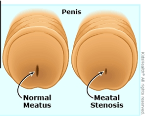
The urethral meatus is the opening through which urine leaves the body. Meatal stenosis means that this opening has become abnormally narrow and is interfering with urinary flow. It is most often discussed in boys and men, although narrowing of the female urethral opening can also occur.
An important point is that a meatus can look small without causing obstruction. Treatment should therefore be based on the complete picture, symptoms, the appearance of the opening, the urinary stream and, where appropriate, objective testing, not appearance alone.
What symptoms can it cause?
Typical symptoms include:
- a thin, forceful or upward-deflected stream;
- spraying or difficulty aiming the urine;
- taking longer to pass urine or needing to strain;
- burning or discomfort during urination;
- a small spot of blood at the meatus;
- urinary frequency, urgency or incomplete emptying; and
- recurrent urinary infection in selected patients.
In toilet-trained boys, an abnormal stream is often the most useful clue. Published patient-reported data show that improvement after meatotomy is most predictable when the preoperative problem is an abnormal or deflected stream. Frequency, urgency, wetting or dysuria may have another cause and should not automatically be attributed to a narrow-looking meatus.
Complete inability to pass urine is uncommon but requires urgent medical attention.
Congenital meatal stenosis
Congenital meatal stenosis is present from birth. True isolated congenital narrowing is uncommon and should be distinguished from normal variation in meatal size. It may also occur as part of another developmental urethral condition, including hypospadias, or following congenital urethral reconstruction.
In a baby or young child, symptoms can be difficult to recognise. The diagnosis becomes more apparent after toilet training, when a persistently narrow, spraying or markedly deflected stream can be observed.
Not every anatomically small meatus needs surgery. An asymptomatic child with a satisfactory stream, no urinary infections and normal bladder emptying can often be observed. Symptomatic obstruction, however, should be assessed by a paediatric urologist.
Acquired meatal stenosis
Acquired stenosis develops after birth. Its causes vary with age.
In boys
Meatal stenosis is recognised after circumcision, although published estimates vary substantially because studies use different definitions and methods of examination. Proposed mechanisms include irritation and inflammation of the exposed meatus, contact with wet nappies, meatal ulceration and subsequent scar formation. It often becomes clinically obvious between early childhood and school age rather than immediately after circumcision.
Other causes include inflammation, trauma, catheterisation and previous surgery, particularly repair of hypospadias. Following hypospadias surgery, narrowing may involve more than the external opening and must be assessed in the context of the reconstructed urethra.
In adolescents and adults
Important causes include:
- lichen sclerosus (also called balanitis xerotica obliterans or BXO), which can scar the foreskin, glans, meatus and more proximal urethra;
- repeated urethral instrumentation, catheterisation or endoscopic surgery;
- prior hypospadias repair or other penile surgery;
- trauma, infection or chronic inflammation; and
- previous radiotherapy or treatment affecting the urethra.
In adults, it is essential to determine whether narrowing is confined to the meatus or extends into the fossa navicularis or penile urethra. Treating only the visible opening will fail if more extensive scar disease has been overlooked.
Female meatal or urethral stenosis is uncommon. Symptoms may resemble recurrent urinary infection or other causes of bladder-outlet obstruction. Diagnosis should be made carefully, as urinary symptoms alone do not prove that the urethra is narrowed.
How is it diagnosed?
Assessment may include:
- A detailed history: including the direction and calibre of the stream, spraying, pain, infections, prior circumcision, catheterisation, surgery, trauma and skin disease.
- Examination: assessing the meatus and surrounding skin for scarring, pallor, inflammation, lichen sclerosus, hypospadias or surgical change.
- Observation of the urinary stream, particularly in a toilet-trained child.
- Uroflowmetry and post-void residual ultrasound when symptoms are unclear, the patient is older, or more extensive obstruction is suspected.
- Urinalysis or urine culture if pain, blood or infection is suspected.
- Urethral calibration, cystoscopy or urethrography selectively, especially in adults, recurrent disease, previous urethral surgery or suspected extension beyond the meatus.
Kidney and bladder ultrasound is not required for every straightforward case, but may be appropriate when there are recurrent infections, incomplete emptying, significant obstruction or concern about the upper urinary tract.
What is the correct management?
Management must match the patient’s symptoms, cause, age and extent of narrowing.
1. Observation
Observation is reasonable when the meatus is merely small in appearance but the patient has no relevant symptoms, passes a satisfactory stream and empties the bladder normally. Treating an incidental finding is unlikely to improve unrelated urgency, frequency or wetting.
2. Treat active skin or inflammatory disease
When lichen sclerosus or another inflammatory disorder is present, the underlying disease must be treated as well as the narrowing. Potent topical corticosteroid treatment is commonly used for genital lichen sclerosus under medical supervision. Circumcision may be indicated when the foreskin is affected, but established meatal or urethral scar may also require surgery.
Persistent or suspicious penile lesions may need biopsy. Long-term review can be appropriate because lichen sclerosus can recur, extend into the urethra and is associated with a small but important risk of penile malignancy.
3. Meatotomy or meatoplasty
For a short, symptomatic stenosis confined to the meatus, meatotomy or meatoplasty is usually the definitive treatment.
- A meatotomy enlarges the opening with a controlled incision.
- A meatoplasty reconstructs and sutures the edges to create a durable, appropriately shaped opening.
Both can provide excellent relief in appropriately selected children. A large paediatric series reported that meatotomy required more early manual spreading and had a higher reoperation rate than meatoplasty; technique and postoperative care therefore matter. In another study, 95% of families reported their child was at least somewhat improved after meatotomy, with the strongest benefit in boys treated for an abnormal stream.
The operation may be performed under local anaesthesia in selected cooperative patients or under general anaesthesia, particularly in younger children. Expected short-term effects include stinging, minor spotting of blood and temporary spraying while swelling settles. The surgeon may advise ointment and gentle separation of the meatal edges during early healing; instructions vary according to the procedure used.
4. Dilatation
Repeated blind dilatation is generally not a durable solution for dense scar-related meatal stenosis. It may cause further tearing and scarring and can commit a patient to repeated procedures. Carefully selected dilation or self-dilatation may have a role as temporary or palliative management, or as part of a specialist regimen for inflammatory disease, but it should not replace an appropriate reconstructive assessment in recurrent or complex disease.
5. Recurrent, adult or extended distal disease
If narrowing recurs, is associated with lichen sclerosus, or extends into the fossa navicularis/distal urethra, a simple repeat incision may not be sufficient. Options include formal meatoplasty or distal urethroplasty using an oral mucosal graft. The European Association of Urology recommends offering open meatoplasty or distal urethroplasty for meatal and distal urethral strictures; the precise operation should be individualised to stricture length, tissue quality and the patient’s priorities.
In lichen-sclerosus-related urethral disease, genital skin should not be used as a graft because the disease may recur in that tissue. Oral mucosa is generally preferred when graft reconstruction is required.
Follow-up and recurrence
Most patients with an isolated, properly treated meatal stenosis do well. Follow-up should assess:
- improvement in stream direction and calibre;
- pain, bleeding, infection or difficulty voiding;
- bladder emptying when clinically indicated; and
- restenosis or progression of an underlying condition such as lichen sclerosus.
Prompt reassessment is advisable if the stream narrows again, spraying persists after healing, voiding becomes painful or difficult, infections recur, or new skin changes appear.
The practical message
Meatal stenosis is not simply “a small hole.” Correct care begins by confirming that the narrowing is clinically important and identifying its cause and extent. A symptomatic, short stenosis confined to the meatus is usually treated successfully with meatotomy or meatoplasty. Recurrent stenosis, adult disease, previous hypospadias repair or lichen sclerosus requires a more detailed urethral assessment and sometimes formal reconstruction.
This article provides general educational information and does not replace an individual assessment. Seek urgent care if you or your child cannot pass urine, develops fever with urinary symptoms, or has significant bleeding or pain.
Selected references
- European Association of Urology. EAU Guidelines on Urethral Strictures: Disease Management in Males. 2026. https://uroweb.org/guidelines/urethral-strictures/chapter/disease-management-in-males
- European Association of Urology. EAU Guidelines on Urethral Strictures: Definition, Epidemiology, Aetiology and Prevention. 2026. https://uroweb.org/guidelines/urethral-strictures/chapter/definition-epidemiology-aetiology-and-prevention
- Wessells H, et al. Urethral Stricture Disease Guideline Amendment (2023). Journal of Urology. 2023. doi:10.1097/JU.0000000000003482.
- Dothan D, et al. Surgical Treatment of Meatal Stenosis: Lessons Learned from the Pediatric Urology Practice. Urology. 2023;171:220–224. PMID: 35981660.
- Varda BK, et al. Minor procedure, major impact: patient-reported outcomes following urethral meatotomy. Journal of Pediatric Urology. 2018;14(2):165.e1–165.e5. doi:10.1016/j.jpurol.2017.11.018.
- Godley SP, et al. Meatal stenosis: a retrospective analysis of over 4000 patients. Journal of Pediatric Urology. 2015;11(1):38.e1–38.e6. doi:10.1016/j.jpurol.2014.09.016.
- Morris BJ, Krieger JN. Does circumcision increase meatal stenosis risk? A systematic review and meta-analysis. Urology. 2017;110:16–26. doi:10.1016/j.urology.2017.07.027.
- Wang MH. Surgical management of meatal stenosis with meatoplasty. Journal of Visualized Experiments. 2010;(45):2213. doi:10.3791/2213.
Prepared for patient education.
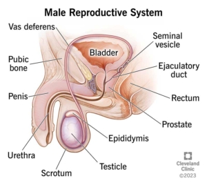
Male Anatomy
Dr Jo Schoeman, Urologist
Suite 46, Level 4
The Wesley Medical Centre
Wesley Hospital
Auchenflower Brisbane
16 DOT ‘Nesbitt’ Plication
Correction of penile chordee / curvature.
Why is it done?
- To treat an acquired deviation of an erect penis.
- Usually occurs in males 55-65 years of age.
- Can be associated with previous penile trauma, usually no associated history.
- A dorsal (up) curvature is more common than a ventral (down) one. Can also deviate to the side.
- Pain is usually the presenting symptom with a gradually worsening curvature.
- The curvature may be so bad that penetration becomes impossible.
- Associated with the connective tissue disorder: Dupuytren’s Contracture, which is an auto-immune disease.
- Worse cases may require a penile prosthesis.
Pre-requirements
- An informed consent is required from the patient/ parents.
- Patients are informed that this may shorten the penis to the length of the shorter side of the penis, usually 2-3 cm.
- In patients who wish to preserve penile length, a lengthening technique using buccal mucosa may be indicated and will be referred to a colleague.
- Patients may not eat or drink from 6-8 hours prior to surgery according to age.
- Any anti-coagulants such as Warfarin or Aspirin must be stopped 7 days prior to surgery. Clexane injections may be substituted.
- Be prepared for an overnight stay.
How is it done?
 This procedure is done under general anesthetic.
- Supine position.
- The foreskin is loosened proximal to the glans with a circumferential incision and the whole penile skin is retracted to the base of the penis.
- An artificial erection will be induced by injecting a sterile saline solution into the penile corpora cavernosa with a tourniquet around the base.
- Non-dissolvable sutures will be placed on the sides opposite to the diseased areas in an attempt to pull the erect penis into a straight alignment.
- Occasionally a circumcision may result due to complications with this technique, yet foreskin preservation is attempted.
- If there is a dorsal curvature, ventral sutures are laced and the penis pulled in upright position, therefore sutures are always placed on the opposite site avoiding vital structures such as.
- An indwelling catheter will be inserted until you are awake.
- A dressing is then applied, which should be removed after 72 hours.
- A local anesthetic is injected at the base of the penis as a penile block thus giving post-operative pain relief for the next 4-6 hours.
What to expect after the procedure?
- Any anesthetic has its risks, and the anesthetist will explain such risks.
- Bleeding is a common complication.
- A hematoma (blood collection under the skin) may form and needs to be reviewed by Dr Schoeman as soon as possible. Bruising is normal.
- Sutures may tear loose with vigorous use of erect penis, and the procedure may then require revision.
- An infection of the wound may occur and requires immediate attention.
- Necrosis of the foreskin and some penile skin can occur in rare circumstances. This may require skin-grafting.
- DANGER SIGNS: A wound that swells immediately, fever, and puss. Contact Dr Schoeman or the hospital immediately as this occurs in up to 15–20% of all cases.

What next?
- Dressings should be kept dry for the initial 72 hours after surgery and soaked off in a bath thereafter.
- The dressing may sometimes adhere to the wound causing slight bleeding on removal. Don’t panic, the bleeding will stop.
- The catheter will be removed as soon as you are awake, or if there are concerns, the following morning.
- On discharge, a prescription may be issued for patients to collect.
- Patients should schedule a follow-up appointment with Dr Schoeman 2 weeks after the procedure.
- There will be signs of bruising for at least 10 days.
- Refrain from using your erect penis for 6 weeks.
- The suture-line will be hard and indurated for at least 8-10 weeks.
- Sick leave will be granted for 10 days.
- Please direct all further queries to Dr Schoeman’s rooms.
- PLEASE CONTACT THE HOSPITAL DIRECTLY WITH ANY POST-OPERATIVE CONCERNS AND RETURN TO THE HOSPITAL IMMEDIATELY SHOULD THERE BE ANY SIGNS OF SEPSIS.
Download Information Sheet
Cauterization of Penile Condylomata
Fulguration of penile condyloma, with penile block
Why is it done?
- One of the non-medical treatment options for condylomas (genital warts)..
How is it done?
 This procedure is done under local or general anaesthetic.
- Supine position.
- The foreskin may be the only affected area and therefore a circumcision is done.
- Otherwise, the affected area is exposed and cleaned.
- The affected lesions are cauterized, including the root of the wart.
- Due to charring, and good hemostasis, no sutures are required.
- Hemostatic dressings are placed.
- Specimen is sent to a histopathologist.
- An indwelling catheter may be inserted if the biopsy area involves the meatus of your urethra.
- A dressing is then applied, which should be removed after 72 hours.
- A local anesthetic is injected at the base of the penis as a penile block thus giving post-operative pain relief for the next 4-6 hours.
What to expect after the procedure?
- Any anesthetic has its risks, and the anesthetist will explain such risks.
- Bleeding is an uncommon complication.
- Eschar may loosen with vigorous use of erect penis and could lead to bleeding.
- An infection of the wound may occur and requires immediate attention.
- DANGER SIGNS: A wound that swells immediately, fever, and puss. Contact Dr Schoeman or the hospital immediately as this occurs in up to 15–20% of all cases.
What next?
- Dressings should be kept dry for the initial 72 hours after surgery and soaked off in a bath thereafter.
- The dressing may sometimes adhere to the wound causing slight bleeding on removal. Don’t panic, the bleeding will stop.
- The catheter will be removed as soon as you are awake, or if there are concerns, the following morning.
- Patients should schedule a follow-up appointment with Dr Schoeman 4-6 weeks after the procedure.
- There will be signs of bruising for at least 10 days.
- Refrain from using your erect penis for 6 weeks.
- Sick leave will be granted for a few days.
- Please direct all further queries to Dr Schoeman’s rooms.
- PLEASE CONTACT THE HOSPITAL DIRECTLY WITH ANY POST-OPERATIVE CONCERNS AND RETURN TO THE HOSPITAL IMMEDIATELY SHOULD THERE BE ANY SIGNS OF SEPSIS.
Download Information Sheet
Caverject Intra-Cavernosal Injections
Intra cavernous injections for erectile dysfunction, demonstrating and teaching self-administration.
Why is it done?
-
- A treatment option for erectile dysfunction.
- Usually when oral techniques have failed ie Viagra, Levitra and Cialis.
- Or where the patient wishes to skip the oral phase for something more effective.
- ED can occur from the age of 40.
- Risk factors:
- Older.
- Overweight.
- Diabetic,
- Hypertensive,
- Cardiac issues.
- An alternative is a Vacuum Pump Device.
- Worse cases may require a penile prosthesis.
How is it done?
 This procedure is done at home.
- You will be given a ‘hands-on’ instruction.
- An effective dose will be determined.
- A pre-made-up syringe with a determined dose is made ready prior to the injection.
- The penis is pulled away from your body with the non-dominant hand.
- The injection site is cleaned with an alcohol wipe.
- Caverject is injected at the base of the corpora cavernosa at an angle of 45 degrees with the shaft. See hand-out.
- Allow 5 minutes for an erection to be obtained.
- The erection lasts approximately 20 minutes.
What next?
- Once the correct the dose has been established.
- You feel comfortable using the injections.
- A repeat script will be issued.
- Your GP will continue with the medication.
- Dr Michael Gillman, Men’s Health Physician is a fantastic alternative.
Complications?
- Dose too high for the individual.
- Prolonged painful erection.
- If erection lasts longer than 4-6 hours, it becomes an emergency, and you are required to visit your local Emergency Department.
- This will be drained with a syringe.
- A surgical bypass my be done with possible erectile dysfunction as complication.
Download Information Sheet
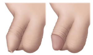
Circumcision
Surgical removal of foreskin with penile block for post-operative pain relief
Why is it done?
- Religious reasons.
- Health reasons.
- Personal reasons.
- Medical reasons:
- Narrowing of foreskin -phimosis.
- Foreskin stuck behind head of penis – paraphimosis.
- Severe infection of the foreskin and head of penis -balanoposthitis.
- Cancer of the foreskin – SCC.
- Trauma.
How is it done?
- This is done under general anesthetic or penile block.
- A cut is made at the level of the glans penis, circumferentially around the penis, through the skin.
- The foreskin is then retracted, and a second incision is made circumferentially around the base of the glans penis.
- The skin between the 2 incisions is then surgically removed.
- Dissolvable sutures are then placed between the 2 remaining edges.
- A Jelonet and Bactroban dressing is then placed tightly around the penis, still allowing urine to pass through the end.
- A local anesthetic is injected into the base of the penis thus giving post-operative pain relief for the next 4-6 hours.
Unfortunately, no infants younger than 12 months can be operated on in any of the private hospitals ONLY at Children’s Hospital.
What to expect after the procedure
- Any anesthetic has its risks, and the anesthetist will explain such risks.
- Bleeding is a common complication.
- With any subsequent erections post operatively, the sutures may pull out causing an opening of the wound with subsequent bleeding.
- An infection of the wound can occur if the dressings are left on too long.
- If the dressing has been applied too tightly, or if there is any discomfort, please remove the dressing immediately. If some hemorrhaging re-occurs, REDO the dressing!
- In very young patients, the foreskin may still be attached to the head of the penis, thus leaving a raw and red glans penis after surgery. Keep the affected area clean and apply ointment as prescribed.
- NB! Each person is unique and for this reason symptoms may vary!
What next?
- The dressing should be removed in a bath 48 hours after the procedure.
- The dressing should be soaked until it comes off with ease.
- The dressing may sometimes adhere to the wound causing slight bleeding on removal. Don’t panic the bleeding will stop.
- As soon as the dressing has been removed, Bactroban (or similar) ointment should be applied on the wound twice a day.
- This may not be required if the foreskin was not adhered to the glans penis.
- On discharge a prescription may be issued for the patient to collect.
- A follow-up appointment should be scheduled to see Dr Schoeman within 2 weeks.
- Please don’t hesitate to direct all pre-operative queries to Dr Schoeman’s rooms.
- PLEASE CONTACT THE HOSPITAL WITH ANY POST-OPERATIVE CONCERNS AND RETURN TO THE HOSPITAL IMMEDIATELY SHOULD THERE BE ANY SIGNS OF SEPSIS.
Download Information Sheet
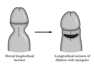
Dorsal Slit Procedure
Opening of foreskin where a phimosis exists, yet foreskin preservation is a requirement. Also done initially with severe septic para-phimosis as interim procedure until sepsis is cleared and a circumcision is possible
Why is it done?
- Paraphimosis: foreskin stuck behind head of penis.
- Foreskin preserving.
How is it done?
 This is done under general anesthetic or a penile block.
- A vertical cut is made through the tightest part of the para-phimosis, and the foreskin is than able to be covered over the meatus.
- The incision is then closed by opposing edges in the horizontal plane.
- Dissolvable sutures are placed between the 2 remaining edges.
- A local anesthetic is injected into the base of the penis thus giving post-operative pain relief for the next 4-6 hours.


What to expect after the procedure
- Any anesthetic has its risks, and the anesthetist will explain such risks.
- Minor bleeding.
- With any subsequent erections post operatively, the sutures may pull out causing an opening of the wound with subsequent bleeding.
- An infection of the wound can occur if the dressings are left on too long.
- If the dressing has been applied too tightly, or if there is any discomfort, please remove the dressing immediately. If some hemorrhaging re-occurs, REDO the dressing!
- In very young patients, the foreskin may still be attached to the head of the penis, thus leaving a raw and red glans penis after surgery. Keep the affected area clean and apply ointment as prescribed.
- NB! Each person is unique and for this reason symptoms may vary!
What next?
- The dressing should be removed in a bath 48 hours after the procedure.
- The dressing should be soaked until it comes off with ease.
- The dressing may sometimes adhere to the wound causing slight bleeding on removal. Don’t panic the bleeding will stop.
- As soon as the dressing has been removed, Bactroban (or similar) ointment should be applied on the wound twice a day.
- This may not be required if the foreskin was not adhered to the glans penis.
- On discharge a prescription may be issued for the patient to collect.
- A follow-up appointment should be scheduled to see Dr Schoeman within 2 weeks.
- Please don’t hesitate to direct all pre-operative queries to Dr Schoeman’s rooms.
- PLEASE CONTACT THE HOSPITAL WITH ANY POST-OPERATIVE CONCERNS AND RETURN TO THE HOSPITAL IMMEDIATELY SHOULD THERE BE ANY SIGNS OF SEPSIS.
Download Information Sheet