
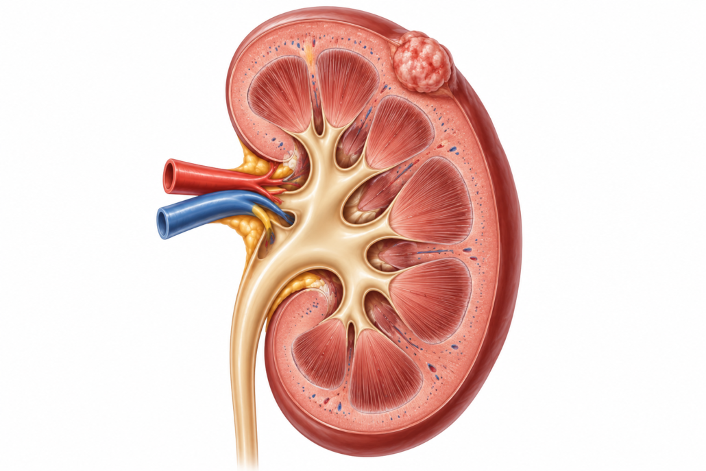
Small renal masses: observation, surgery and image-guided ablation
An unexpected spot on a kidney scan can be alarming. A small renal mass usually means a solid, enhancing growth measuring 4 cm or less and confined to the kidney. It is not automatically cancer: some are benign, and many small kidney cancers grow slowly. The right approach depends on the scan, any biopsy result, kidney function, other health conditions and what matters to you. [1,2]
How do we investigate it?
I review the original imaging and usually arrange a dedicated, multiphase contrast CT or MRI to establish whether the lesion enhances, its size and position, and whether it looks like a simple cyst or another benign condition. Blood tests assess kidney function. The need for chest imaging depends on the findings and clinical circumstances. If contrast is unsuitable because of impaired kidney function, we plan an alternative with the radiologist. [1,2]
A needle biopsy can help when knowing whether a mass is benign or what type of cancer it contains would change the decision. A biopsy can occasionally be inconclusive. If ablation is planned, the European Association of Urology (EAU) recommends biopsy before a separate ablation session to avoid treating benign lesions unnecessarily. [1]
What are the choices?
| Approach | When it may suit | Main consideration |
|---|---|---|
| Active surveillance | A very small mass, particularly under 2 cm; slow growth; or a patient who prefers to defer treatment | Planned scans and a clear trigger to reconsider treatment |
| Partial nephrectomy | A mass requiring treatment when kidney-sparing surgery is technically suitable | Removes the tumour and supplies a complete specimen, but requires an operation |
| Percutaneous ablation | A selected small mass when treatment is indicated, especially if surgery carries greater risk or preserving kidney tissue is important | Less invasive, but imaging follow-up is essential and repeat treatment may be needed |
| Radical nephrectomy | A tumour that cannot reasonably be treated with a kidney-sparing approach | Removes the whole kidney and may have a greater effect on kidney function |
For masses under 2 cm, surveillance is often a sensible first option. For masses 2–4 cm, surveillance, partial nephrectomy and ablation may all deserve discussion. If surgery is appropriate, guidelines generally favour partial nephrectomy for a treatable small cancer. Frailty or limited life expectancy may make symptom-focused observation preferable. [1,2]
Surveillance is an active plan, not neglect. Scans are often repeated every 3–6 months initially and then every 6–12 months if stable, with timing adapted to the individual. Growth beyond 4 cm, repeated growth above approximately 5 mm a year, new concerning features or patient preference can prompt reconsideration; growth rate alone does not prove that a mass is dangerous. [2]
What is radiofrequency ablation (RFA)?
RFA destroys tissue by passing an electrical current through a thin probe, generating heat inside the mass. An interventional radiologist usually places the probe through the skin using CT or ultrasound guidance. Anaesthesia or sedation, patient positioning and sometimes fluid injected to protect nearby bowel are planned around the tumour’s location. The radiologist treats the mass and a small surrounding margin, then checks for immediate complications. Many patients have a short hospital stay, but arrangements vary. [1–3]
RFA is most established for small, accessible masses, particularly those up to 3 cm. A mass deep in the kidney, next to the urine-drainage system or bowel, or near major vessels can be harder to treat safely or completely. EAU guidance advises against routinely offering RFA for masses over 3 cm, because control becomes less reliable. A mass between 3 and 4 cm needs an individual discussion rather than an automatic exclusion. [1]
What other ablation methods are available?
- Cryoablation uses probes to freeze the tumour. The visible ice ball can help the operator plan the treatment margin. It is an established alternative to RFA; the EAU advises against its routine use for tumours over 4 cm. [1,2]
- Microwave ablation uses microwave energy to heat tissue. Experience is growing and the EAU describes encouraging results, though long-term comparative evidence remains less certain than for surgery. Availability and expertise vary. [1]
- Irreversible electroporation delivers electrical pulses to disrupt cell membranes. Evidence for routine treatment of small renal cancers remains limited. [2]
- Stereotactic ablative radiotherapy (SABR/SBRT) directs highly focused radiation without inserting a probe. It can be discussed for selected patients who need treatment but are unsuitable for surgery; long-term comparisons remain limited. [1]
The choice depends on tumour size and position, prior treatment, kidney function, local expertise and the patient’s preferences. No technique is right for every lesion.
Benefits, limitations and risks
Percutaneous ablation can avoid a surgical incision, usually involves less immediate recovery than surgery and can preserve functioning kidney tissue. Risks include bleeding, infection, pain, injury to the urine-collecting system or nearby organs, and incomplete treatment or local recurrence. Risks depend strongly on where the tumour sits. A second ablation or surgery may be possible if follow-up imaging shows persistent disease. [1,2]
Evidence comparing ablation with partial nephrectomy largely comes from observational studies involving patients with different health risks. Ablation offers good local control for carefully selected small tumours, but its long-term cancer control relative to surgery is less certain; it should not be described as a guaranteed equivalent. [1,2]
What happens after ablation?
Treatment destroys the lesion in place, so there is no whole-tumour surgical specimen. Contrast-enhanced CT or MRI checks for remaining enhancing tissue and later recurrence. One published guideline suggests imaging at 3, 6 and 12 months, then annually after ablation, adjusted to biopsy findings, renal function and the treating team’s protocol. We also monitor kidney function. A new enhancing focus requires specialist review and may lead to repeat biopsy or treatment. [1,2]
My role in your care
I can review the findings and discuss surveillance, biopsy and the treatment choices with you. I no longer perform nephrectomies. If an operation is the best option, I will refer you to a colleague who performs kidney surgery. If ablation appears suitable, I will arrange assessment by an experienced interventional radiology team; the final decision follows review of the images and your overall health.
Seek urgent medical attention for heavy visible bleeding, inability to pass urine, fever with flank pain, or severe worsening pain after a procedure.
This article provides general information. Individual advice requires assessment of your imaging, biopsy findings where relevant, kidney function and medical history.
Dr Jo Schoeman | Patient information | September 2026
References
- European Association of Urology. EAU Guidelines on Renal Cell Carcinoma: Disease Management, current online edition, accessed September 2026.
- Richard PO, et al. Canadian Urological Association guideline: Management of small renal masses. Canadian Urological Association Journal. 2022;16(2)–E75.
- Park BK, et al. Asian Conference on Tumor Ablation guidelines for renal cell carcinoma. Investigative and Clinical Urology. 2021.
- European Association of Urology. EAU Guidelines on Renal Cell Carcinoma: Follow-up, current online edition, accessed September 2026.





Leave a Reply
Want to join the discussion?Feel free to contribute!