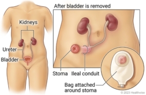
Conduitoscopy, Retrograde Pyelogram, Stent Management

A diagnostic procedure under general anesthetic where a rigid / flexible cystoscope is placed in your ileal conduit (stoma), ureteric catheters are placed to enable imaging of the upper tracts with/without insertion or removal of ureteric stents.

Why is it done?

To investigate:

  • Hematuria (blood in the urine)
  • Recurrent upper urinary tract infections
  • Space occupying lesions in the kidneys and ureters
  • Abnormal cells suggestive of urothelial carcinoma, on urine cytology
  • Surveillance of previous bladder cancer

Risk factors:

  • Previous bladder cancer
  • Upper tract urothelial carcinoma
  • Ureteric structuring
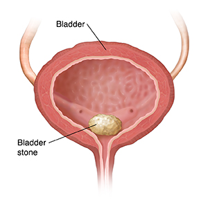
  • Stone disease


How is it done?

  • This is done under General anesthesia.
  • A cystoscopy is performed by placing a   camera in the conduit
  • The conduit is then distended with saline.
  • The inside of the conduit is viewed for pathology.
  • A retrograde pyelogram is done at the same time, (placement of small silicone catheters up the kidney pipes). Through this iodine contrast is injected up into the kidney collecting systems. This facilitates the viewing of kidney pipes and kidney collecting systems on X-ray to exclude any upper tract pathology.
  • If any abnormalities are found in the kidney/ ureters, a flexible ureteroscopy (which is the placement of a long thin camera up the ureter) will be performed.
  • If any suspicious lesions are seen, a biopsy will be taken.
  • A ureteric stent may be placed
  • Urine would have been sent for cytology, to rule out the existence of cancer.
  • Antibiotics may be given to prevent infection.

What to expect after the procedure?

  • Pain on initial passing of urine
  • Infection ranging from a burning sensation to, fever, to puss (rare)
  • Bloodstained urine
  • Lower abdominal discomfort which will persist for a few days
  • Pain radiating from bladder to renal angle associated with urinating.
  • An infection could present with a stent being present.

.

Wes Conduitogram and stent

Copyright 2019 Dr Jo Schoeman

Cysto-Lithopaxy

Endoscopic procedure used for breaking up a bladder stone. Either with a stone crusher or laser

Why is it done?

  • To break up a bladder calculus (stone).


Risk factors:

  • Bladder outflow obstruction.
  • BPH with chronic retention.
  • Urethral stricture.
  • Neurogenic bladder.
  • Renal calculi disease.
  • Metabolic disorders.
  • Malnutrition.
  • Chronic infections.
  • Foreign objects in bladder.

How is it done?

  • A cystoscopy is performed by placing a camera in the urethra with the help of a lubricant jelly and an irrigate (fluid).
  • The bladder is then distended with fluid (saline).
  • The inside of the bladder is viewed for pathology.
  • If any suspicious lesions are seen, a biopsy will be taken.
  • Stone crushing is attempted with a lithotrite (a crushing device).
  • If the calculus is too large, laser will be utilized to fragment the stone and the smaller stones evacuated.
  • Antibiotics may be given to prevent infection.

What to expect after the procedure?

  • Hematuria (blood in your urine)
  • You will have a n indwelling catheter (IDC), which will remain in your bladder until your urine is clear.
  • You may have a continuous bladder irrigation with Saline to help clear the bleeding.
  • Pain on initial passing of urine when the catheter is removed.
  • Bladder infection ranging from a burning sensation to, fever, to pus (rare).
  • Lower abdominal discomfort which will persist for a few days.
  • NB! Each person is unique and for this reason symptoms vary.

What next?

  • This all depends on what is found during the procedure. All the options will be discussed in detail.
  • You may require further attention to your prostate or bladder outlet to prevent further stone formation.
  • There may be some blood in the urine. This can be remedied by drinking plenty of fluids until it clears.
  • Patients should schedule a follow-up appointment within 1 month to discuss the etiology of the calculus as well as what other procedures may be involved to prevent this from occurring again.
  • Please don’t hesitate to direct all further queries to Dr Schoeman.


Download Information Sheet

Wes Cysto-Lithopaxy Laser

Cystocoele Repair (Natural)

Repair of an anterior / bladder prolapse using natural tissue and repair of introitis


Why is it done?

  • The aim of surgery is to relieve the symptoms of vaginal bulge and / or laxity.
  • Improve bladder function without interfering with sexual function.

How is it done?

  • This procedure is done under a spinal / general anesthetic, as decided by the anesthetist.
  • An incision is made along the center of the front wall of the vagina starting near the vaginal entrance and finishing near the top of the vagina.
  • The vaginal skin is then separated from the underlying supportive fascial layer.
  • The weakened fascia is then repaired using absorbable stitches, which will absorb over 4 weeks to 5 months depending on the type of stitch (suture) material used.
  • Sometimes excessive vaginal skin is removed, and the vaginal skin is closed with absorbable sutures, these usually take 4 to 6 weeks to fully absorb.
  • Reinforcement material in the form of biological (absorbable) may be used to repair the anterior vaginal wall.
  • Mesh is no longer used
  • A cystoscopy may be performed to confirm that the appearance inside the bladder is normal and that no injury to the bladder or ureters has occurred during surgery.
  • A pack may be placed into the vagina and a catheter into the bladder at the end of surgery.
  • If so, this is usually removed after 3-48 hours. The pack acts like a compression bandage to reduce vaginal bleeding and bruising after surgery.
  • You will have a vaginal pack to reduce any bleeding.
  • Both the pack and the catheter are usually removed within 48 hours of the operation.

How successful is the surgery?

  • Quoted success rates for anterior vaginal wall repair are 70-90%.
  • There is a chance that the prolapse may come back in the future, or another part of the vagina may prolapse for which you need further surgery.
  • Recurrence rates are as much as 50% in the next 3 years.

No Intercourse for 6 weeks following surgery!

Complications?

  • With any surgery there is always a small risk of complications.
  • Anesthetic problems. With modern anesthetics and monitoring equipment, complications due to anesthesia are very rare.
  • Bleeding. Serious bleeding requiring blood transfusion is unusual following vaginal surgery (less than 1%).
  • Post operative infection. Although antibiotics are often given just before surgery and all attempts are made to keep surgery sterile, there is a small chance of developing an infection in the vagina or pelvis.
  • Bladder infections (cystitis) occur in about 6% of women after surgery and are more common if a catheter has been used. Symptoms include burning or stinging when passing urine, urinary frequency and sometimes blood in the urine. Cystitis is usually easily treated by a course of antibiotics.
  • Constipation is a common postoperative problem.
  • Pain with intercourse (dyspareunia). Some women develop pain or discomfort with intercourse.
  • Damage to the bladder or ureters during surgery is an uncommon complication which can be repaired during surgery.
  • Incontinence. After a large anterior vaginal wall repair some women develop stress urinary incontinence due to the unkinking of the urethra.
  • This is usually simply resolved by placing a supportive sling under the urethra section.


Download Information Sheet

Wes Cystoscoele Repair Natural

Drainage Renal Abscess

To drain a large abscess causing low grade to high temperatures. Percutaneous or open procedure for the drainage of abscess.

Why is it done?

  • Patients presenting with low grade persistent fevers, even high fevers requiring admission to High Dependency Unit for septicemia.
  • Usually immune compromised patients: Diabetics, Corticosteroid users, Viral immune-deficiency states etc.
  • This condition requires urgent drainage.
  • The patients’ need to be resuscitated first by an emergency team with appropriate fluids and antibiotics and placed in an area where all systems can be supported (HDU).
  • As soon as the patient is stable, this abscess needs to be drained, either with open surgery or percutaneous drain placement.
  • If it is a large pyonephrosis with a non-functioning kidney, a nephrectomy should be considered.

How is it done?

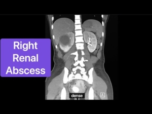
  • Patients will receive a general anesthesia.
  • Appropriate resuscitation would have been started.
  • Prophylactic anti-biotics is given.
  • An indwelling catheter is placed.
  • The correct kidney is identified and marked while you are awake.
  • If it is a small abscess, an ultrasound guided needle is placed through your back or side into the fluid collection. A guidewire will be placed through the cannula and a drain fed in over the guidewire. All the pus will be drained.
  • If it is a large loculated abscess, an incision will be made over the area closest to the skin. The cavity will be opened, drained and rinsed, after which a drain will be placed.
  • If you have a non-functioning kidney associated with this, your kidney may be removed at the same time.
  • A drain is left post-operatively.

What next?

  • You will spend up to 7 or more nights in hospital.
  • You may be on life support depending on the degree of sepsis.
  • You will have intravenous fluids, antibiotics and circulatory supporting drugs being administered. Either a central venous line for monitoring, an arterial line and a peripheral infusion line.
  • You will have a catheter for that time.
  • A drain for 2-3 days.
  • Your drain will be removed with minimal drainage present.
  • You will a trial without the catheter as soon as you are back in the ward.
  • You will be discharged as soon as your renal function has stabilized, and you have opened your bowels.
  • Allow for 6 weeks for stabilization of symptoms.
  • A follow-up appointment will be scheduled for 6 weeks.
  • DON’T SUFFER IN SILENCE, OR YOU WILL SUFFER ALONE!

Risks

  • This a potentially dangerous condition, which could result in death. It requires urgent management!
  • May lose your kidney in serious cases.
  • May risk dialysis when in septic shock.
  • Wound Infection.
  • Prolonged stay in HDU.
  • Post-operative hernia formations especially associated in the elderly with atrophic abdominal muscles.
  • NB! Each person is unique and for this reason symptoms vary


Download Information Sheet

Wes Drainage renal abcess

Dorsal Slit Procedure

Opening of foreskin where a phimosis exists, yet foreskin preservation is a requirement. Also done initially with severe septic para-phimosis as interim procedure until sepsis is cleared and a circumcision is possible

Why is it done?

  • Paraphimosis: foreskin stuck behind head of penis.
  • Foreskin preserving.

How is it done?

  • This is done under general anesthetic or a penile block.
  • A vertical cut is made through the tightest part of the para-phimosis, and the foreskin is than able to be covered over the meatus.
  • The incision is then closed by opposing edges in the horizontal plane.
  • Dissolvable sutures are placed between the 2 remaining edges.
  • A local anesthetic is injected into the base of the penis thus giving post-operative pain relief for the next 4-6 hours.

What to expect after the procedure

  • Any anesthetic has its risks, and the anesthetist will explain such risks.
  • Minor bleeding.
  • With any subsequent erections post operatively, the sutures may pull out causing an opening of the wound with subsequent bleeding.
  • An infection of the wound can occur if the dressings are left on too long.
  • If the dressing has been applied too tightly, or if there is any discomfort, please remove the dressing immediately. If some hemorrhaging re-occurs, REDO the dressing!
  • In very young patients, the foreskin may still be attached to the head of the penis, thus leaving a raw and red glans penis after surgery. Keep the affected area clean and apply ointment as prescribed.
  • NB! Each person is unique and for this reason symptoms may vary!

What next?

  • The dressing should be removed in a bath 48 hours after the procedure.
  • The dressing should be soaked until it comes off with ease.
  • The dressing may sometimes adhere to the wound causing slight bleeding on removal. Don’t panic the bleeding will stop.
  • As soon as the dressing has been removed, Bactroban (or similar) ointment should be applied on the wound twice a day.
  • This may not be required if the foreskin was not adhered to the glans penis.
  • On discharge a prescription may be issued for the patient to collect.
  • A follow-up appointment should be scheduled to see Dr Schoeman within 2 weeks.
  • Please don’t hesitate to direct all pre-operative queries to Dr Schoeman’s rooms.
  • PLEASE CONTACT THE HOSPITAL WITH ANY POST-OPERATIVE CONCERNS AND RETURN TO THE HOSPITAL IMMEDIATELY SHOULD THERE BE ANY SIGNS OF SEPSIS.


Download Information Sheet

Wes Dorsal Slit procedure

Endoscopic vesico-ureteric reflux surgery (STING)

Indication:

  • Vesical-ureteric reflux. It is a minimally invasive procedure performed with endoscopy.
  • A synthetic material (Bulkamid) is injected at the ureteric opening to prevent reflux
  • Grade 3-4 Vesical-Ureteric Reflux where conservative management has failed with a progressive deterioration in renal function.

How is it done?

  • Patients will receive a general anaesthesia.
  • Prophylactic antibiotics is given.
  • The correct ureteric system is identified and marked while you are awake.
  • This will be an endoscopic procedure.
  • A cystoscopy will be done with injection of Bulkamid just under the affected ureteric orifice.
  • Enough Bulkamid will be injected to partially close the ureteric opening yet not obstructing the orifice.
  • An indwelling catheter is placed.

What next?

  • You may be in hospital the day or overnight.
  • As soon as you are comfortable with no signs of pain and emptying your bladder sufficiently, you will be discharged.
  • Review in 6 weeks.
  • DON’T SUFFER IN SILENCE, OR YOU WILL SUFFER ALONE!

Download Information Sheet

Wes Cystoscopy RGP and STING anti-reflux VUR procedure

Epididymectomy

Surgical excision of epididymis with spermatic cord block

Why is it done?

  • To remove a symptomatic painful epididymis post vasectomy.


How is it done?

  • This procedure is performed under general anesthetic.
  • A single incision is made on the midline raphe of the scrotum.
  • The affected testis and vas deference is then extracted through this incision.
  • The epididymis is exposed.
  • The epididymis is carefully removed off the testis without disrupting the blood supply to the testis.
  • The cord is checked for hemorrhaging.
  • A drain may be placed.
  • A catheter may be left over night.
  • A dressing is then applied, which should be removed after 72 hours.
  • No strenuous movements are permitted for at least 14 days.

NB! You are required to bring 2 pairs of tight new undies for post-operative scrotal support.

What to expect after the procedure?

  • Any anesthetic has its risks, and the anesthetist will explain all such risks.
  • The drain will be removed the next morning.
  • The catheter will be removed 6-8 hours after the procedure.
  • A hematoma (blood collection under the skin or in the scrotal cavity) may form and needs to be reviewed by Dr Schoeman as soon as possible. This may require drainage. Bruising is normal.
  • An infection of the wound can occur and requires immediate attention.
  • Owing to the nature of the surgery and the soft skin of the scrotum, bruising may appear to be much worse than it actually is and is no cause for alarm.
  • DANGER SIGNS: A scrotum that swells immediately to size of a football, fever, puss. Contact Dr Schoeman or the hospital immediately as this may occur in up to 15% of all cases.

What next?

  • The dressing should be kept dry for the initial 72 hours after surgery and then soaked in a bath until the dressing comes off with ease.
  • The dressing may sometimes adhere to the wound causing slight bleeding on removal. Don’t panic, the bleeding will stop.
  • Do not tug at the sutures!
  • Sutures will dissolve after 10-14 days
  • On discharge a prescription may be issued for patients to collect.
  • Please direct all further queries to Dr Schoeman’s rooms.
  • PLEASE CONTACT THE HOSPITAL DIRECTLY WITH ANY POST OPERATIVE CONCERNS AND RETURN TO THE HOSPITAL IMMEDIATELY SHOULD THERE BE ANY SIGNS OF SEPSIS.


Download Information Sheet

Wes Epididymectomy

Excision of Epididymal Cyst – Spermatocelectomy

What is it 

  • spermatocele (spermatic cyst) is a common, noncancerous, fluid-filled sac that develops in the epididymis,
  • The cyst usually contains milky or clear fluid that may contain sperm. 

Why is it done?

  • Enlarged scrotum
  • Could be uncomfortable
  • The cyst can become so big that the enlarged scrotum buries the penis making usual functions difficult, ie urination and sexual function
  • May contribute to infertility


How is it done?

  • This procedure is done under general anesthetic.
  • Supine position.
  • A midline scrotal incision is done.
  • The intact spermatocele/epididymal cyst with the testis is delivered through the skin incision.
  • The epididymis cyst is carefully surgical resected off the spermatic cord or epididymis.
  • A hemostatic running suture is placed around the raw edge of resection if required
  • Hemostasis is actively chased.
  • A drain is left overnight.
  • An Indwelling catheter is left for 6-8 hours to prevent acute urinary retention.
  • The scrotum is closed in 2 layers with dissolvable sutures.
  • You would be required to bring 2 pairs of tight new undies for post-operative scrotal support; these will be placed post-operatively


Complications

Side–effects

  • Any anesthetic has its risks, and the anesthetist will explain such risks.
  • Bleeding is a possible complication, therefore, the scrotal drain/s overnight.
  • Your catheter will be removed the next
  • You will have scrotal swelling and bruising for the next 2-6 weeks
  • Any sudden, increased swelling needs urgent attention!
  • Any symptoms of fever and signs of infection require urgent attention!


Download Information Sheet

Wes Spermatocoelectomy

Copyright 2019 Dr Jo Schoeman

Exision of Urethral Caruncle Prolapsed Urethral Mucosa

Why is it done?

  • Prolapsed urethral mucosa causing pain and bleeding
  • Occurs from childhood to old age


How is it done?

  • This procedure is done under a spinal/general anesthetic, as decided by the anesthetist.
  • The legs will be elevated into the lithotomy position.
  • This procedure is done with cystoscopy.
  • Your bladder and urethra are inspected with cystoscopy
  • The prolapsed mucosa will then be excised at the external meatus.
  • Dissolvable sutures will be placed for hemostasis
  • A catheter will be placed until you are awake for some compression.
  • Prophylactic antibiotics will be given to prevent infection.

Complications

Side–effects

  • Any anesthetic has its risks, and the anesthetist will explain all such risks.
  • Complications: hemorrhaging, and urine retention
  • Patients’ catheter will be removed the next morning.
  • If you cannot urinate after 2-3 attempts, a catheter may be inserted to empty your bladder.
  • You may be required to keep the catheter for a few days if you have persistent bleeding or urinary retention.
  • NB! Each person is unique and for this reason, symptoms may vary!


Download Information Sheet  

Wes Urethral Caruncle – Prolapse Excision

Copyright 2019 Dr Jo Schoeman

Excision of Urethral Diverticulum – Female

Why is it done?

  • Usually, an infected peri-urethral gland blocks and becomes infected
  • Causes a bulge which interferes with urination
  • Can mimic a vaginal prolapse
  • Usually, an MRI of the urethra delineates this beautifully.

How is it done?

  • This procedure is done under a general anaesthetic, as decided by the anaesthetist.
  • The legs will be elevated into the lithotomy position.
  • This procedure is done both cystoscopically and with an incision over the urethra.
  • The urethra is evaluated. endoscopically and a catheter placed
  • The vaginal mucosa will then be incised over the urethrocele.
  • A Fogarty catheter will be placed inside the diverticulum and the balloon inflated to delineate the borders of the diverticulum.
  • The diverticulum will be dissected out with injuring adjacent structures.
  • The neck will be tied off at the level of the adjoining urethra.
  • Dissolvable vaginal closure sutures will be placed for hemostasis
  • A cystoscopy confirms no injury to the urethra.
  • A catheter will be placed until you are awake for some compression.
  • Prophylactic antibiotics will be given to prevent infection.

Complications

Side–effects

  • Any anaesthetic has its risks, and the anaesthetist will explain all such risks.
  • Complications: hemorrhaging, and urine retention
  • Patients’ catheter will be removed the next morning
  • If you cannot urinate after 2-3 attempts, a catheter may be inserted to empty your bladder.
  • You may suffer temporary incontinence
  • You may suffer permanent incontinence as advised by Jo, depending on the extent of the diverticulum. Make sure you have discussed this with Jo.

NB! Each person is unique and for this reason, symptoms may vary


Download Information Sheet

Wes Urethral Diverticulum Excision Female

Copyright 2019 Dr Jo Schoeman