‘Bag-of-Worms” Varicocele: Causes, Symptoms, Fertility and Treatment Options

A varicocele is an enlargement of the veins surrounding the testicle. It is similar to a varicose vein in the leg, although it occurs within the scrotum.

Varicoceles are common and often harmless. However, in some men they may cause discomfort, reduce the size or function of the affected testicle, or contribute to fertility problems. Most varicoceles do not require treatment, and the decision to intervene depends on symptoms, testicular development, semen quality and plans for future fertility.

What causes a varicocele?

Blood leaves each testicle through a network of small veins called the pampiniform plexus. These veins normally carry blood upwards towards larger veins in the abdomen.

A varicocele develops when blood pools within these veins. This is usually related to poorly functioning venous valves, together with the anatomy and pressure within the testicular veins.

Around 80–90% of clinically detected varicoceles occur on the left side. This is because the left testicular vein is longer and enters the left renal vein at a right angle, making venous drainage less efficient. A varicocele may occur on both sides, while an isolated right-sided varicocele is much less common.

Most varicoceles develop gradually during puberty and have no dangerous underlying cause.

A new varicocele in an older man, particularly one that is right-sided, develops suddenly or does not disappear when lying down, may require abdominal imaging to exclude obstruction of the vein by an abdominal or retroperitoneal condition. This is uncommon but important to recognise.

How common are varicoceles?

A varicocele is found in approximately 15% of adolescent and adult men. Many men never know that they have one.

Varicoceles are more frequently identified among men being investigated for infertility. They may be present in approximately:

  • 35–40% of men with primary infertility
  • Up to 70–80% of men with secondary infertility, where a previous pregnancy has occurred, but the couple is now having difficulty conceiving

Importantly, most men with a varicocele remain fertile. Finding a varicocele does not automatically mean that it is causing infertility.

What are the symptoms?

Many varicoceles cause no symptoms and are discovered during a routine examination, fertility assessment or scrotal ultrasound.

When symptoms occur, they may include:

  • A dull ache, dragging sensation or heaviness in the scrotum
  • Discomfort that becomes worse after standing, exercising or later in the day
  • Improvement in discomfort when lying down
  • Visible or enlarged veins above the testicle
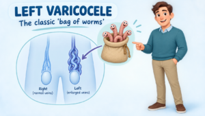
  • A soft swelling sometimes described as feeling like a “bag of worms”
  • Reduced size or slower growth of the affected testicle
  • Abnormal semen-analysis results
  • Difficulty conceiving with a partner

A varicocele generally does not cause erectile dysfunction or difficulty urinating. Its relationship with testosterone production remains under investigation, although some men with a clinically significant varicocele may have impaired testicular function.

Sudden severe testicular pain is not typical of a varicocele and requires urgent medical assessment to exclude testicular torsion or another acute scrotal condition. An abdominal ultrasound is also required to exclude a possible lesion in the abdomen obstructing the veins.

How is a varicocele diagnosed?

Diagnosis usually begins with a physical examination. The scrotum is examined while the patient is standing and lying down. You may be asked to take a breath and gently bear down, the Valsalva manoeuvre, to make enlarged veins easier to feel.

Clinical varicoceles are commonly graded as:

  • Grade 1: Felt only during the Valsalva manoeuvre
  • Grade 2: Felt while standing without straining
  • Grade 3: Easily visible through the scrotal skin

A scrotal ultrasound may be recommended if the examination is uncertain, the testicles differ in size, pain is present, or another scrotal problem needs to be excluded. Ultrasound can measure the veins, assess backward blood flow and examine the testicles.

For fertility assessment, investigations may include:

  • One or more semen analyses
  • Testicular volume measurement
  • Hormone tests when indicated
  • Assessment of both partners, because fertility is a shared consideration

An ultrasound-only or “subclinical” varicocele that cannot be felt on examination is not usually treated for infertility.

How can a varicocele affect fertility?

The testicles work best at a temperature slightly below core body temperature. The pampiniform veins help cool the arterial blood entering the testicle.

Pooling of warm blood in a varicocele may interfere with this cooling system. Increased temperature, venous pressure, oxidative stress and altered blood flow may affect sperm production and testicular function.

Possible semen changes include:

  • Reduced sperm concentration
  • Reduced sperm movement or motility
  • Increased abnormal sperm forms
  • Increased sperm DNA damage

The degree of impact varies considerably. A large varicocele can be associated with normal fertility, while a smaller palpable varicocele may be significant in another man. The diagnosis must therefore be interpreted together with semen results, the couple’s fertility history, the female partner’s age and other fertility factors.

Does every varicocele require treatment?

No. Observation is appropriate when a varicocele:

  • Causes no troublesome symptoms
  • Is not affecting testicular growth
  • Is associated with normal semen parameters
  • Is found incidentally on ultrasound but cannot be felt
  • Is unlikely to be the main cause of the couple’s fertility difficulty

Simple measures such as supportive underwear, avoiding activities that consistently aggravate discomfort and occasional appropriate pain relief may help mild symptoms. Regular review may be recommended for adolescents with a difference in testicular size.

When should treatment be considered?

Treatment may be considered when there is:

Infertility with abnormal semen parameters

Repair is most likely to benefit a couple when the man has:

  • A varicocele that can be felt on examination
  • Difficulty conceiving
  • One or more abnormal semen parameters
  • No better explanation for the infertility
  • A reasonable opportunity for natural conception following treatment

Both the AUA/ASRM and European guidance support considering repair in men attempting conception who have a palpable varicocele and abnormal semen parameters. Treatment is not routinely recommended for a varicocele detected only by ultrasound. AUA/ASRM male infertility guideline and EAU male infertility guideline

Persistent scrotal pain

Repair may be reasonable when typical varicocele-related discomfort persists despite conservative measures and other causes of scrotal pain have been excluded.

Reduced testicular growth in an adolescent

Treatment may be discussed if the affected testicle is persistently smaller, growth is impaired, the varicocele is large or semen parameters are abnormal in an appropriately selected older adolescent.

Progressive testicular dysfunction

Selected men with deteriorating semen quality or evidence of impaired testicular function may benefit from repair after individual assessment.

What treatment procedures are available?

Treatment works by blocking or dividing the abnormal veins. Blood then returns through healthy alternative veins while the testicular artery and lymphatic drainage are preserved.

Management option How it is performed Advantages Disadvantages and risks
Observation Clinical review, with semen analysis or testicular measurements when appropriate No procedure or anaesthetic; suitable for most asymptomatic men; avoids unnecessary treatment Does not correct the varicocele; symptoms or testicular changes may require future review
Conservative symptom management Scrotal support, activity modification and appropriate pain relief Simple and non-invasive; may adequately control mild discomfort Does not remove the varicocele or improve abnormal semen parameters; long-term anti-inflammatory medication may not be suitable for everyone
Microsurgical subinguinal varicocelectomy A small incision is made near the groin. An operating microscope helps identify and divide abnormal veins while protecting the testicular artery and lymphatics Usually the lowest recurrence and hydrocele rates; no abdominal incision; effective for unilateral or bilateral disease; commonly preferred for fertility treatment Requires an anaesthetic and microsurgical expertise; temporary bruising, swelling or discomfort; uncommon risks include infection, recurrence, hydrocele or testicular artery injury
Microsurgical inguinal varicocelectomy Similar microsurgical repair through a slightly higher groin incision Good visualisation and low recurrence when performed microsurgically Requires opening the external oblique fascia; recovery may involve slightly more groin discomfort
Laparoscopic varicocelectomy Veins are clipped or divided through small abdominal keyhole incisions Both sides can be treated during the same procedure; useful in selected cases General anaesthetic; entry into the abdomen; generally higher hydrocele risk than lymphatic-sparing microsurgery; rare abdominal or vascular complications
Open high ligation The testicular vein is divided through an abdominal or groin incision without microsurgical magnification Established technique; may be effective where microsurgery is unavailable Higher recurrence and hydrocele rates than modern microsurgical repair; less precise preservation of small arteries and lymphatics
Radiological embolisation or sclerotherapy An interventional radiologist passes a catheter into the testicular vein and blocks it using coils, plugs or a sclerosant No surgical incision in the scrotum or groin; often performed with local anaesthesia or sedation; relatively quick recovery; useful for recurrence after surgery The vein cannot always be accessed; radiation and contrast exposure; recurrence or persistence may occur; coils can rarely migrate; availability depends on local expertise

How effective is treatment?

The results depend on why treatment is being performed, the technique used and whether other fertility or pain-related factors are present.

Improvement in semen quality

Following repair of a clinically significant varicocele, approximately 60–80% of appropriately selected men demonstrate improvement in at least one semen parameter. Improvement is not guaranteed, and some men show little or no measurable change.

Because sperm production takes approximately three months, semen analysis is usually repeated about three months after treatment and may be reassessed again at six months.

Pregnancy and fertility outcomes

Varicocele repair may increase the chance of natural pregnancy in selected infertile couples, but it does not guarantee conception. Studies commonly report spontaneous pregnancy in roughly 25–40% of couples after repair, although rates vary substantially according to female partner factors, duration of infertility, baseline semen quality and study design.

Treatment may also improve semen quality sufficiently to allow a couple to use a less invasive assisted-reproduction option. In other cases, IVF or intracytoplasmic sperm injection may still be required.

The couple’s overall fertility timeline is important. If the female partner has reduced ovarian reserve or advancing reproductive age, waiting several months for semen improvement may not be appropriate. Varicocele repair and assisted reproductive treatment should therefore be considered as part of a shared plan.

Relief of pain

When pain is typical of a varicocele, a dull ache or heaviness made worse by standing or activity, approximately 70–90% of appropriately selected men experience meaningful improvement after repair.

Surgery is less predictable when pain is sharp, constant or atypical, and pain may occasionally persist despite technically successful treatment.

Testicular growth

In adolescents with a persistently smaller affected testicle, repair may allow “catch-up” growth. Decisions should be based on repeated measurements rather than a single ultrasound result.

What are the possible complications?

Varicocele procedures are generally safe, but potential complications include:

  • Bruising, swelling or wound discomfort
  • Infection or bleeding
  • Persistence or recurrence of the varicocele
  • Hydrocele, caused by fluid collecting around the testicle
  • Ongoing or occasionally altered scrotal pain
  • Injury to the testicular artery, which is uncommon but potentially serious
  • Testicular shrinkage, which is rare
  • Anaesthetic complications
  • Technical failure, contrast reaction, radiation exposure or coil-related problems following embolisation

Using microsurgical magnification and preserving the lymphatic vessels usually reduces the risks of recurrence, hydrocele and arterial injury.

Recovery after varicocele repair

Most men return home on the day of treatment. Bruising, mild swelling and groin or scrotal discomfort are expected during the early recovery period.

Patients are commonly advised to:

  • Wear supportive underwear
  • Keep the wound clean and dry
  • Avoid heavy lifting and strenuous exercise for the period advised by the surgeon
  • Resume sexual activity when comfortable and medically cleared
  • Attend follow-up to assess healing and symptom improvement
  • Repeat semen analysis after approximately three months when fertility is the reason for treatment

Specific recovery instructions vary according to the procedure and the individual patient.

When should you seek medical advice?

Arrange a medical assessment if you notice:

  • A new lump or swelling in the scrotum
  • Persistent scrotal discomfort or heaviness
  • A difference in testicular size
  • Difficulty conceiving
  • A new right-sided varicocele
  • A swelling that remains when lying down

Seek urgent medical attention for sudden severe testicular pain, rapid swelling, nausea or a high-riding testicle. These features may indicate testicular torsion, which is an emergency.

The bottom line

A varicocele is common and, for most men, causes no significant harm. Treatment is not based on the ultrasound appearance alone. The most important considerations are symptoms, testicular development, semen quality and the couple’s overall fertility situation.

For men with persistent typical pain, impaired testicular growth or a palpable varicocele associated with infertility and abnormal semen parameters, treatment can be worthwhile. Microsurgical varicocelectomy generally offers the most favourable balance between effectiveness, recurrence and complication risk, while embolisation is an effective minimally invasive alternative in selected patients.

A consultation with a urologist allows the varicocele—and the patient rather than simply the scan—to be assessed before deciding whether observation, surgery or embolisation is the most appropriate approach.

This article provides general information and does not replace an individual medical assessment. Treatment recommendations depend on examination findings, semen results, symptoms, age and the fertility circumstances of both partners.

So, if this is something that you have found on your regular scrotal examinations, and you have concerns, come see your Brisbane urologist, Dr Jo Schoeman for advice.

Hydrocoele in Younger Men: Causes, Fertility and Treatment Options

A hydrocoele is a collection of clear fluid around the testicle. It usually causes a painless swelling on one side of the scrotum, although both sides can occasionally be affected.

Hydrocoeles are common in babies and older men, but they can also develop during adolescence or young adulthood. In a younger man, the important question is not only “Is this a hydrocoele?” but also “Why has it developed?”

Most hydrocoeles are benign and do not threaten the testicle. Nevertheless, any new scrotal swelling should be examined because conditions such as an inguinal hernia, infection, testicular torsion or testicular tumour can sometimes produce a similar appearance or cause a secondary hydrocoele.

What causes a hydrocoele?

The testicle is partly surrounded by a thin membrane called the tunica vaginalis. This membrane normally produces a small amount of lubricating fluid, which is continually reabsorbed. A hydrocoele develops when fluid production exceeds absorption.

In younger men, possible causes include:

  • Idiopathic hydrocoele: No specific cause is identified. This is common.
  • Persistent communication with the abdomen: A small channel through which the testicle descended before birth may remain open. This is more typical in children but can occasionally persist into young adulthood and may be associated with an inguinal hernia.
  • Inflammation or infection: Epididymitis, orchitis and some sexually transmitted infections may cause fluid to collect around the testicle.
  • Trauma: A sporting injury, direct blow or previous scrotal injury can lead to a reactive hydrocoele.
  • Previous surgery: Hydrocoeles may occasionally follow groin, hernia, varicocele or testicular surgery.
  • Testicular torsion: Twisting of the spermatic cord can produce a reactive hydrocoele, although severe sudden pain is normally the dominant symptom.
  • Testicular tumour: A tumour may occasionally be accompanied by a hydrocoele. The hydrocoele itself is not cancer, but the fluid can make examination of the underlying testicle difficult.

What does a hydrocoele feel like?

A typical hydrocoele produces a smooth, soft or tense swelling around the testicle. It may fluctuate in size and often feels heavier as it enlarges.

Symptoms can include:

  • painless scrotal enlargement;
  • a dragging or heavy sensation;
  • discomfort when walking, running or exercising;
  • irritation from clothing;
  • difficulty examining the testicle;
  • embarrassment or concern about appearance; and
  • discomfort during sexual activity.

A very large hydrocoele can become inconvenient and may interfere with sport, work or everyday activities.

When is urgent assessment needed?

A hydrocoele usually develops gradually and is not an emergency. Seek urgent medical care, however, if there is:

  • sudden or severe testicular pain;
  • rapid swelling;
  • nausea or vomiting with scrotal pain;
  • redness, fever or increasing tenderness;
  • a hard lump within the testicle;
  • scrotal swelling following a significant injury; or
  • a swelling that cannot be pushed back and is associated with groin or abdominal pain.

Sudden testicular pain may represent torsion, in which the blood supply to the testicle becomes twisted. This requires emergency assessment and should never be attributed to “just a hydrocoele.”

How is a hydrocoele diagnosed?

Assessment usually begins with a discussion about how and when the swelling developed, followed by examination of the abdomen, groin and scrotum.

A hydrocoele may transmit light when a torch is placed behind it, a finding called transillumination. This can support the diagnosis, but it does not replace a proper examination.

A scrotal ultrasound is commonly arranged in a younger man, particularly when:

  • the hydrocoele is new;
  • the testicle cannot be examined clearly;
  • there is pain or tenderness;
  • the swelling developed after injury;
  • the diagnosis is uncertain; or
  • an underlying testicular abnormality needs to be excluded.

Ultrasound can confirm that the swelling contains fluid and assess the testicle, epididymis, blood flow and surrounding structures. The European Association of Urology recommends high-frequency ultrasound when investigating a suspected testicular mass because it can determine whether a lesion is inside or outside the testicle and assess the opposite testis. EAU Testicular Cancer Guideline

Urine tests, STI testing or blood tests may also be appropriate when infection or inflammation is suspected.

Can a hydrocoele affect fertility?

For most younger men, a small or moderate uncomplicated hydrocoele does not cause infertility. It does not normally block sperm transport and does not automatically mean that sperm production is abnormal.

There are, however, several important qualifications:

  1. The underlying cause may matter more than the fluid.
    Previous torsion, testicular trauma, severe infection, an undescended testicle, testicular cancer or another testicular disorder may affect fertility independently of the hydrocoele.
  2. A very large or tense hydrocoele may theoretically affect the testicle.
    Prolonged pressure or increased local temperature has been proposed as a possible mechanism for impaired testicular function. Evidence in adults remains limited, and clinically significant infertility from an isolated hydrocoele appears uncommon.
  3. Surgery carries a small fertility-related risk.
    Hydrocoele repair is performed close to the epididymis, vas deferens and testicular blood supply. Injury to one of these structures is uncommon but could affect fertility, particularly when operating on both sides or when the opposite testicle is abnormal. The British Association of Urological Surgeons lists inadvertent injury to these structures as an uncommon recognised complication of surgery. BAUS hydrocoele repair information

A semen analysis is not routinely required for every man with a hydrocoele. It may be appropriate when:

  • a couple has been unable to conceive;
  • the hydrocoele is very large or present on both sides;
  • the opposite testicle is small or abnormal;
  • there is a history of undescended testis, torsion, chemotherapy, testicular surgery or significant infection; or
  • fertility preservation is an important concern before surgery.

The EAU recommends that the assessment of male infertility include a reproductive and medical history, physical examination, semen analysis and, where indicated, hormonal tests and imaging. It also stresses that semen analysis must be interpreted as part of the couple’s overall reproductive assessment rather than as a simple “fertile or infertile” test. EAU Male Infertility Guideline

Does every hydrocoele need treatment?

No. Treatment depends on the cause, size, symptoms and effect on quality of life.

Observation

A small, painless hydrocoele with a normal underlying testicle can often be monitored. It does not need to be removed merely because it is present.

Observation may include:

  • self-awareness of any change in size or consistency;
  • periodic clinical review when appropriate; and
  • reassessment if pain, rapid enlargement or a new lump develops.

The British Association of Urological Surgeons supports observation when an adult hydrocoele is small or not bothersome. BAUS

Treating an underlying cause

If the fluid is secondary to infection, inflammation or another condition, treatment should address that cause. The hydrocoele may reduce once the underlying problem settles, although established hydrocoeles do not always disappear.

Aspiration

Aspiration involves inserting a needle and draining the fluid. Although this can provide temporary relief, the fluid usually returns because the lining that produces it remains in place.

Aspiration also carries risks of bleeding and infection. BAUS does not regard aspiration alone as curative or standard treatment because recurrence is common. It may occasionally be considered for someone who is unsuitable for surgery. In selected circumstances, a sclerosant may be injected after aspiration, but recurrence and inflammation remain concerns.

Hydrocoelectomy

Hydrocoelectomy is the most reliable definitive treatment for a bothersome adult hydrocoele. Surgery may be considered when the swelling:

  • is large or progressively enlarging;
  • causes discomfort, heaviness or skin irritation;
  • interferes with exercise, work or sexual activity;
  • causes significant cosmetic or psychological concern; or
  • makes examination of the testicle difficult.

The operation is generally performed through a small scrotal incision under general or spinal anaesthesia. The fluid is drained, and the fluid-producing sac is folded, turned behind the testicle or partly removed to reduce the likelihood of recurrence.

Most procedures are performed as day surgery.

What should you expect after surgery?

Bruising, swelling and discomfort are expected and may initially make the scrotum look larger rather than smaller. Supportive underwear, simple pain relief and protected ice packs can help during the early recovery period.

Strenuous exercise and heavy lifting are generally avoided for several weeks. The testicle may continue to feel somewhat bulkier after repair because of the folded or thickened tissues around it.

Possible complications include:

  • bleeding or a scrotal haematoma;
  • wound or testicular infection;
  • persistent swelling;
  • recurrence of the hydrocoele;
  • chronic scrotal discomfort;
  • anaesthetic complications; and
  • uncommon injury to the epididymis, vas deferens or testicular blood supply.

The current BAUS patient information notes that temporary bruising and swelling are very common, while infection, haematoma, recurrence and injury to adjacent reproductive structures occur much less often. Individual risks depend on the size and complexity of the hydrocoele, previous surgery, infection and general health. BAUS hydrocoele repair leaflet

The important message for younger men

A hydrocoele is usually harmless, and many do not require treatment. However, a new scrotal swelling in a younger man should not be self-diagnosed or ignored. Clinical examination and often an ultrasound can confirm the diagnosis and ensure that the testicle underneath is healthy.

Most isolated hydrocoeles do not impair fertility. When fertility is a concern, the entire reproductive history and both testicles should be assessed rather than assuming that the hydrocoele is responsible.

Management should be individualised. For one man, reassurance and observation may be entirely appropriate. For another, surgery may provide worthwhile relief from heaviness, discomfort or embarrassment.

This article provides general patient information and does not replace individual medical assessment. Seek urgent care for sudden testicular pain, rapid swelling, fever or a hard testicular lump.

So, if you have noted a scrotal swelling and this is concerning you and need to have this checked out, see your GP or obtain a referral to see you Brisbane Urologist, Uro-Jo, for peace of mind.

Persistent sperm after vasectomy: does this mean the procedure has failed?

Finding sperm in a semen sample after vasectomy can be worrying. However, a positive result does not always mean that the vasectomy has failed.

Sperm may remain in the reproductive tract for several weeks or months after the procedure. The most important questions are:

  • How long has it been since the vasectomy?
  • How many sperm are present?
  • Are any of the sperm still moving?
  • Is the sperm count decreasing on repeat testing?

A vasectomy does not work immediately

During a vasectomy, the two vas deferens, the tubes carrying sperm from the testicles, are divided and sealed. Sperm already present beyond the site of the vasectomy may remain in the seminal tract and appear in subsequent ejaculations.

It can take several months and approximately 20 ejaculations to clear these remaining sperm. Australian patient guidance commonly recommends the first post-vasectomy semen analysis at approximately three months. International guidelines generally allow testing within about 8–16 weeks, depending on the surgeon’s protocol.

Another form of contraception must be used until your surgeon has reviewed the semen result and formally confirmed clearance.

What is a post-vasectomy semen analysis?

A post-vasectomy semen analysis, or PVSA, examines the semen for:

  • The presence or absence of sperm
  • The number of sperm present
  • Whether the sperm are motile or non-motile

The desired result is azoospermia, meaning that no sperm are detected. However, complete azoospermia is not always necessary for a vasectomy to be considered successful.

When should azoospermia occur?

Many men are azoospermic by approximately three months after vasectomy, particularly if they have ejaculated regularly. Others take longer to clear their residual sperm.

Delayed clearance may be associated with:

  • A relatively low number of ejaculations
  • Individual differences in reproductive-tract anatomy
  • Older age
  • A long interval between ejaculations
  • Laboratory technique and the timing of sample examination
  • Temporary early recanalisation of the vasectomy site

Persistent sperm at the first test should therefore not automatically be interpreted as surgical failure. The type of sperm and the trend on repeat testing are more informative.

What does an “immotile sperm count” mean?

Immotile or non-motile sperm are sperm that are present but show no movement when examined under the microscope.

Following vasectomy, this commonly represents old residual sperm that have not yet been completely cleared. Non-motile sperm have extremely limited capacity to cause pregnancy, particularly when present in very small numbers.

The generally accepted categories are:

Semen-analysis result Usual interpretation
No sperm detected Azoospermia, vasectomy clearance can usually be given
No motile sperm and ≤100,000 non-motile sperm/mL Rare non-motile sperm, generally regarded as successful vas occlusion
More than 100,000 non-motile sperm/mL Repeat testing and assessment of the trend are required
Any motile sperm Clearance should not be given; repeat testing is required

The report should be interpreted carefully because some laboratories report the concentration per millilitre, while others may report a total number or simply state that “occasional sperm” were seen.

A fresh sample is important when assessing motility. If examination is substantially delayed, sperm that were initially moving may have stopped, potentially producing a misleading “non-motile” result.

What are rare non-motile sperm?

Rare non-motile sperm, often abbreviated as RNMS, usually means that no moving sperm are seen and the concentration is no more than 100,000 non-motile sperm per millilitre.

Current Australian and American guidance generally regards either azoospermia or RNMS at or below this threshold as evidence of successful vas occlusion. Depending on the laboratory method and local protocol, your surgeon may provide clearance after one satisfactory sample or request another confirmatory sample.

The estimated risk of pregnancy after clearance based on azoospermia or RNMS is approximately 1 in 2,000. Vasectomy is therefore extremely reliable, but no method of contraception is completely infallible.

What happens if sperm are detected at three months?

Management depends on the result.

A small number of non-motile sperm

This is usually reassuring. If the count is no more than 100,000/mL and no motile sperm are present, clearance may be appropriate according to the treating surgeon’s protocol.

More than 100,000 non-motile sperm/mL

Continue contraception and repeat the semen analysis. A steadily decreasing count suggests delayed clearance rather than vasectomy failure.

Motile sperm

Continue contraception and arrange another semen analysis. Motile sperm early after vasectomy do not invariably mean that a repeat operation will be required, particularly if the number is low and decreasing. Persistent or increasing motile sperm are more concerning for incomplete occlusion or recanalisation.

What is recommended at six months?

Six months is an important decision point.

Motile sperm persisting at six months

If any motile sperm remain at six months, the vasectomy should generally be regarded as an occlusive failure. A repeat vasectomy should be discussed.

Possible explanations include:

  • One vas deferens was not successfully divided
  • An unusual or duplicated vas deferens was present
  • The divided ends have reconnected
  • A small channel has developed across the vasectomy site

More than 100,000 non-motile sperm/mL at six months

This result requires individual assessment rather than an automatic decision.

The surgeon will consider:

  • Whether the count is decreasing, stable or increasing
  • Whether motile sperm were present in earlier samples
  • The reliability and timing of sample collection
  • The vasectomy technique used
  • The couple’s tolerance for even a very small pregnancy risk
  • Whether further semen analyses are likely to provide clarity

Repeat vasectomy, continued contraception or further surveillance may be considered through shared decision-making.

Fewer than 100,000 non-motile sperm/mL at six months

If no motile sperm are present and the count is at or below 100,000/mL, most contemporary guidance considers this compatible with successful vasectomy. Formal clearance must nevertheless come from the treating surgeon.

When should a repeat vasectomy be considered?

A repeat procedure is usually considered when:

  • Motile sperm persist at six months
  • Motile sperm counts increase on consecutive tests
  • There is no meaningful reduction in the sperm count
  • More than 100,000 non-motile sperm/mL persist beyond six months and the results remain concerning
  • A pregnancy occurs after the vasectomy
  • Clinical or operative findings suggest that one vas deferens may not have been successfully occluded

Repeat vasectomy is required in no more than approximately 1% of procedures when an effective occlusion technique has been used.

Is repeat vasectomy the same as vasectomy reversal?

No. A repeat vasectomy aims to divide and seal the vas deferens again because the first procedure has not produced reliable contraception.

A vasectomy reversal is a different microsurgical procedure that attempts to restore fertility by reconnecting the vas deferens.

What should I do while waiting for another test?

Until formal clearance has been provided:

  • Continue using reliable contraception
  • Ejaculate regularly if comfortable
  • Follow the laboratory’s collection instructions carefully
  • Collect the entire sample
  • Record the collection time accurately
  • Deliver the sample within the laboratory’s required timeframe
  • Do not assume that “non-motile” automatically means that clearance has been granted

The important message

Persistent sperm after vasectomy does not necessarily mean that the procedure has failed.

A small number of non-motile sperm is common and may be compatible with successful vasectomy. Motile sperm, a persistently high non-motile count or a count that is increasing requires continued contraception and further assessment.

At six months, persistent motile sperm generally prompts consideration of repeat vasectomy. Persistent non-motile sperm above 100,000/mL requires review of serial results and an individual discussion with the treating urologist.

Do not stop contraception until your surgeon has confirmed in writing that your post-vasectomy semen analysis meets the required clearance criteria.

This information is intended for general education and does not replace individual medical advice or interpretation of your laboratory result.

References and further information

So, if you are having issues, come see your Brisbane Urologist to discuss management options.

Testicular Cancer: Symptoms, Self-Examination, Diagnosis, Treatment and Prognosis

One of the most curable cancers in men

A lump in the testicle can be frightening, particularly because testicular cancer often occurs in younger men. The reassuring news is that testicular cancer is one of the most successfully treated solid cancers.

When detected early, cure rates approach 100%. Even when the cancer has spread to lymph nodes, lungs or other parts of the body, modern chemotherapy and surgery can still cure many men.

The most important message is therefore:

Know what is normal for you, and if something changes, have it checked.

Most testicular lumps are not cancer, but a new lump, enlargement, hardness or persistent change in a testicle deserves medical assessment and usually an ultrasound.


The importance of checking your testicles

There is no population screening program for testicular cancer in Australia.

This makes testicular awareness particularly important.

Cancer Council Australia recommends that men become familiar with the normal shape, size and feel of their testicles and seek medical attention if they notice a lump, swelling, heaviness, aching or another change.

This is slightly different from recommending a rigid population-wide screening program. There is currently insufficient evidence that formal scheduled self-examination reduces mortality from testicular cancer.

Nevertheless, knowing your own anatomy makes sense.

How do I check my testicles?

A convenient time is during or after a warm shower or bath when the scrotal skin is relaxed.

Gently examine one testicle at a time between your fingers and thumb.

Become familiar with:

  • The usual size of each testicle
  • Its shape
  • Its firmness
  • The fact that one testicle commonly hangs slightly lower
  • The epididymis, which feels like a soft cord-like structure behind the testicle

You are not searching for microscopic abnormalities. You are simply learning what is normal for you.

Seek medical advice if you discover:

  • A new hard lump
  • Enlargement of one testicle
  • A change in shape
  • Increasing firmness
  • Persistent swelling
  • A heavy or dragging sensation
  • Persistent testicular discomfort
  • An unexplained difference from how the testicle normally feels

How often?

There is some variation between international recommendations.

The NHS in the United Kingdom advises checking the testicles regularly, approximately monthly, whereas Cancer Research UK emphasises awareness of what is normal rather than insisting upon a rigid monthly examination schedule.

In Australia, the practical message is best described as testicular awareness: become familiar with your testicles and investigate a persistent change rather than waiting to see whether it disappears.

Men at increased risk, including those with a previous undescended testicle, previous testicular cancer or significant family history, should discuss their individual surveillance with their doctor.


What is testicular cancer?

Approximately 90–95% of primary testicular cancers are germ-cell tumours.

They are divided into two broad groups:

Seminoma

Seminoma tends to behave in a relatively predictable manner and is extremely sensitive to both chemotherapy and radiotherapy.

Non-seminomatous germ-cell tumour (NSGCT)

This group includes:

  • Embryonal carcinoma
  • Yolk sac tumour
  • Choriocarcinoma
  • Teratoma
  • Mixed germ-cell tumours

A tumour containing both seminoma and non-seminomatous components is treated as a non-seminomatous germ-cell tumour.

An important clinical rule is:

Pure seminoma should not produce AFP.

If AFP is significantly elevated, the tumour is managed clinically as a non-seminomatous germ-cell cancer even if seminoma is reported in the pathological specimen.


Other testicular tumours

Less common tumours include:

Leydig cell tumours

Most are benign, although malignant variants occur.

Sertoli cell tumours

Again, most are benign but malignant variants are recognised.

Testicular lymphoma

Lymphoma becomes particularly important in older men and is managed quite differently from conventional germ-cell cancer.

Occasionally another cancer can metastasise to the testicle.


How does testicular cancer present?

The classic presentation is a:

Painless testicular lump

A man may notice:

  • Enlargement of one testicle
  • A hard area
  • A new lump
  • Alteration in shape
  • Increasing firmness
  • Scrotal heaviness

Testicular cancer can also cause discomfort or pain.

Therefore:

Pain does not rule cancer in, and absence of pain does not rule cancer out.


Symptoms of more advanced disease

When cancer has spread beyond the testicle, symptoms can include:

  • Back pain
  • Abdominal discomfort
  • Persistent cough
  • Shortness of breath
  • Chest symptoms
  • Enlarged lymph nodes
  • Weight loss
  • Fatigue
  • Breast tenderness or enlargement

Back pain can result from enlarged retroperitoneal lymph nodes behind the abdominal organs.


Diagnosis in Australia

The Australian diagnostic pathway is broadly consistent with European and British practice.

Step 1: Examination

Both testicles should be examined.

The abdomen and lymph-node regions may also be assessed.

Step 2: Testicular ultrasound

A high-resolution scrotal ultrasound with Doppler assessment is usually the first imaging investigation.

It determines whether a lesion is:

  • Within the testicle
  • Outside the testicle
  • Solid
  • Cystic
  • Vascular
  • Potentially malignant

A solid intratesticular mass should generally be considered malignant until proven otherwise.


Step 3: Tumour markers

Blood should ideally be collected before orchidectomy for:

AFP, Alpha-fetoprotein

May be elevated with:

  • Yolk sac tumour
  • Embryonal carcinoma
  • Mixed germ-cell tumours

β-hCG, Beta human chorionic gonadotropin

Can be elevated in:

  • Choriocarcinoma
  • Embryonal carcinoma
  • Mixed germ-cell tumours
  • Some seminomas

LDH, Lactate dehydrogenase

LDH is less specific but provides information regarding tumour burden and prognosis.

Importantly:

Normal tumour markers do not exclude testicular cancer.

Markers are repeated after orchidectomy because the rate at which AFP and β-hCG fall provides valuable information regarding whether active cancer remains elsewhere.


Step 4: Staging

Staging commonly involves CT imaging of the:

  • Chest
  • Abdomen
  • Pelvis

The retroperitoneal lymph nodes are particularly important because they represent the characteristic first lymphatic landing zone for many testicular cancers.

MRI can be used in selected circumstances.

PET scanning is not routinely recommended for initial staging.

FDG-PET has a specialised role after chemotherapy in selected patients with seminoma and a persistent residual mass.


Should the testicular lump be biopsied?

Usually no.

A needle biopsy through the scrotum is generally avoided when a germ-cell malignancy is suspected.

The standard procedure is:

Radical inguinal orchidectomy

The testicle and spermatic cord are removed through an incision in the groin.

This provides both treatment of the primary tumour and the tissue required for an accurate pathological diagnosis.


Fertility before treatment

This is particularly important because many patients are diagnosed while young.

The possibility of sperm banking should be discussed before chemotherapy, radiotherapy or other treatments that may impair fertility.

Ideally this conversation begins at diagnosis rather than after treatment has started.

One healthy remaining testicle will usually produce adequate testosterone and sperm, but some men with testicular cancer already have impaired sperm production before treatment.


Management of testicular cancer in Australia

Australian treatment is generally delivered through a multidisciplinary cancer team involving:

  • Urologists
  • Medical oncologists
  • Radiation oncologists
  • Radiologists
  • Pathologists
  • Fertility specialists when required

Complex metastatic, recurrent and post-chemotherapy disease is particularly suited to treatment through centres experienced in germ-cell cancer.

Australian practice broadly follows international evidence-based principles and is closely aligned with European and British practice, while incorporating Australian multidisciplinary cancer-care pathways.


Stage I seminoma

After radical inguinal orchidectomy, approximately 80% of men with unselected Stage I seminoma are cured by surgery alone.

For most reliable patients:

Active surveillance is generally preferred

This avoids exposing the majority of men who have already been cured to unnecessary chemotherapy or radiotherapy.

Surveillance involves scheduled:

  • Clinical review
  • Imaging
  • Tumour markers where appropriate

If recurrence occurs, treatment is usually extremely successful.

Adjuvant carboplatin

A single cycle of carboplatin may be considered for selected patients who prefer adjuvant treatment or for whom surveillance is unsuitable.

Radiotherapy

Radiotherapy is extremely effective against seminoma but is now used much less frequently for Stage I disease because of concern regarding long-term:

  • Secondary malignancies
  • Cardiovascular effects
  • Other radiation-related complications

Thus, in contemporary Australian practice, routine adjuvant radiotherapy for uncomplicated Stage I seminoma has largely moved into the background.


Stage I non-seminomatous germ-cell cancer

Approximately 70% of patients overall are cured by orchidectomy alone.

Management is influenced particularly by the presence or absence of lymphovascular invasion – LVI.

Without lymphovascular invasion

Surveillance is generally preferred for a reliable patient who is able to comply with follow-up.

With lymphovascular invasion

The risk of recurrence is considerably greater.

Options include:

  • Surveillance
  • One cycle of BEP chemotherapy

BEP consists of:

Bleomycin
Etoposide
Platinum – cisplatin

The advantages of avoiding unnecessary chemotherapy must be balanced against the increased likelihood of requiring several cycles of chemotherapy if metastatic recurrence subsequently occurs.


Retroperitoneal lymph-node dissection, RPLND

RPLND involves removal of lymph nodes from the retroperitoneum at the back of the abdomen.

It has an important but selective role.

In contemporary European and Australian-style practice, RPLND may be considered particularly for:

  • Selected marker-negative Stage II NSGCT
  • Residual masses after chemotherapy
  • Teratoma
  • Selected recurrent disease
  • Particular patients in whom chemotherapy is undesirable

Modern nerve-sparing RPLND attempts to preserve the sympathetic nerves responsible for normal antegrade ejaculation.

These procedures should ideally be performed in experienced high-volume centres.


Stage II seminoma

For limited Stage IIA or IIB seminoma, treatment options can include:

  • Radiotherapy
  • Cisplatin-based chemotherapy
  • In highly selected cases, specialist nerve-sparing RPLND

The precise choice depends upon lymph-node size, disease distribution, patient factors and the potential long-term consequences of each treatment.

For more extensive Stage IIB and Stage IIC disease, chemotherapy becomes increasingly favoured.


Metastatic seminoma and non-seminoma

Cisplatin-based combination chemotherapy transformed testicular cancer from a frequently fatal metastatic disease into one of the most curable metastatic solid cancers.

Common treatment includes:

BEP

Bleomycin + etoposide + cisplatin.

Depending upon the IGCCCG prognostic classification, treatment commonly consists of three or four cycles.

EP

Etoposide + cisplatin can be used in selected good-prognosis patients when bleomycin is unsuitable.

VIP

Etoposide + ifosfamide + cisplatin has a role in selected circumstances.


Surgery after chemotherapy

Surgery can remain crucial even after successful chemotherapy.

This is particularly important in non-seminomatous germ-cell cancer.

If tumour markers normalise but a residual retroperitoneal mass remains, surgery may reveal:

  • Fibrosis or necrosis
  • Mature teratoma
  • Persistent viable cancer

Teratoma is particularly important because it can be relatively resistant to chemotherapy and radiotherapy.

The EAU recommends surgical resection of visible residual NSGCT masses greater than 1 cm when serum tumour markers are normal or normalising.


What about immunotherapy?

Immunotherapy has revolutionised treatment for several urological cancers.

Unfortunately, germ-cell cancer has not followed the same script.

Checkpoint inhibitors targeting PD-1, PD-L1 and related pathways have been investigated in chemotherapy-resistant germ-cell tumours, but responses have generally been disappointing.

Therefore:

Immunotherapy is not standard first-line treatment for conventional seminoma or NSGCT in Australia, Europe, Britain or the United States.

Its role is currently largely confined to highly selected refractory disease and clinical trials.


European, British, Australian and American guidelines – are they different?

The reassuring answer is:

The major principles are remarkably similar.

All emphasise:

  1. Prompt ultrasound of a suspicious testicular mass
  2. AFP, β-hCG and LDH assessment
  3. Radical inguinal orchidectomy
  4. Appropriate CT staging
  5. Histological distinction between seminoma and non-seminoma
  6. Fertility discussion and sperm banking
  7. Surveillance for many Stage I cancers
  8. Cisplatin-based chemotherapy for metastatic germ-cell cancer
  9. Specialist surgery for appropriate residual or retroperitoneal disease

There are, however, some interesting differences in emphasis.


European approach: EAU

The European Association of Urology guidelines strongly favour avoiding unnecessary treatment.

For Stage I seminoma, surveillance is preferred when the patient can comply with follow-up.

Routine adjuvant radiotherapy is not recommended.

For Stage I NSGCT, the EAU uses lymphovascular invasion prominently for risk-adapted counselling:

  • LVI negative → surveillance generally preferred
  • LVI positive → surveillance or one cycle of BEP

Primary RPLND has a relatively limited role in Stage I NSGCT.

For marker-negative Stage IIA NSGCT, however, the 2026 EAU guideline supports nerve-sparing RPLND in an experienced specialised centre.


British approach

British practice is broadly similar to European practice.

The NHS pathway centres around:

Ultrasound → tumour markers → inguinal orchidectomy → staging → multidisciplinary oncology review.

Surveillance is commonly used following orchidectomy for appropriate Stage I disease.

Carboplatin remains an option for Stage I seminoma, while BEP chemotherapy is used for appropriate non-seminomatous and metastatic disease.

Radiotherapy retains a role predominantly in selected seminoma rather than non-seminomatous cancer.

British cancer services place considerable emphasis on:

  • Specialist germ-cell cancer multidisciplinary teams
  • Fertility preservation
  • Long-term follow-up
  • Minimising unnecessary treatment toxicity

This is very similar to contemporary Australian practice.


How does the American approach differ?

American management follows the same oncological principles, but the American Urological Association – AUA – gives RPLND somewhat greater prominence as an acceptable primary treatment option in selected early-stage disease.

For Stage IA NSGCT, the AUA recommends surveillance but recognises:

  • RPLND
  • One cycle of BEP

as alternatives for appropriate patients who decline surveillance or may not comply reliably.

For Stage IB NSGCT, American guidance recognises:

  • Surveillance
  • RPLND
  • One or two cycles of BEP

as options following shared decision-making.

This differs subtly from the EAU approach, where primary RPLND in Stage I NSGCT has a considerably narrower role.


Another evolving difference: RPLND for Stage II seminoma

Traditionally seminoma involving retroperitoneal lymph nodes was treated with either:

Radiotherapy or chemotherapy.

American guidelines have increasingly recognised primary RPLND as an option for carefully selected Stage IIA/IIB seminoma with limited retroperitoneal disease, particularly for patients wishing to avoid the potential long-term toxicity of chemotherapy or radiotherapy.

European recommendations are also evolving in this direction.

The 2026 EAU guideline now incorporates nerve-sparing RPLND and newer de-escalation strategies into the management discussion for selected Stage IIA/B seminoma.

This is an excellent example of why testicular cancer management continues to evolve.


Australia: where do we sit?

Australian practice sits comfortably between these international approaches.

For most Australian patients:

Stage I seminoma

Surveillance is generally preferred, with carboplatin available for selected patients.

Stage I NSGCT without LVI

Surveillance is generally preferred.

Stage I NSGCT with LVI

Surveillance or one cycle of BEP following individualised discussion.

Stage II seminoma

Radiotherapy or cisplatin-based chemotherapy depending upon disease volume, with increasingly selective consideration of surgical strategies in specialist centres.

Marker-positive metastatic disease

Cisplatin-based chemotherapy according to IGCCCG prognostic classification.

Residual NSGCT after chemotherapy

Surgical resection when indicated, ideally through an experienced germ-cell cancer service.

Australian cancer care increasingly emphasises multidisciplinary decision-making and treatment that achieves cure while reducing unnecessary long-term toxicity.


Why avoiding unnecessary treatment matters

A 25-year-old cured of testicular cancer may live another 60 years.

That changes the treatment equation.

The question is no longer simply:

“Which treatment will cure the cancer?”

It is also:

“Which treatment will cure this cancer while leaving the smallest possible footprint over the next several decades?”

This is why surveillance has become so important.

Chemotherapy and radiotherapy are extraordinarily effective, but they should be used when their benefit justifies their immediate and long-term risks.


Prognosis

The prognosis for testicular cancer is excellent.

Stage I disease has survival approaching 100%.

Even metastatic disease is frequently curable.

Prognosis depends upon:

  • Seminoma versus non-seminoma
  • Stage
  • Tumour-marker levels
  • Sites of metastatic disease
  • Response to chemotherapy
  • Tumour-marker decline
  • Presence of residual disease
  • Ability to completely resect appropriate residual masses

Importantly, doctors genuinely use the word cure when discussing metastatic testicular cancer.


After treatment: don’t forget the other testicle

Having had one testicular cancer increases the risk of developing cancer in the remaining testicle.

Men should therefore remain familiar with the remaining testicle and report any new abnormality promptly.

Long-term survivorship care may also address:

  • Testosterone levels
  • Fertility
  • Cardiovascular health
  • Kidney function
  • Hearing
  • Peripheral neuropathy
  • Lung health following bleomycin
  • Psychological wellbeing
  • Sexual health
  • Risk of late treatment complications

The take-home message

Testicular cancer tends to arrive at an inconvenient age, when most men are thinking about careers, relationships, families and weekend plans rather than cancer.

Fortunately, it is also one of medicine’s most impressive cancer success stories.

Know your testicles.

Become familiar with what is normal for you.

Don’t ignore a change.

A lump, enlargement, hardness, heaviness or persistent discomfort deserves examination.

Don’t be embarrassed.

Your urologist has quite literally made a career out of discussing these things.

And don’t assume a diagnosis of testicular cancer means the worst.

With modern surveillance, surgery, chemotherapy and selective radiotherapy, the overwhelming majority of men diagnosed with testicular cancer can expect to be cured.

So, if you have felt a testis lump and you are concerned, come see me your local Brisbane Urologist, Dr Jo, to chat to you about treatment. This is URGENT and I will squeeze you in!

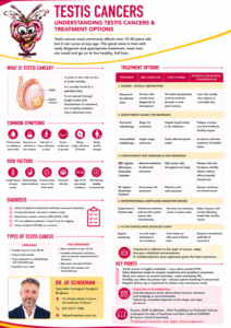
Information Sheet: Testis Cancer

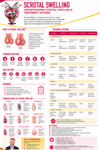
Information Sheet: Scrotal Swelling

Testicular Torsion: When a Testicle Takes a Very Wrong Turn

Sudden testicular pain is an emergency. This is not a “wait until tomorrow and see how it goes” situation.

There are few occasions in life when being twisted is definitely not a good thing. Testicular torsion is one of them.

Testicular torsion occurs when a testicle rotates around the spermatic cord, twisting the blood vessels that supply it. Think of a garden hose being twisted until the water stops flowing, except this particular hose supplies a rather important piece of anatomy.

The result can be sudden, severe pain and, if the blood supply is not restored quickly enough, permanent damage or loss of the testicle.

The good news is that prompt treatment can often save the testicle. The important word here is prompt.

Sudden severe testicular pain should be treated as testicular torsion until proven otherwise. Go directly to an Emergency Department.


What exactly is testicular torsion?

Normally, each testicle has enough mobility to accommodate everyday movement without being able to rotate freely.

Some boys and men have an anatomical arrangement commonly called a “bell-clapper” deformity, in which the testicle is more mobile within the scrotum than usual. This allows it to rotate around the spermatic cord.

When the cord twists, venous drainage is obstructed first, followed by arterial blood flow. The testicle becomes swollen, increasingly painful and eventually starved of oxygen.

This is why testicular torsion is a time-critical surgical emergency.

Although torsion is particularly common in adolescents, it can occur at almost any age, including infancy and adulthood.


What does testicular torsion feel like?

The classic presentation is difficult to ignore:

  • Sudden onset of severe pain in one testicle
  • Rapidly increasing scrotal pain or swelling
  • A testicle sitting higher than usual or lying at an unusual angle
  • Lower abdominal or groin pain
  • Nausea and vomiting
  • Marked tenderness of the affected testicle
  • Occasionally pain that wakes someone from sleep

Some patients experience previous episodes of sudden testicular pain that disappear spontaneously. This may represent intermittent torsion, where the testicle twists and then untwists itself.

A disappearing pain does not necessarily mean that the problem has disappeared.


The clock starts ticking immediately

With torsion, time matters.

The two major factors determining whether a testicle survives are how long it has been twisted and how tightly the spermatic cord has rotated. Current European Association of Urology guidance recommends surgical exploration as soon as possible and notes that the best outcomes occur with very early intervention, ideally within approximately 4–6 hours of symptom onset.

There isn’t a magical cliff at six hours where the testicle suddenly gives up and packs its bags. Salvage may still be possible later, which is precisely why patients should seek emergency treatment regardless of how long the pain has been present.

Don’t wait to see whether it gets better.

And perhaps more importantly:

Don’t be embarrassed.

Emergency doctors and urologists have seen testicles before. Quite a few of them.


How is testicular torsion diagnosed?

The diagnosis begins with the history and examination.

A doctor will examine the abdomen, groin and scrotum and assess the position, swelling and tenderness of the affected testicle.

A Doppler ultrasound can assess blood flow to the testicle and is extremely useful when the diagnosis is uncertain.

However, there is an important catch.

An ultrasound should not delay surgery when torsion is strongly suspected.

The EAU specifically recommends that Doppler ultrasound be used as an adjunct without delaying intervention.

In other words, when the clinical picture is shouting “torsion”, we shouldn’t spend precious time politely asking the ultrasound machine for a second opinion.


Can the testicle simply be untwisted?

Sometimes a doctor can attempt manual detorsion while arrangements for surgery are being made.

This involves carefully rotating the testicle in the direction that releases the twist. Relief of pain may be dramatic when successful.

But this is important:

Successful manual detorsion does not replace surgery.

Residual twisting may remain even when the pain improves, and the testicle can twist again. EAU guidance therefore states that manual detorsion may be attempted while awaiting surgery but must not delay surgical exploration.


Emergency surgery: scrotal exploration

Definitive treatment is an operation called scrotal exploration.

Under anaesthesia, the surgeon makes an incision in the scrotum and examines the affected testicle.

The spermatic cord is untwisted and the testicle is allowed time to recover its blood supply.

Sometimes a rather alarming blue or purple testicle gradually becomes reassuringly pink again. Few colour changes make a urologist happier.

If the testicle appears viable, it is preserved.


Orchiopexy: preventing another twist

If the testicle can be saved, it is secured inside the scrotum using an operation called an orchiopexy or orchidopexy.

The testicle is fixed so that it cannot freely rotate around the spermatic cord again.

But there is another important part of the operation.

We usually fix the other testicle too.

The anatomical predisposition that allowed one testicle to twist may also be present on the opposite side. Therefore, when torsion is confirmed, the unaffected testicle is commonly fixed during the same operation.

Current guidance recommends contralateral orchiopexy at the time of surgery rather than returning later for an elective procedure.

Think of it as discovering that one wheel on the car was loose and sensibly checking the other one while you’re already holding the spanner.


What if the testicle cannot be saved?

Unfortunately, sometimes the blood supply has been absent for too long and the testicular tissue is irreversibly damaged.

If the testicle is clearly non-viable or necrotic, it may need to be removed.

This operation is called an orchidectomy or orchiectomy.

Losing a testicle can understandably be upsetting, particularly for an adolescent or young man. Concerns about appearance, masculinity, testosterone, sexual function and fertility are extremely common.

These concerns deserve a proper conversation rather than a hurried reassurance.

The encouraging news is that one healthy testicle is generally sufficient to maintain normal testosterone production, sexual function and fertility.


What about fertility after testicular torsion?

This subject is more complicated than simply asking whether the testicle survived.

Torsion can damage sperm-producing tissue through the original loss of blood supply and potentially through ischaemia-reperfusion injury when circulation returns.

Long-term studies have therefore reported abnormalities in semen parameters in some men following torsion. The EAU notes reported rates of subfertility of approximately 36–39%, although study results vary considerably and long-term paternity may remain comparable with control populations.

Importantly, having had testicular torsion does not automatically mean that a man will be infertile.

Many men subsequently have normal fertility, particularly when the other testicle is healthy. Studies have also reported normal pregnancy rates following unilateral torsion, whether the affected testicle was preserved or ultimately removed.

For patients concerned about future fertility, particularly after severe torsion, testicular atrophy or problems involving the remaining testicle, subsequent assessment may include:

  • Testicular examination and ultrasound
  • Measurement of testicular volume
  • Semen analysis when appropriate
  • Testosterone and reproductive hormone testing
  • Fertility counselling where indicated

Can a rescued testicle shrink later?

Unfortunately, yes.

A testicle may look viable during surgery and still subsequently undergo testicular atrophy, becoming smaller over the following months.

Current EAU guidance notes that testicular atrophy can occur even following timely detorsion and fixation, which is why appropriate follow-up is important.

This does not mean the operation failed. The initial period without adequate blood supply may simply have caused microscopic damage that wasn’t visible during surgery.


What about testosterone and sexual function?

For most patients with a normal opposite testicle, testosterone production remains adequate.

Long-term studies suggest that hormonal measurements may differ somewhat following torsion, but overall endocrine testicular function generally remains within the normal range.

Removal of one testicle therefore does not normally cause erectile dysfunction, loss of masculinity or the need for testosterone replacement.

The remaining testicle usually quietly gets on with the job.


Testicular prosthesis: replacing what has been lost

Some patients are bothered by the cosmetic asymmetry following removal of a testicle.

A testicular prosthesis can be placed inside the scrotum to restore a more natural appearance and feel.

The prosthesis does not produce sperm or testosterone. Its purpose is cosmetic and psychological rather than functional.

Depending upon the circumstances, a prosthesis may be inserted during the original operation or at a later date. Prosthesis placement is an established option following orchidectomy, and current EAU guidance recommends that patients undergoing removal of a testicle be offered the opportunity to discuss a testicular prosthesis.

The decision is entirely personal.

Some men want one.

Some don’t.

Both decisions are perfectly reasonable.


Recovery after torsion surgery

Most patients recover relatively quickly following uncomplicated orchiopexy.

There will usually be some swelling, bruising and discomfort for several days. Supportive underwear can be surprisingly valuable during this period. The humble pair of supportive briefs finally gets its moment of glory.

Patients are generally advised to avoid strenuous activity, heavy lifting, cycling and contact sports until healing is satisfactory.

Timing for returning to work, school, sport and sexual activity should be discussed with the treating surgeon because this varies according to the operation and individual recovery.


Could torsion happen again after orchiopexy?

It is unusual, but recurrent torsion after orchiopexy is possible.

The EAU guideline describes recurrence after fixation as rare, but documented.

Therefore, even someone who has previously undergone orchiopexy should seek urgent medical attention if they develop sudden severe testicular pain.

Previous surgery doesn’t grant a testicle diplomatic immunity.


The message worth remembering

Testicular torsion is one of the true emergencies in urology.

If you or your son develops sudden severe pain in one testicle, particularly when associated with swelling, nausea, vomiting or an abnormally positioned testicle:

Don’t wait.

Don’t Google for three hours.

Don’t go to bed hoping it will disappear.

Go to an Emergency Department.

When it comes to testicular torsion, hours matter.

Early surgery may mean the difference between fixing a testicle in place and having to remove it.

And in this particular corner of medicine, keeping both passengers on board is very much the preferred destination.

So, get to your local Emergency Department urgently. I operate out of The Wesley and St Andrews War Memorial hospitals where I am on the on call roster. Ask for your local Brisbane Urologist, Dr Jo Schoeman.


Medical disclaimer

This information is intended for general patient education and does not replace individual medical assessment. Acute or sudden testicular pain requires urgent medical evaluation. If testicular torsion is suspected, attend an Emergency Department immediately. Your local Brisbane Urologist is on an on-call roster at both the Wesley Hospital and St Andrews War Memorial Hospital. Don’t delay!

Vasectomy: A Guide to Permanent Male Contraception

Vasectomy is a safe, effective and permanent method of male contraception. It is a relatively minor surgical procedure that prevents sperm from entering the semen by interrupting the vas deferens, the tubes that carry sperm from the testicles.

For men and couples who are confident that their family is complete, vasectomy provides highly reliable contraception without the need for ongoing medication or contraceptive devices.

What Happens During a Vasectomy?

Sperm are produced in the testicles and mature within the epididymis. During ejaculation, sperm normally travel through a tube called the vas deferens, where they ultimately mix with fluid produced by the prostate and seminal vesicles to form semen.

During a vasectomy, each vas deferens is divided and sealed so that sperm can no longer reach the ejaculate.

Importantly, vasectomy does not stop the testicles from producing sperm. The sperm that continue to be produced are naturally broken down and absorbed by the body.

Who Should Consider a Vasectomy?

Vasectomy may be appropriate for men who:

  • Have completed their family and do not want more children.
  • Do not wish to have children in the future.
  • Want a highly effective permanent form of contraception.
  • Have a partner for whom pregnancy would represent a significant health risk.
  • Prefer male sterilisation rather than their partner undergoing a more invasive sterilisation procedure.
  • Wish to avoid the need for long-term hormonal or barrier contraception.

The most important consideration is that vasectomy should be regarded as permanent.

Although vasectomy reversal is possible, successful reversal cannot be guaranteed. Men who are uncertain about future fertility should carefully consider alternative contraception or, in selected circumstances, sperm banking before proceeding.

How Is Vasectomy Performed?

Vasectomy is usually performed as a day procedure, commonly under local anaesthetic, although sedation or general anaesthesia may occasionally be appropriate.

A conventional or no-scalpel technique may be used.

The vas deferens is identified through a small opening in the scrotal skin. A short segment of the vas may be removed, and the divided ends are then sealed using techniques such as cautery, ligation and/or fascial interposition.

The procedure generally takes approximately 15–30 minutes.

Patients are usually able to return home shortly afterwards.

What Should I Expect After the Procedure?

Mild discomfort, bruising and swelling of the scrotum are common during the first few days.

Simple measures can help recovery, including:

  • Wearing supportive underwear.
  • Using simple analgesia as recommended.
  • Avoiding strenuous physical activity and heavy lifting for several days.
  • Keeping the wound clean and dry according to your surgeon’s instructions.
  • Avoiding sexual activity until discomfort and swelling have settled.

Most men can return to sedentary work relatively quickly, while men involved in heavy physical work may require a longer recovery period.

Vasectomy Does Not Work Immediately

This is one of the most important facts to understand about vasectomy.

A man is not sterile immediately after the procedure.

Sperm that were already present beyond the site of the vasectomy may remain within the reproductive tract for a period after surgery.

For this reason, another reliable method of contraception must be used until a post-vasectomy semen analysis confirms that the procedure has been successful.

Your urologist will advise when the semen test should be performed according to the protocol being followed.

Do not stop contraception simply because a certain number of weeks or ejaculations have passed. Wait until appropriate semen testing has confirmed success.

How Effective Is Vasectomy?

Vasectomy is one of the most effective forms of contraception available.

However, no contraceptive procedure can be regarded as absolutely 100% effective. Rarely, the divided ends of the vas deferens can reconnect, a process known as recanalisation.

Recanalisation may occur early after surgery and be detected on semen testing, or very rarely occur later after an initially successful vasectomy.

This is one reason why post-vasectomy semen analysis is an essential part of the procedure rather than an optional extra.

Does Vasectomy Affect Testosterone?

No.

Vasectomy does not remove or damage the testicles, and testosterone production continues normally.

A vasectomy should therefore not cause:

  • Loss of masculinity.
  • Reduced testosterone levels.
  • Loss of facial or body hair.
  • Reduced muscle mass.
  • Premature ageing.

Does Vasectomy Affect Erections or Sexual Function?

Vasectomy does not normally affect the ability to obtain or maintain an erection.

It also does not interfere with orgasm.

The nerves and blood vessels responsible for erections are not interrupted during a routine vasectomy.

Will Ejaculation Be Different?

Most of the volume of semen comes from the prostate and seminal vesicles, rather than from sperm.

Consequently, ejaculation continues after vasectomy and the appearance and volume of the semen are generally essentially unchanged.

The difference is microscopic: once the vasectomy has been successful, sperm are no longer present in the ejaculate.

What Are the Possible Complications?

Vasectomy is generally a low-risk procedure, but complications can occur.

Bleeding and Haematoma

Bleeding into the scrotum can result in swelling, bruising or a collection of blood known as a haematoma.

Small haematomas usually settle with conservative management. Large or expanding haematomas occasionally require further treatment.

Infection

Infection of the skin or deeper scrotal tissues is uncommon but may occur. Symptoms can include increasing pain, redness, swelling, discharge or fever.

Pain and Swelling

Some degree of discomfort and swelling is expected during the initial recovery period and usually settles.

Sperm Granuloma

Sperm may occasionally leak from the divided end of the vas and produce a small inflammatory lump known as a sperm granuloma.

These are often harmless but can occasionally be tender.

Epididymal Congestion

Some men develop aching or discomfort around the epididymis following vasectomy, possibly related to pressure within the sperm-carrying system.

This is usually temporary but can occasionally persist.

Post-Vasectomy Pain Syndrome

A small proportion of men experience persistent or recurrent testicular or scrotal pain lasting for months or longer after vasectomy.

This is known as chronic post-vasectomy pain or post-vasectomy pain syndrome.

Treatment depends upon severity and may include:

  • Anti-inflammatory or pain medication.
  • Pelvic floor or other targeted therapy where appropriate.
  • Nerve-directed treatment.
  • Spermatic cord procedures.
  • Epididymectomy in selected cases.
  • Vasectomy reversal in carefully selected patients.

Further surgery is rarely required, but persistent pain is an important complication to discuss before deciding on vasectomy.

Vasectomy Failure and Recanalisation

Occasionally sperm remain present because the vas deferens has reconnected or the procedure has otherwise failed to achieve sterility.

Persistent sperm on semen analysis may require further testing and, occasionally, a repeat vasectomy.

Very rarely, late recanalisation can occur after a previously satisfactory semen analysis, meaning that pregnancy remains possible even after initial clearance.

Does Vasectomy Increase the Risk of Prostate Cancer?

Vasectomy has been extensively studied in relation to prostate cancer and other long-term health concerns.

Current evidence has not established vasectomy as a cause of prostate cancer, and vasectomy is not generally considered a reason to alter routine prostate cancer screening or assessment.

Men should continue appropriate prostate health checks based on their age, family history, symptoms and individual risk factors.

Can a Vasectomy Be Reversed?

Yes, but reversal is considerably more complex than the original vasectomy.

Vasectomy reversal involves microsurgically reconnecting the reproductive tract, usually with a vasovasostomy or, when necessary, a vasoepididymostomy.

Success depends on several factors, including:

  • The length of time since vasectomy.
  • The condition of the vas deferens and epididymis.
  • The surgical technique required.
  • The experience of the microsurgeon.
  • The fertility and age of both partners.

Successful return of sperm to the semen does not necessarily guarantee pregnancy.

For this reason, a vasectomy should never be undertaken on the assumption that it can simply be reversed later.

Alternatives to Vasectomy

Men who are uncertain about permanent contraception should consider reversible alternatives.

Depending upon individual circumstances, these may include condoms or contraceptive methods used by the female partner, including hormonal contraception, intrauterine devices and contraceptive implants.

The choice should take into account effectiveness, side effects, medical history, personal preference and future fertility plans.

When Should I Seek Medical Attention After Vasectomy?

Contact your doctor or urologist if you develop:

  • Increasing rather than improving scrotal pain.
  • Significant or rapidly increasing swelling.
  • Heavy bleeding.
  • Increasing redness or discharge from the wound.
  • Fever or feeling systemically unwell.
  • Persistent testicular pain.
  • A concerning new scrotal lump.

The Bottom Line

Vasectomy is a highly effective, relatively simple and permanent form of male contraception.

For appropriately selected men who have completed their families, it can remove the need for ongoing contraception while leaving testosterone production, erections, orgasm and ejaculation essentially unchanged.

The key points are to understand that vasectomy should be considered permanent, complications such as chronic scrotal pain and failure can occur, and the procedure does not provide immediate contraception.

Most importantly, continue contraception after vasectomy until your post-vasectomy semen analysis has confirmed that it is safe to stop.

This information is intended for general education and does not replace individual medical advice. Your urologist can discuss whether vasectomy is appropriate for you, the technique used, expected recovery and the post-vasectomy semen-testing protocol.

So, if you have made the careful and considered decision to cease your ability to father another kid and you are a sensible adult, come see your local Brisbane Urologist, Dr Jo Schoeman to discuss this option. Your partner needs to be in on this as this will also affect her!

Orchidectomy – Radical

Why is it done?

  • For testis cancer.
  • For testis lesions highly suspicious of testis cancer.

How is it done?

  • This is done under general anesthetic.
  • A single incision is made in the groin. The underlying muscle layers are split for good oncological control.
  • The affected testis and spermatic cord are then surgically removed through this incision.
  • A metal clip may be left right at the internal inguinal ring, as a future marker, should radiotherapy be required.
  • Subcutaneous sutures (which need not be removed) are used, unless stated otherwise.
  • A dressing is then applied, which should be removed after 72 hours.
  • A local anesthetic is injected into the wound, thus giving post-operative pain relief for the next 4-6 hours.
  • A drain may also be left for 24-48 hours to prevent the collection of serous fluids


What to expect after the procedure

  • Any anesthetic has its risks, and the anesthetist will explain such risks.
  • Bleeding is a common complication. If   concerned call the hospital.
  • A hematoma (blood collection under the skin) may form and needs to be reviewed as soon as possible.
  • An infection of the wound may occur and requires immediate attention.
  • Owing to the nature of the surgery and the soft skin of the scrotum, bruising may appear much worse than it is and is no cause for alarm.
  • DANGER SIGNS: A scrotum that swells immediately to the size of a football, fever, or pus. Contact Dr Schoeman or the hospital immediately as this occurs in up to 15 % of all     cases.


What next?

  • The dressing should be kept dry for the initial 72 hours after surgery.
  • The dressing should then be removed in a bath. It should be soaked until it comes off with ease.
  • The dressing may sometimes adhere to the wound causing slight bleeding on removal. Don’t panic, the bleeding will stop.
  • Patients should schedule a follow-up appointment with Dr Schoeman within 2 weeks to review pathology and arrange subsequent management.
  • There will be signs of bruising for at least 10 days.
  • The suture-line will be hard and indurated for at least 8-10 weeks.
  • PLEASE CONTACT THE HOSPITAL DIRECTLY WITH ANY POST-OPERATIVE CONCERNS AND RETURN TO THE     HOSPITAL IMMEDIATELY SHOULD THERE BE ANY SIGNS OF SEPSIS.


NB! Regular self-examination highly recommended.

Wes Orchidectomy Radical