
Spina Bifida and the Bladder: Protecting the Kidneys and Improving Continence
Spina bifida is a congenital condition in which the spine and spinal cord do not develop normally. Although it is often associated with reduced leg movement or sensation, it can also affect the nerves controlling the bladder, urethral sphincter and bowel.
This is called neurogenic lower urinary tract dysfunction, or more simply a neurogenic bladder.
The bladder problem is not always obvious from the person’s mobility or the appearance of the spinal abnormality. Someone with a relatively small or “closed” spinal defect may still have significant bladder dysfunction. Conversely, urinary leakage does not necessarily mean that bladder pressures are safe.
The principal goals of urological care are to:
- Protect the kidneys from high bladder pressures and infection.
- Ensure that the bladder empties safely.
- Improve urinary continence and independence.
- Minimise infections, stones and catheter-related complications.
- Provide a management plan that remains practical throughout childhood and adult life.
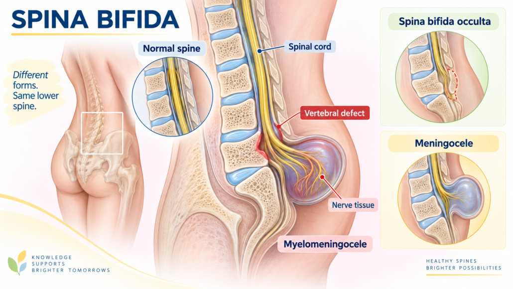
What are the different types of spina bifida?
Spina bifida occulta
This is the mildest anatomical form. One or more vertebrae have not completely closed, but there may be no visible sac or exposed spinal tissue.
Many people have no neurological symptoms. Others may have an associated tethered spinal cord, fatty lesion or other form of closed spinal dysraphism that affects bladder, bowel or leg function.
Meningocele
The coverings around the spinal cord protrude through a spinal defect and form a fluid-filled sac. The spinal cord itself may remain relatively preserved, although neurological and bladder dysfunction can still occur.
Myelomeningocele
In myelomeningocele, both the spinal cord or nerves and their coverings protrude through the spinal defect. This is the form most commonly associated with leg weakness, altered sensation, bowel dysfunction and significant neurogenic bladder problems.
Closed spinal dysraphism and tethered cord
A lipoma, abnormal attachment or other congenital spinal abnormality may tether the spinal cord. Symptoms can appear or worsen as a child grows or later in adult life.
New urinary leakage, recurrent infections, difficulty catheterising, increasing residual urine, leg weakness, back pain or changes in bowel function may indicate tethering and require further assessment.
How does spina bifida affect the bladder?
Normal bladder function requires coordination between the bladder muscle, the detrusor, and the urinary sphincter.
During filling, the bladder should remain relaxed and at low pressure while the sphincter stays closed. When it is time to urinate, the bladder should contract and the sphincter should relax.
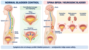
Spina bifida can disrupt these signals in several ways.
An overactive bladder
The bladder may contract unexpectedly during filling. This can cause:
- Urgency.
- Frequent urination.
- Urge incontinence.
- Leakage between catheterisations.
- Bedwetting.
- A small functional bladder capacity.
A poorly compliant, high-pressure bladder
A stiff bladder does not stretch normally as it fills. Pressure rises even when the volume is relatively small.
This is sometimes called a hostile bladder. It is particularly important because high pressure may be transmitted to the ureters and kidneys, causing:
- Vesicoureteric reflux.
- Hydronephrosis.
- Recurrent infections.
- Kidney scarring.
- Progressive loss of kidney function.
A high-pressure bladder may be dangerous even when the patient has few symptoms.
Failure of bladder emptying
The bladder muscle may be weak or unable to contract effectively. This can result in:
- Difficulty starting urination.
- A weak or intermittent stream.
- Straining to pass urine.
- A sensation of incomplete emptying.
- A large residual volume.
- Overflow leakage.
- Recurrent urinary infections.
Some people have very little bladder sensation and may not recognise that the bladder is full.
Detrusor-sphincter dyssynergia
In this situation, the bladder contracts while the urinary sphincter remains closed or tight. The bladder must generate high pressure against the obstruction.
This combination can cause incomplete emptying, leakage, infections and upper urinary tract damage.
A weak bladder outlet
If the bladder neck or sphincter does not close adequately, stress-type leakage may occur with coughing, transfers, exercise or movement. Some patients have both a high-pressure bladder and a weak outlet, which makes treatment more complex.
Bladder dysfunction is reported in up to 96% of people with spina bifida, while more than half experience urinary incontinence. Importantly, open and closed spinal defects can produce equally significant bladder dysfunction. European Association of Urology Neuro-urology Guideline
How is the bladder investigated?
Investigation is tailored to the patient’s age, symptoms, previous surgery and risk to the kidneys.
Clinical assessment
The consultation should consider:
- The person’s usual method of passing urine.
- Daytime and night-time leakage.
- Urinary urgency and frequency.
- Catheterisation frequency and volumes.
- Recurrent symptomatic urinary infections.
- Previous kidney or bladder stones.
- Bowel function and constipation.
- Mobility, hand function and available carer support.
- Previous spinal, bladder or continence surgery.
- Changes that may suggest a tethered spinal cord.
A bladder and catheterisation diary can document fluid intake, voided volumes, catheter volumes and episodes of leakage.
Urine testing
Urinalysis and urine culture may be required when there are symptoms of infection. Bacteria are frequently found in the urine of people who catheterise, but bacteriuria without symptoms does not always require antibiotic treatment.
Symptoms may be less typical than in someone with normal bladder sensation. Warning signs can include fever, new leakage, pelvic or back discomfort, cloudy or offensive urine, malaise or an unexplained change in bladder behaviour.
Blood tests
Kidney function and electrolytes should be monitored. Serum creatinine can underestimate kidney impairment in people with low muscle mass, so an alternative measurement such as cystatin C or measured renal clearance may occasionally be helpful.
Vitamin B12, acid–base balance and other blood tests may be required following urinary reconstruction using bowel.
Ultrasound
Renal and bladder ultrasound can assess:
- Kidney size and appearance.
- Hydronephrosis.
- Bladder wall thickening.
- Stones.
- Residual urine after voiding.
Ultrasound is important for surveillance, but a normal ultrasound does not prove that bladder pressures are safe.
Urodynamic studies
Urodynamics measures how the bladder stores and empties urine. It may identify:
- Neurogenic detrusor overactivity.
- Reduced bladder capacity.
- Poor compliance and unsafe filling pressures.
- Stress incontinence.
- Detrusor-sphincter dyssynergia.
- A weak or non-contractile bladder.
- Incomplete emptying.
Video urodynamics combines pressure measurements with imaging and can also demonstrate reflux, bladder shape and the level of outlet obstruction.
Urodynamic testing is particularly useful when there are changes in continence, recurrent infections, hydronephrosis, kidney deterioration or consideration of major treatment changes.
People with spina bifida require lifelong urological surveillance, because bladder behaviour can change with growth, ageing, tethering, surgery or reduced ability to catheterise. Spina Bifida Association Urology Guideline
Treatment options
There is no single treatment suitable for every neurogenic bladder. Management depends on bladder pressure, emptying, continence, kidney health, dexterity, mobility and personal goals.
Clean intermittent self-catheterisation
Clean intermittent catheterisation, often abbreviated to CIC or CISC, is a central part of treatment when the bladder does not empty safely.
A catheter is passed through the urethra at regular intervals and removed after the bladder has drained. Depending on age and physical ability, catheterisation may be performed by the patient, a parent or a carer.
Potential benefits include:
- Reliable bladder emptying.
- Lower residual urine volumes.
- Reduced overflow leakage.
- Better control of bladder pressure when combined with medication.
- Greater independence and predictability.
- Protection of the kidneys.
The frequency is individualised according to fluid intake, bladder capacity, pressures and catheterised volumes. Proper education, catheter selection and ongoing review are important.
If urethral catheterisation is difficult because of mobility, body shape, poor access, pain or urethral damage, a continent catheterisable channel may be considered.
Bladder medication
Antimuscarinic medicines such as oxybutynin, solifenacin or other agents may reduce involuntary bladder contractions and improve storage at lower pressures.
A beta-3 agonist such as mirabegron may be considered in selected patients. Combination treatment is sometimes used, although medication choice and licensing differ between children and adults.
Possible adverse effects include dry mouth, constipation, blurred vision and, with some medications, cognitive effects or changes in blood pressure. Constipation should be treated actively because a loaded bowel can worsen bladder pressure, leakage and infection risk.
Botulinum toxin: Botox
Botulinum toxin A can be injected through a cystoscope into the bladder muscle. It temporarily reduces involuntary bladder contractions and may improve capacity, continence and storage pressures.
Botox is generally considered when oral or intravesical medication has not provided sufficient control or has caused unacceptable side effects.
Important considerations include:
- Its effect is temporary, so repeat treatment is usually required.
- Intermittent catheterisation may still be necessary and may become necessary in someone who previously emptied without a catheter.
- Urinary infection, temporary retention and blood in the urine can occur.
- Follow-up urodynamics may be required to confirm that bladder pressure has improved, rather than relying only on reduced leakage.
The evidence supporting Botox is strongest across neurogenic detrusor overactivity populations generally. Individual assessment remains particularly important in congenital spina bifida. The EAU recommends detrusor Botox when antimuscarinic therapy has been ineffective. EAU Neuro-urology Guideline
Sacral neuromodulation
Sacral neuromodulation uses an implanted lead near a sacral nerve root, usually S3, to modify communication between the bladder, sphincter and nervous system.
It may be considered for selected patients with:
- Urgency and urge incontinence.
- Urinary frequency.
- Non-obstructive urinary retention.
- Some forms of neurogenic bladder dysfunction.
However, its role in spina bifida is more selective than in non-neurogenic overactive bladder. Abnormal sacral anatomy, previous spinal surgery, tethering and the exact neurological pattern may influence feasibility and response.
A test or staged phase is generally used before permanent implantation. Sacral neuromodulation should not be expected to correct a severely fibrotic, very small or dangerously high-pressure bladder. Evidence in neuro-urological patients is growing, but it remains uncertain which neurological conditions respond best. EAU Neuro-urology Guideline
Surgery to improve the bladder outlet
If bladder pressures and emptying have been made safe but significant stress leakage remains, surgery may be considered to increase outlet resistance.
Options may include:
- A bladder-neck reconstruction.
- An autologous fascial sling.
- An artificial urinary sphincter in selected patients.
- Bladder-neck closure in complex circumstances.
Any procedure that tightens the outlet can make catheterisation essential. The ability to catheterise reliably through the urethra or a catheterisable channel, must therefore be established before surgery.
Mitrofanoff catheterisable channel
A Mitrofanoff is a narrow channel, commonly created using the appendix, that connects the bladder to a small opening on the abdominal wall. The opening is often placed at or near the umbilicus.
A catheter is passed through this channel to empty the bladder. There is no continuously draining external bag.
A Mitrofanoff may be helpful when:
- Urethral catheterisation is difficult or painful.
- Wheelchair positioning makes urethral access impractical.
- Greater independence is desired.
- Urethral damage or reconstruction prevents reliable catheterisation.
It is often created at the same time as bladder augmentation, although it can also be performed without augmentation when bladder capacity and pressure are already satisfactory.
Possible complications include stomal narrowing, leakage, difficulty catheterising, false passages, stones and the need for revision surgery.
Clam augmentation cystoplasty
A clam cystoplasty, also called augmentation cystoplasty or ileocystoplasty, enlarges the bladder using a patch of bowel. The bladder is opened widely, rather like opening a clam, and the bowel patch is added to create a larger, lower-pressure reservoir.
It may be considered when there is:
- Persistently poor bladder compliance.
- A small, high-pressure bladder.
- Refractory neurogenic detrusor overactivity.
- Continuing risk to the kidneys despite catheterisation, medication and Botox.
- Severe incontinence caused by inadequate bladder storage.
The operation can be highly effective in improving capacity and lowering pressure, but it is major lifelong reconstructive surgery rather than a simple continence procedure.
Most patients need lifelong intermittent catheterisation after augmentation. A Mitrofanoff may be added if catheterisation through the urethra is difficult.
Long-term considerations include:
- Mucus production from the bowel patch.
- The need for regular bladder irrigation in some patients.
- Urinary infections and stones.
- Metabolic and electrolyte abnormalities.
- Vitamin B12 deficiency, depending on the bowel segment used.
- Bladder perforation, which is uncommon but potentially life-threatening.
- Possible need for further surgery.
- A small long-term risk of malignancy.
- Lifelong specialist follow-up.
The EAU recommends augmentation for low bladder compliance or neurogenic detrusor overactivity that remains refractory to less invasive treatment. EAU Neuro-urology Guideline
Mitrofanoff versus ileal conduit: an important distinction
These procedures are sometimes confused, but they work differently.
A Mitrofanoff is a continent catheterisable channel connected to the bladder or an augmented bladder. The person remains dry between catheterisations and empties the reservoir periodically with a catheter.
An ileal conduit is an incontinent urinary diversion. The ureters are connected to a short segment of bowel that drains continuously through a stoma into an external appliance.
An ileal conduit may be considered when:
- Safe catheterisation is not possible.
- Previous reconstruction has failed.
- Severe incontinence cannot otherwise be controlled.
- The bladder or urethra is badly damaged.
- The upper urinary tract is threatened.
- A simpler continuously draining diversion is more practical for the patient or carers.
Therefore, the usual surgical comparison is not “a conduit with or without a Mitrofanoff.” More commonly, the alternatives are:
- Native bladder with urethral catheterisation.
- Native or augmented bladder with a Mitrofanoff channel.
- A continent urinary reservoir.
- An incontinent diversion such as an ileal conduit.
The best reconstruction depends on kidney function, bowel health, previous surgery, hand function, mobility, cognition, family or carer support and the patient’s own priorities.
The importance of bowel management
Bladder and bowel problems frequently coexist in spina bifida. Constipation can increase bladder pressure, worsen leakage, make catheterisation more difficult and contribute to urinary infections.
A coordinated bladder and bowel programme may include dietary measures, fluids, oral medication, suppositories, enemas, transanal irrigation or in selected patients, a surgical catheterisable bowel channel.
Warning signs requiring medical review
Prompt assessment is advisable if there is:
- Fever or suspected symptomatic urinary infection.
- New flank, pelvic or back pain.
- Blood in the urine.
- New or worsening urinary leakage.
- Increasing catheterisation volumes or difficulty passing a catheter.
- Reduced urine output.
- Recurrent catheter blockage.
- New hydronephrosis or deterioration in kidney function.
- New leg weakness, sensory changes or bowel dysfunction.
- Sudden abdominal pain in someone with an augmented bladder.
The key message
In spina bifida, a dry bladder is not necessarily a safe bladder and a leaking bladder is not always a high-pressure bladder. The most important questions are whether the bladder stores urine at a safe pressure, empties reliably and protects the kidneys.
With regular surveillance and an individualised combination of catheterisation, medication, Botox, selected neuromodulation or reconstructive surgery, many people can achieve good kidney protection, improved continence and greater independence.
Management is best coordinated through an experienced multidisciplinary team involving urology, rehabilitation, neurosurgery, continence nursing and bowel-management services.
This information is general and does not replace individual medical assessment. Treatment decisions should be based on symptoms, kidney function, imaging, urodynamic findings, physical ability and the patient’s personal goals.


Leave a Reply
Want to join the discussion?Feel free to contribute!