Urodynamic Studies: Understanding How Your Bladder Works

Bladder problems are not always as straightforward as they appear.

Two people may both complain of urinary urgency, leakage or difficulty emptying their bladder, yet the underlying causes can be completely different. One bladder may be contracting when it should be relaxing, another may not contract strongly enough, and sometimes the bladder is behaving perfectly well while the outlet is causing all the trouble.

This is where urodynamic studies, often simply called urodynamics, can help.

Think of urodynamics as a stress test for the bladder. Rather than simply looking at its anatomy, we observe how the bladder fills, stores urine and empties while simultaneously measuring pressures, urine flow and sometimes pelvic floor activity.

The aim is not merely to produce a collection of colourful graphs. The important question is:

What is the bladder actually doing, and will knowing this change the treatment?


What Are Urodynamic Studies?

Urodynamics refers to a group of tests that assess the function of the bladder, urethra and urinary sphincter.

Depending on the clinical situation, testing may evaluate:

  • How much urine the bladder can comfortably hold
  • Bladder sensation during filling
  • Whether the bladder remains relaxed while filling
  • Whether involuntary bladder contractions occur
  • Whether urinary leakage occurs and why
  • The pressure required to store urine
  • The strength of the bladder muscle during urination
  • Whether there is obstruction to urinary flow
  • Whether the bladder empties completely
  • How the urinary sphincter and pelvic floor behave during filling and voiding

Urodynamics therefore investigates function rather than simply structure.

An ultrasound or cystoscopy may show us what the urinary tract looks like. Urodynamics tells us what it is doing.


Who May Need Urodynamic Testing?

Not every patient with urinary symptoms requires urodynamics.

For many straightforward urinary problems, the diagnosis can be made from the history, examination, bladder diary, urine testing, flow studies and ultrasound.

Urodynamics becomes particularly useful when the diagnosis is uncertain, symptoms are complex, previous treatments have failed, neurological disease is present, or an invasive treatment is being considered where understanding bladder function may alter the choice of treatment.

Common indications include:

Urinary Incontinence

Urodynamics may be useful when assessing:

  • Stress urinary incontinence
  • Urgency urinary incontinence
  • Mixed urinary incontinence
  • Persistent leakage following previous continence surgery
  • Incontinence following prostate surgery
  • Complex or unexplained urinary leakage

In straightforward female stress urinary incontinence, urodynamics is not necessarily required before treatment. It becomes more useful when symptoms and clinical findings do not agree or when there are complicating factors.


Overactive Bladder

Patients with overactive bladder may experience:

  • Urinary urgency
  • Frequent urination
  • Nocturia
  • Urgency urinary incontinence

Urodynamics can sometimes demonstrate detrusor overactivity, where the bladder muscle contracts involuntarily during filling.

Importantly, a normal urodynamic study does not necessarily exclude overactive bladder. Symptoms and urodynamic findings do not always travel together.


Difficulty Emptying the Bladder

Patients may report:

  • Poor urinary flow
  • Hesitancy
  • Straining
  • Intermittent flow
  • Incomplete emptying
  • Urinary retention

The challenge is determining why.

Is there an obstruction?

Or is the bladder muscle simply not contracting strongly enough?

The symptoms can look remarkably similar, but the treatments can be very different.


Men With Lower Urinary Tract Symptoms

In men with prostate enlargement, urodynamics may help distinguish between:

Bladder outlet obstruction

and

Detrusor underactivity, where the bladder muscle has insufficient strength to empty effectively.

This distinction may be particularly valuable before prostate surgery when there is uncertainty about whether removing the obstruction will improve urinary flow.


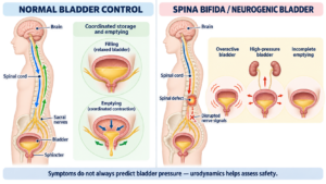
Neurological Bladder Dysfunction

Urodynamics has an especially important role in patients with neurological conditions such as:

  • Spinal cord injury
  • Multiple sclerosis
  • Parkinson’s disease
  • Spina bifida
  • Stroke
  • Certain spinal disorders

In these patients, we are interested not only in urinary symptoms but also in whether bladder pressures could potentially damage the kidneys.


Before or After Certain Urinary Procedures

Urodynamics may also be considered:

  • Before complex continence surgery
  • Following unsuccessful continence surgery
  • Before some bladder outlet procedures
  • Following prostate surgery when symptoms persist
  • When considering treatments such as bladder Botox or sacral neuromodulation in selected complex patients

What Happens During a Urodynamic Study?

The test is usually performed as an outpatient procedure.

It generally takes approximately 30–60 minutes, although this varies depending on the type of study being performed.

You can usually eat, drink and take your normal medications unless specifically instructed otherwise.


Step 1: Arriving With a Comfortably Full Bladder

You may be asked to arrive with a reasonably full bladder.

You will urinate into a special toilet called a uroflowmeter.

This measures:

  • Urine volume
  • Maximum urinary flow rate
  • Average flow rate
  • The shape of the urinary flow curve
  • How long urination takes

An ultrasound may then measure how much urine remains in the bladder.

This is called the post-void residual.


Step 2: Placement of Small Catheters

A very fine catheter is gently passed through the urethra into the bladder.

A second small pressure catheter is usually placed into the rectum, or occasionally the vagina.

Why two pressure measurements?

Because pressure inside the abdomen affects pressure inside the bladder.

By measuring both, the computer can calculate the pressure generated specifically by the bladder muscle:

Detrusor pressure = bladder pressure − abdominal pressure

This simple equation sits at the heart of conventional urodynamics.


Step 3: Filling the Bladder

The bladder is slowly filled with sterile fluid.

During filling, you will be asked to describe what you feel.

We may record:

  • First sensation of bladder filling
  • First desire to urinate
  • Strong desire to urinate
  • Maximum bladder capacity

The pressure inside the bladder is continuously monitored.

Normal filling cystometrogram

https://images.openai.com/static-rsc-4/Hgriy5E9FGtRilFNQh3tQE53yrlbWRzHlZE-lg73YC5P10G0EFIrVgMREqgOL5uEui9m4vTZQa3aZdbm2IQos6eTVRAdTIXlg1d841nyByIJ3GZk8QFyf9avtCBCjESBk_UcSEGbJGzOSIvZvPI6wZtNpz7LHnbB3kQIYDHsibqiwXmWpERNixUhOeW6Tm2B?purpose=fullsize
https://images.openai.com/static-rsc-4/BXwVAcldUDjh2tAaV2En87FX0ONS1oomHAkm4Z6BLZyoexIiDmaACUh8t4rB7TDfao-guZf76ULaTYX-VsV554nagvCAPRpNzs7HNQvKKZYuX8N6oPO5c_n_74de2tABnoEt69kwxCY_fJyF86M5rduxVEOG1zrNzm0SXMSrnXPiadRMzOfo1hPUnKvP1ahH?purpose=fullsize
https://images.openai.com/static-rsc-4/ySZcZmtN_5IZCgz4scVLbu2KqbZD3OVebnVIjG7F41-xqaWl2-D1RqW5T2NMWjugPRIkt7H-S3JB2UGWLPXIYYyURXURsMwzaASQ1zMpc7f1yB8UJcFDYMc1HW3qPkSw7rWCM7V8cMPh-oI2CVWQYD1HhlPxcJ6eJY6HJvYEz3QSK5VgEbfQgqejsiCVUfkK?purpose=fullsize
4

Normal filling cystometrogram. During filling, the bladder accommodates increasing volume while detrusor pressure (Pdet) remains relatively stable. Coughs cause corresponding rises in Pves and Pabd without a true detrusor contraction. Sensations such as first sensation, first desire and strong desire can be marked on the tracing.


Step 4: Provocative Testing

You may be asked to:

  • Cough
  • Strain
  • Stand
  • Change position
  • Listen to running water

These manoeuvres are not designed to make the test unnecessarily theatrical. They help reproduce the circumstances under which your symptoms normally occur.

If urinary leakage occurs during coughing without an involuntary bladder contraction, this may demonstrate urodynamic stress incontinence.


Step 5: Voiding

Once the bladder is comfortably full, you will be asked to urinate.

During urination we simultaneously measure:

  • Urinary flow
  • Bladder pressure
  • Abdominal pressure
  • Detrusor pressure

This is called a pressure-flow study.

It can be extremely useful when determining whether poor urinary flow is caused by obstruction or weak bladder contraction.


What Does Urodynamics Measure?

Bladder Sensation

We assess when you first become aware that the bladder is filling and when the desire to urinate becomes stronger.

Sensation may be:

  • Normal
  • Increased
  • Reduced
  • Absent

Abnormal bladder sensation may occur in several neurological and functional bladder disorders.


Bladder Capacity

The amount of urine the bladder can comfortably hold is measured.

A small functional capacity may occur with conditions such as severe urgency or bladder hypersensitivity, although capacity must always be interpreted in clinical context.


Bladder Compliance

A healthy bladder should behave rather like a flexible reservoir.

As it fills, its volume should increase without a major rise in pressure.

Poor bladder compliance means pressure rises excessively as the bladder fills.

This is particularly important in some neurological bladder disorders because persistently elevated storage pressures can potentially threaten the upper urinary tract.


Detrusor Overactivity

If the bladder muscle contracts involuntarily during filling, this is called:

Detrusor overactivity.

It may be associated with:

  • Urgency
  • Urgency incontinence
  • Frequency
  • Nocturia

When associated with an underlying neurological condition, it may be described as neurogenic detrusor overactivity.

Examples of terminal, phasic, wet and dry detrusor overactivity.

https://images.openai.com/static-rsc-4/Snzbj3YLBffWTq3-7c6jgZQWHTjwV03Siqh8rQZor7nWrcUTSujo5I4h9H6QmPWIw5TQO8oSYgzzugdooybbroYfbxONiyj0mx1Qe1uBjCwkL4K0AR5hGhULd9PKwFrw7LhUIrL8ZxpKQO1sQjk0qtqph3By5IOK30vdjd9Iahom6SUGG32ajdo2KW48nbhP?purpose=fullsize
https://images.openai.com/static-rsc-4/kx0pZHTOwdE7kDcSE1Awe3tnf1oB9kJizr_DFgQV0kdb9o6N2KxN9PW2lGCO4Rxcyx__zDzECnRwDhOrCXcXYbHZKkw4yASruDH4PtFNhlq6LqHzLF1d6NmcZkyOnoMHG_lNoKxw1Y5hCVuog5TIJqiN5O_zT8m7FtEhZkE-uHlwJMGAWpqcUhXKZdvq-h6c?purpose=fullsize
https://images.openai.com/static-rsc-4/9Fkv2MrdLw18QItSBrvBMWrFM_bRYOzwvod8KcVnSNQcBgQhcEl7m90lhNqlnqb3sX1pkEmdrx3XvCJxwi0gyzVvPse-T2lyODyK7BrW5xC3GJ2ZMggrrj7i-iArmLIQ05FXaE_4Cl8FYZ8yrU3dqXP5YN2gEVqQjCLOqH_xcbXAxuyLiYNLNijADJM9eUNR?purpose=fullsize

Detrusor overactivity. During bladder filling there is an involuntary increase in Pdet that cannot be explained by a corresponding increase in abdominal pressure. The contraction may be associated with urgency and, when leakage occurs, urgency urinary incontinence.

View the LABORIE-based detrusor-overactivity tracings


Stress Urinary Incontinence

If urine leaks when coughing or straining without a detrusor contraction, this supports a diagnosis of:

Urodynamic stress incontinence.

Additional measurements such as leak-point pressures may occasionally contribute to assessment in selected patients.


Bladder Outlet Obstruction

During urination, a pattern of:

High bladder pressure + low urinary flow

may suggest obstruction.

In men, benign prostate enlargement is a common cause.

Other causes include:

  • Urethral stricture
  • Bladder neck obstruction
  • Previous continence surgery
  • Dysfunctional voiding

Bladder outlet obstruction: pressure-flow study

https://images.openai.com/static-rsc-4/Efx8v2pSLdy0yriNx_qrcdTAHacUkb8mu_Sb7IjS9JvC0wxMQnPCPhcGQqAt0MQUNQ5jvnXe4yfU0qsIfsNRYrR7VLV4HhGFH6Ko01-kkE8GfSflqXD25Q6gkF3bmxyKYN9iYNHRqCJ0bV-igYyNfPP-21Nuvrls3kleV1a2fqsUDDQTALKE8mVkjEwglUbr?purpose=fullsize
https://images.openai.com/static-rsc-4/tQjdaSX6-MApQNNeYZuCDcT--H0Z_StPvpdtjnAdqL29pMWjRBGXsV_1KAe9iBU0kahuMqsxvNOoXDeZHFfSLBLxXg9w9uWzWnQxuEBsHfk3yjNkiDLedV-AQMJTHLPoif8aUAghyfhiv605EryWDjmBxVWKeBTNK3ky_okFHyRdz9rN1G-HNnhJBUnvnQhG?purpose=fullsize

Bladder outlet obstruction. During voiding the bladder generates a relatively high detrusor pressure, but urinary flow remains reduced. This high-pressure, low-flow relationship is characteristic of increased bladder outlet resistance.

In men, pressure-flow studies allow calculation of the Bladder Outlet Obstruction Index (BOOI):

BOOI = PdetQmax − 2(Qmax)

For men, BOOI >40 is generally classified as obstructed, 20–40 as equivocal and <20 as unobstructed.


Detrusor Underactivity

Sometimes the urinary flow is poor because the bladder muscle does not generate sufficient pressure.

This is known as:

Detrusor underactivity.

This distinction matters.

Operating on the prostate of a patient whose main problem is a very weak bladder may not produce the expected improvement in urinary flow.

Urodynamics can therefore occasionally prevent us from treating the wrong end of the plumbing.


Dysfunctional Voiding

Some patients involuntarily contract their pelvic floor or urinary sphincter while attempting to urinate.

Instead of the outlet opening smoothly, it intermittently closes.

Pelvic floor electromyography may help identify this pattern in selected patients.


Understanding the Urodynamic Tracing

A urodynamic report typically contains several pressure and flow curves.

At first glance, the tracing can resemble an enthusiastic seismograph.

The important measurements usually include:

Pves: pressure measured inside the bladder.

Pabd: pressure measured within the abdomen.

Pdet: pressure generated by the bladder muscle.

Flow: the rate at which urine leaves the bladder.

The relationship between these measurements allows us to understand what is happening during bladder filling and emptying.


Common Findings and What They May Mean for Treatment

Urodynamic Finding Possible Interpretation Potential Treatment Direction
Normal storage and emptying Normal urodynamic study Treatment guided by symptoms and other investigations
Detrusor overactivity Overactive bladder physiology Bladder training, medication, Botox, neuromodulation
Stress leakage Stress urinary incontinence Pelvic floor therapy, bulking agents, sling or other continence surgery
High pressure + low flow Bladder outlet obstruction Treatment of obstruction where appropriate
Low pressure + low flow Detrusor underactivity Emptying strategies, intermittent catheterisation or selected treatments
Poor compliance High-pressure storage Pressure-reducing treatment and upper urinary tract surveillance
Pelvic floor activation during voiding Dysfunctional voiding Pelvic floor physiotherapy/biofeedback
Significant residual urine Incomplete bladder emptying Identify obstruction versus impaired bladder contraction

The urodynamic result is not a treatment plan by itself.

It must be interpreted alongside symptoms, examination findings, bladder diaries, ultrasound, cystoscopy and other investigations where appropriate.


How Urodynamics Can Guide Treatment

If Detrusor Overactivity Is Found

Initial treatment may include:

  • Bladder training
  • Fluid modification
  • Pelvic floor physiotherapy
  • Antimuscarinic medication
  • Beta-3 agonist medication

For persistent symptoms, options may include:

  • Intravesical Botox injections
  • Sacral neuromodulation
  • Other specialist treatments in selected patients

If Stress Incontinence Is Demonstrated

Treatment depends on severity, sex, previous surgery and individual circumstances.

Options may include:

  • Pelvic floor rehabilitation
  • Continence devices
  • Peri-urethral bulking agents
  • Sling procedures
  • Colposuspension in selected women
  • Male sling following prostate surgery
  • Artificial urinary sphincter in appropriate men

If Bladder Outlet Obstruction Is Demonstrated

Treatment depends on the underlying cause.

For benign prostate enlargement, options may include:

  • Medication
  • Minimally invasive prostate treatments
  • Laser prostate surgery
  • Transurethral prostate surgery
  • Other surgical procedures appropriate to prostate size and anatomy

A urethral stricture or bladder neck obstruction requires a different treatment strategy.


If the Bladder Muscle Is Weak

Treatment may involve:

  • Double voiding
  • Timed voiding
  • Reviewing medications
  • Treating reversible causes
  • Intermittent self-catheterisation when necessary
  • Long-term catheterisation in selected circumstances
  • Sacral neuromodulation in carefully selected patients with non-obstructive urinary retention

The key issue is avoiding unnecessary outlet surgery when obstruction is not actually the problem.


If Bladder Storage Pressures Are High

This is particularly important in neurological bladder disease.

Treatment may include:

  • Medication
  • Intermittent catheterisation
  • Intravesical Botox
  • Regular renal imaging
  • Monitoring kidney function
  • Repeat urodynamic assessment
  • Reconstructive bladder surgery in uncommon severe cases

Here the objective extends beyond improving symptoms. We may also be trying to protect kidney function over the long term.


Are Urodynamic Studies Uncomfortable?

Most patients tolerate the investigation well.

Passing the small catheter can cause brief discomfort, and there may be some mild burning when urinating afterwards.

Having an audience while your bladder performs under laboratory conditions is admittedly not anyone’s preferred afternoon entertainment, but the clinical staff performing these studies do this routinely and will make the process as private and comfortable as possible.


Possible Side Effects and Complications

Urodynamic testing is generally safe.

Possible side effects include:

  • Temporary burning during urination
  • Urinary urgency
  • Minor urethral discomfort
  • A small amount of blood in the urine
  • Urinary tract infection
  • Temporary difficulty urinating

Significant complications are uncommon.

Patients should seek medical advice if they develop fever, chills, worsening urinary symptoms, significant bleeding or an inability to urinate following the test.


Does Everyone With Bladder Symptoms Need Urodynamics?

No.

This is an important point.

Urodynamics should generally be performed when the result is likely to answer a clinically relevant question or potentially alter management.

The investigation is particularly valuable when:

  • Symptoms are complex
  • The diagnosis remains uncertain
  • Symptoms and examination findings disagree
  • Previous treatment has failed
  • Neurological bladder dysfunction is suspected
  • There is significant urinary retention
  • Complex surgery is being considered
  • Understanding bladder pressure or contractility could change treatment

Testing simply because “we haven’t done one yet” is not a particularly compelling indication.


Urodynamics: Turning Symptoms Into Physiology

Patients often arrive describing urgency, leakage, poor flow or difficulty emptying their bladder.

These symptoms tell us what is happening.

Urodynamics can sometimes tell us why.

By measuring how the bladder stores and releases urine, we can distinguish between problems involving the bladder muscle, urinary sphincter, pelvic floor and bladder outlet.

Most importantly, urodynamics can help ensure that treatment is directed at the underlying problem rather than simply the symptom.

The Bottom Line

Urodynamics is not necessary for every bladder problem. But when the diagnosis is uncertain or treatment decisions depend on understanding bladder function, it can provide exceptionally useful information.

A good urodynamic study does more than generate graphs.

It helps answer the question that matters most:

What treatment is most likely to work for this particular bladder?


This information is intended for general patient education and does not replace individual medical assessment. The need for urodynamic testing and interpretation of results should be discussed with your treating urologist or continence specialist.

So, if you have incontinence symptoms and they are bothersome and you have tried pelvic floor rehabilitation and at least 2 of the available drugs for overactive bladder with no results, come see you friendly Brisbane Urologist, Uro-Jo to assist you in regaining continence and confidence.

Intravesical Botox for Overactive Bladder: A Patient’s Guide

Overactive bladder can have a significant impact on everyday life. The sudden need to find a toilet, frequent trips to the bathroom, waking repeatedly at night and episodes of urinary leakage can interfere with work, travel, exercise, sleep and social activities.

When bladder training, pelvic floor therapy and medications have not provided adequate relief, Botox injections into the bladder can be an effective treatment option.

What is overactive bladder?

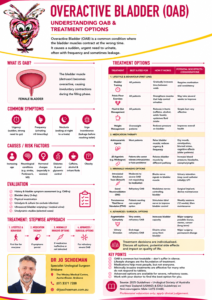
Overactive bladder (OAB) is a condition characterised by urinary urgency, usually accompanied by increased urinary frequency and waking at night to pass urine (nocturia), with or without urgency urinary incontinence.

Typical symptoms include:

  • A sudden, difficult-to-defer urge to urinate
  • Passing urine more frequently than expected
  • Waking several times during the night to urinate
  • Leakage of urine before reaching the toilet
  • Planning activities around access to toilets

Importantly, similar symptoms can sometimes be caused by urinary infection, bladder stones, bladder obstruction or other urinary tract conditions. Appropriate assessment is therefore important before treatment.


What is bladder Botox?

Botox® is the trade name for onabotulinumtoxinA, a purified form of botulinum toxin type A.

Although Botox is widely known for cosmetic treatments, it has several established medical applications, including treatment of bladder dysfunction.

When injected into the bladder muscle, Botox reduces excessive nerve signalling to the bladder and decreases involuntary bladder contractions. The bladder becomes less “trigger-happy”, allowing it to store urine for longer before producing an urgent need to empty.

Clinical studies demonstrate that intradetrusor onabotulinumtoxinA improves urgency urinary incontinence, urinary frequency, urgency and nocturia compared with placebo.

Who may benefit from bladder Botox?

Botox may be considered for patients with troublesome overactive bladder symptoms, particularly when conservative or medication-based treatments have been unsuccessful, poorly tolerated or are not desired.

Current guidelines recommend intradetrusor botulinum toxin as an established minimally invasive treatment for appropriately selected patients with OAB.

It may be particularly useful for patients experiencing:

  • Severe urinary urgency
  • Urgency urinary incontinence
  • Frequent urination
  • Significant nocturia
  • Persistent symptoms despite bladder training
  • Persistent symptoms despite pelvic floor rehabilitation
  • An inadequate response to medications
  • Unacceptable medication side effects

Some patients choose procedural treatment because they would prefer not to take long-term medication.

Other minimally invasive options for refractory OAB include sacral neuromodulation and tibial nerve stimulation.


Assessment before Botox treatment

Before proceeding, your urologist will usually assess the nature and severity of your bladder symptoms.

Depending upon the circumstances, this may include:

  • Medical and urinary history
  • Urinalysis and/or urine culture
  • Bladder or voiding diary
  • Measurement of urinary flow
  • Ultrasound assessment of the bladder
  • Measurement of the post-void residual (PVR) to determine how well the bladder empties
  • Urodynamic testing in selected patients

AUA/SUFU guidelines specifically recommend measuring the post-void residual before intradetrusor Botox treatment. Particular caution may be appropriate when the residual urine is already significantly elevated because Botox can temporarily reduce the strength of bladder emptying.

How is Botox injected into the bladder?

Bladder Botox is administered using a procedure called cystoscopy.

A fine telescope is passed through the urethra into the bladder. A specialised injection needle is then passed through the cystoscope.

Small quantities of Botox are injected at multiple sites across the bladder wall.

For idiopathic overactive bladder, onabotulinumtoxinA 100 units is a commonly used treatment regimen and is the dose recommended in European guidelines for refractory OAB/urgency urinary incontinence.

The procedure may be performed using:

  • Local anaesthetic placed into the bladder
  • Sedation, or
  • General anaesthesia

The most appropriate option depends on the patient, the clinical setting and individual preference.

The procedure itself is generally relatively short, and most patients can go home on the same day.


What happens after the procedure?

Some patients notice an improvement within several days, although the full effect may take a little longer to become apparent.

As Botox begins to work, patients may experience:

  • Less urgency
  • Fewer visits to the toilet
  • Fewer episodes of urgency incontinence
  • Less nocturia
  • Improved bladder capacity
  • Improved confidence when away from a toilet

Quality-of-life improvement following treatment can be substantial in appropriately selected patients.

How long does bladder Botox last?

Botox is not permanent.

The nerve endings gradually recover and bladder symptoms may eventually return. The duration of benefit varies considerably between individuals, but treatment commonly provides symptom control for a number of months.

When symptoms return, the injections can usually be repeated.

Evidence suggests that repeat treatment generally remains effective, although some patients eventually choose an alternative treatment.


What are the side effects of bladder Botox?

Most patients tolerate the procedure well, but Botox treatment is not without risk.

Urinary tract infection

Urinary tract infection (UTI) is one of the most important and common complications following bladder Botox.

Symptoms can include:

  • Burning when passing urine
  • Increased frequency
  • Cloudy or offensive-smelling urine
  • Lower abdominal discomfort
  • Fever or feeling unwell

Botox treatment is associated with an increased incidence of UTI.

Difficulty emptying the bladder

Botox deliberately reduces bladder muscle activity. Occasionally it works a little too effectively.

Some patients consequently develop an increased amount of urine remaining in the bladder after urination, known as an increased post-void residual.

This may cause:

  • Difficulty starting urination
  • A weak urinary stream
  • A feeling of incomplete emptying
  • Abdominal discomfort
  • Recurrent urinary infection

For this reason, bladder emptying may be reassessed following treatment, particularly if symptoms suggest incomplete emptying.

Temporary need for self-catheterisation

A small proportion of patients may be unable to empty their bladder adequately after Botox treatment.

If this occurs, clean intermittent self-catheterisation (CISC) may temporarily be required until the effect decreases and normal bladder emptying returns.

Patients should therefore understand this possibility and be willing and physically able to perform catheterisation if necessary before proceeding with treatment. Both EAU and AUA/SUFU guidance emphasise counselling patients about this risk.

Blood in the urine

A small amount of blood in the urine can occur following cystoscopy and the injections themselves. This is usually temporary.

Discomfort passing urine

Mild burning, urgency or bladder discomfort can occur for a short period following the procedure.

Rare systemic effects

Botulinum toxin can very rarely produce effects away from the injection site, potentially causing muscle weakness or other neurological symptoms.

Patients should seek medical attention if they develop significant weakness, difficulty swallowing or breathing difficulties following treatment.


Who should not have bladder Botox?

Intravesical Botox is not suitable for everybody.

Important contraindications include:

  • An active urinary tract infection
  • Acute urinary retention in a patient who is not routinely catheterising
  • Inability or unwillingness to perform intermittent catheterisation if it becomes necessary
  • Known hypersensitivity to Botox or its components
  • Certain neuromuscular disorders, including myasthenia gravis and Eaton-Lambert syndrome

These contraindications are reflected in Australian Botox product information.

Additional caution may be required in patients with:

  • Significant pre-existing incomplete bladder emptying
  • Recurrent urinary tract infections
  • Bladder outlet obstruction
  • Previous urinary retention
  • Certain neurological conditions
  • Frailty or significant medical comorbidity

Patients with recurrent UTIs require particularly careful consideration because Botox may further increase the risk of urinary infection.


Botox in men with overactive bladder

Men can also experience overactive bladder, but it is important to establish whether urinary symptoms are being caused or aggravated by bladder outlet obstruction from an enlarged prostate.

A man with urgency, frequency and nocturia may therefore require assessment of his prostate, urinary flow and residual urine before bladder Botox is considered.

In some men, treatment of prostate obstruction may need to be considered before or alongside treatment directed at the bladder.


Botox versus other treatments for overactive bladder

Botox forms part of a broader range of OAB treatments rather than being the right treatment for every patient.

Management may include:

Conservative treatment

Bladder training, modification of fluid intake, reducing caffeine, weight management where appropriate and pelvic floor rehabilitation.

Medication

Antimuscarinic medications and beta-3 adrenergic agonists can reduce urgency, frequency and urgency incontinence.

Intravesical Botox

An effective minimally invasive treatment that does not require a permanent implant, but needs to be repeated when its effect wears off.

Tibial nerve stimulation

Electrical stimulation of the tibial nerve can modify the nerve pathways involved in bladder control.

Sacral neuromodulation

A small implanted device stimulates the sacral nerves controlling bladder function and can provide longer-term treatment for selected patients.

Current OAB guidelines recognise Botox, tibial nerve stimulation and sacral neuromodulation as established minimally invasive treatment options when conservative or pharmacological therapy has provided inadequate results or unacceptable side effects.


When should I consider bladder Botox?

You may wish to discuss Botox with your urologist if overactive bladder is having a significant effect on your quality of life and:

  • Bladder training and conservative measures have not provided adequate control
  • Medications have not worked sufficiently
  • Medication side effects are unacceptable
  • You do not wish to continue long-term medication
  • You would like to consider a minimally invasive treatment

The decision should take into account both the potential benefits and the possibility of urinary infection, incomplete bladder emptying and temporary catheterisation.

The bottom line

Intravesical Botox is an effective and well-established treatment for overactive bladder and urgency urinary incontinence.

For appropriately selected patients, it can significantly reduce urinary urgency, frequency and leakage and improve quality of life. Its main limitations are that the effect is temporary and repeat injections are usually required.

The two most important complications to understand are urinary tract infection and incomplete bladder emptying, with a small proportion of patients requiring temporary intermittent catheterisation.

A consultation with a urologist can help determine whether Botox, medication, tibial nerve stimulation, sacral neuromodulation or another treatment is most appropriate for your individual bladder symptoms.


This information is intended for general patient education and does not replace individual medical advice. Treatment recommendations should be based on assessment by an appropriately qualified healthcare professional.

So. if this sounds like an option for your overactive bladder, come see your friendly Brisbane Urologist, Uro-Jo, in either Brisbane, Caboolture or on Bribie Island

Sacral Neuromodulation for Urinary Incontinence and Bladder Dysfunction

Urinary incontinence and bladder control problems can have a significant effect on everyday life. Frequent trips to the toilet, sudden urgency, leakage, waking repeatedly at night or being unable to empty the bladder properly can interfere with work, exercise, travel, sleep and social activities.

For men and women whose symptoms have not responded adequately to conservative treatment or medication, sacral neuromodulation (SNM) can provide another treatment option.

Sacral neuromodulation is particularly useful because it can treat two apparently opposite bladder problems:

  • an overactive bladder, where the bladder signals too frequently or contracts when it should not; and
  • an underactive bladder or non-obstructive urinary retention, where the bladder does not empty effectively.

Rather than operating directly on the bladder, sacral neuromodulation works by modifying the nerve signals involved in bladder storage and emptying.


What is sacral neuromodulation?

The bladder, urethral sphincter, pelvic floor and brain communicate through a complex network of nerves. The sacral nerves, particularly those arising from the S3 region, form an important part of this communication system.

Sacral neuromodulation uses a small, implanted device to deliver mild electrical impulses to a sacral nerve. These impulses help modify abnormal signaling between the bladder, pelvic floor, spinal cord and brain.

A useful way of thinking about the treatment is that it attempts to recalibrate the communication system controlling the bladder, rather than simply forcing the bladder to contract or relax.

International Continence Society guidance recognises sacral neuromodulation as an established therapy for refractory urinary urgency and frequency, urgency urinary incontinence and non-obstructive urinary retention.

Importantly, SNM does not “cure” every patient with urinary incontinence. The aim is to achieve a meaningful and sustained reduction in symptoms and improvement in quality of life.


What conditions can sacral neuromodulation treat?

1. Overactive bladder

Overactive bladder (OAB) is characterised by urinary urgency, usually accompanied by increased frequency and nocturia, with or without urgency urinary incontinence.

Typical symptoms include:

  • suddenly needing to pass urine;
  • difficulty postponing urination;
  • frequent urination during the day;
  • waking several times at night to urinate;
  • urinary leakage associated with urgency; and
  • needing to know where the nearest toilet is whenever leaving home.

Some patients experience urgency and frequency without leakage, while others develop urgency urinary incontinence, where the urge is followed by involuntary loss of urine.

Sacral neuromodulation can be considered when symptoms remain troublesome despite appropriate conservative and/or medical treatment. Current AUA/SUFU guidance includes SNM among the minimally invasive treatment options that clinicians should offer to appropriately selected patients with OAB who have not achieved adequate improvement with behavioural or pharmacological therapy.


2. Urgency urinary incontinence

Urgency urinary incontinence is leakage associated with a sudden compelling desire to urinate.

This is different from stress urinary incontinence, where leakage occurs with coughing, sneezing, exercise or physical exertion.

Sacral neuromodulation is principally a treatment for urgency-related leakage. It is not a standard treatment for isolated stress urinary incontinence.

This distinction is particularly important in women who may have both stress and urgency incontinence and in men who develop stress incontinence following prostate surgery.

When both types of leakage are present, treatment needs to be tailored to determine which component is causing the greatest problem.


What about an underactive bladder?

The bladder does not always misbehave by being too active. Sometimes the problem lies at the other end of the spectrum.

Underactive bladder and urinary retention

An underactive bladder describes symptoms associated with inadequate bladder emptying, often related to reduced bladder contractility or impaired coordination between the bladder and urinary sphincter.

Symptoms can include:

  • difficulty starting urination;
  • a slow or interrupted urinary stream;
  • prolonged urination;
  • straining to urinate;
  • feeling that the bladder has not emptied;
  • recurrent urinary tract infections;
  • a large residual volume of urine after voiding; and
  • complete inability to empty the bladder without a catheter.

Some patients need to perform clean intermittent self-catheterisation several times each day.

However, poor bladder emptying does not automatically mean that sacral neuromodulation is appropriate.


Obstruction must first be excluded

In men, urinary retention may be caused by:

  • benign prostate enlargement;
  • urethral stricture;
  • bladder neck obstruction; or
  • scarring following previous prostate or urethral surgery.

In women, causes can include:

  • previous continence surgery;
  • urethral obstruction;
  • pelvic organ prolapse; or
  • functional outlet obstruction.

Sacral neuromodulation is primarily used for non-obstructive urinary retention rather than retention caused by a mechanical blockage.

The distinction may require urine flow testing, measurement of the post-void residual, cystoscopy and, in selected patients, urodynamic studies.

The AUA notes that sacral neuromodulation can be used in appropriately selected men and women with non-obstructive urinary retention, including patients with decreased bladder contractility.


Who may benefit from sacral neuromodulation?

SNM may be considered in appropriately selected men and women with:

  • refractory overactive bladder;
  • urinary urgency and frequency;
  • urgency urinary incontinence;
  • non-obstructive urinary retention;
  • impaired bladder emptying in selected patients; or
  • troublesome bladder dysfunction that has not responded adequately to other treatments.

In Australia, sacral neuromodulation is an established treatment option. The TGA describes implantable sacral nerve stimulation systems as devices used in conditions including overactive bladder, urinary retention and urinary incontinence.

It is generally not a first-line treatment. Australian TGA information relating to the InterStim X system specifically notes that the treatment is not first-line therapy and is intended for appropriately selected patients after conservative and medical management have been attempted.


What treatments are usually tried first?

The treatments used before considering SNM depend upon the underlying bladder problem.

For overactive bladder these may include:

  • bladder retraining;
  • modification of fluid and caffeine intake;
  • pelvic floor physiotherapy;
  • management of constipation and other contributing factors;
  • antimuscarinic medication;
  • beta-3 agonist medication;
  • posterior tibial nerve stimulation; and
  • bladder injections with botulinum toxin (Botox).

For an underactive bladder or urinary retention, treatment may include:

  • treating an identifiable obstruction;
  • reviewing medications that may impair bladder emptying;
  • timed or double voiding in selected patients; and
  • intermittent self-catheterisation.

The appropriate pathway varies considerably between patients.


Assessment before sacral neuromodulation

Successful treatment begins with establishing the correct diagnosis.

Assessment may include:

Medical history

Your urologist will establish whether the predominant problem is urgency, frequency, leakage, difficulty emptying the bladder or a combination of symptoms.

Bladder diary

A bladder diary can record:

  • how frequently you urinate;
  • urine volumes;
  • fluid intake;
  • urgency episodes;
  • leakage episodes; and
  • night-time urination.

Urine testing

Urinary infection should be excluded where appropriate.

Post-void residual measurement

An ultrasound can measure how much urine remains in the bladder after urination.

Uroflowmetry

Urinary flow testing can provide useful information about the strength and pattern of the urinary stream.

Cystoscopy

A cystoscopy may be required when obstruction, urethral narrowing, prostate-related obstruction or another bladder abnormality is suspected.

Urodynamic studies

Urodynamic testing is not required in every patient, but it can be particularly valuable when the diagnosis is uncertain, symptoms are complex, previous treatments have failed or poor bladder emptying needs further investigation.


How is sacral neuromodulation performed?

One of the most attractive features of sacral neuromodulation is that the treatment can usually be tested before committing to a permanent implant.

The procedure therefore generally takes place in two stages.

Stage 1: The test phase

A thin electrode or tined lead is positioned through a small opening in the sacrum adjacent to a sacral nerve, most commonly the S3 nerve root.

The position is confirmed using anatomical landmarks, imaging and appropriate physiological responses during the procedure.

The lead is then connected to an external stimulator.

The patient goes home and uses the system during a trial period while recording symptoms in a bladder diary.

Depending upon the reason for treatment, we look for improvements such as:

  • fewer urgency episodes;
  • fewer episodes of urinary leakage;
  • reduced urinary frequency;
  • fewer night-time toilet visits;
  • improved bladder emptying;
  • lower residual urine volumes; or
  • reduced need for intermittent catheterisation.

A meaningful improvement during the test phase, commonly around 50% or greater improvement in the target symptoms, is generally used to determine whether proceeding to permanent implantation is worthwhile.


Stage 2: Permanent implantation

If the trial is successful, a small pulse generator is implanted beneath the skin, usually in the upper buttock.

The device is connected to the previously positioned sacral lead.

Once the incision has healed, the device remains beneath the skin and is generally not visible, although a small contour may sometimes be noticeable.

The stimulator is programmed to provide electrical impulses tailored to the individual patient.

Patients are provided with a programmer or controller that allows appropriate adjustments within parameters established by the treating team.


What does the stimulation feel like?

Patients may notice a mild:

  • tingling;
  • tapping;
  • pulling; or
  • vibration-like sensation

in the pelvic, perineal, vaginal, scrotal or anal region.

The stimulation should not normally be painful.

The device can be reprogrammed during follow-up if symptoms change or stimulation becomes uncomfortable.


How successful is sacral neuromodulation?

Success depends on the condition being treated, patient selection and how success is defined.

The major advantage of SNM is that each patient effectively undergoes their own therapeutic trial before receiving the permanent implant.

For refractory overactive bladder, clinical studies demonstrate substantial improvement in appropriately selected patients. The 2024 AUA/SUFU guideline cites a randomised study in which 86% of patients allocated to SNM initially responded and 61% met the study’s definition of therapeutic success at six months.

For non-obstructive urinary retention, treatment may substantially improve bladder emptying and can reduce or sometimes eliminate the need for intermittent catheterisation in responders. AUA evidence reviewing SNM for non-obstructive retention reported a significant reduction in post-void residual urine following treatment.

Individual results vary, and improvement rather than complete cure is a more realistic treatment goal.


Sacral neuromodulation versus Botox

Both sacral neuromodulation and intravesical botulinum toxin are established advanced treatments for refractory overactive bladder.

Botox is injected into the bladder wall during cystoscopy and temporarily reduces excessive bladder contractions.

SNM instead modifies the neural pathways controlling bladder function.

Both can be effective. One practical difference is that Botox usually needs to be repeated as its effect wears off, whereas SNM uses an implanted device designed to provide ongoing treatment.

Botox also carries a risk of urinary retention and urinary tract infection. In the ROSETTA trial, women treated with botulinum toxin had higher rates of urinary tract infection and temporary catheterisation than those treated with SNM, although both treatments produced substantial improvement in urgency incontinence.

The best option depends upon the individual patient’s priorities and bladder function.


Advantages of sacral neuromodulation

Potential advantages include:

  • treatment can be tested before permanent implantation;
  • no major bladder reconstruction is required;
  • it can reduce urgency and urgency incontinence;
  • it can improve frequency and nocturia in selected patients;
  • it may improve bladder emptying in non-obstructive retention;
  • it may reduce dependence on intermittent catheterisation;
  • stimulation can be individually programmed;
  • treatment is adjustable over time; and
  • the system can be removed if necessary.

Perhaps most importantly, SNM offers another option when conventional treatment has failed but major reconstructive surgery is undesirable.


What are the risks and possible complications?

Sacral neuromodulation is a surgical procedure and complications can occur.

These include:

  • pain or discomfort around the implant;
  • wound infection;
  • bleeding or bruising;
  • lead movement or migration;
  • loss of effective stimulation;
  • uncomfortable stimulation;
  • change in the location of the stimulation sensation;
  • failure to achieve adequate symptom improvement;
  • technical problems with the lead or pulse generator;
  • need for reprogramming;
  • battery depletion; and
  • need for revision, replacement or removal of the device.

In studies of SNM for overactive bladder, recognised device-related adverse events have included changes in stimulation, implant-site pain and infection.

Modern systems have improved considerably, but patients should understand that implantation begins a long-term relationship with the device rather than being a one-off operation.


What happens if the treatment stops working?

A reduction in benefit does not necessarily mean that the treatment has permanently failed.

The first step is often to check the device and change its programming.

If this does not restore benefit, further investigation may determine whether there has been:

  • lead migration;
  • lead damage;
  • battery depletion;
  • a change in bladder function; or
  • another underlying urinary problem.

Occasionally, surgical revision or replacement of the lead or pulse generator is required.


Battery life and rechargeable devices

Different sacral neuromodulation systems and pulse generators are available, including rechargeable and non-rechargeable technologies.

Battery longevity depends on the particular device and the stimulation settings required.

A non-rechargeable generator eventually requires surgical replacement when its battery reaches the end of its useful life.

Rechargeable systems can potentially reduce the frequency of generator replacement but require the patient to recharge the device periodically.

The most appropriate system depends on factors such as age, lifestyle, dexterity, expected stimulation requirements and patient preference.


What about MRI scans?

MRI compatibility has historically been an important issue for patients with implanted neuromodulation devices.

Many modern SNM systems now provide substantially broader MRI access than older devices. However, MRI eligibility depends on the exact pulse generator, lead and implanted system.

Patients should therefore keep their implant identification information and tell radiology staff that they have a sacral neuromodulation device before undergoing an MRI.


Is sacral neuromodulation suitable for both men and women?

Yes.

Although much of the early research into urgency urinary incontinence involved women, sacral neuromodulation can be used in appropriately selected men and women.

In men, it is particularly important to distinguish non-obstructive bladder dysfunction from obstruction caused by prostate enlargement, urethral stricture or previous prostate surgery.

In women, pelvic organ prolapse and previous continence or pelvic surgery may also need to be considered.

The treatment therefore depends much more on the type of bladder dysfunction than on the patient’s sex.


Overactive versus underactive bladder: one treatment, two different problems

It can initially seem strange that the same treatment can help a bladder that is too active and one that does not empty adequately.

This is because sacral neuromodulation is not simply an electrical switch telling the bladder to contract or relax.

Instead, it modifies the complex sensory and motor signalling involved in bladder storage, awareness, pelvic floor coordination and emptying.

For an overactive bladder, this can reduce inappropriate urgency and bladder activity.

For non-obstructive urinary retention or selected underactive bladder dysfunction, neuromodulation may improve the coordination of the bladder outlet and neural pathways involved in effective voiding.

This ability to influence both storage and emptying disorders makes sacral neuromodulation unusual among treatments for lower urinary tract dysfunction.


When should sacral neuromodulation be considered?

Sacral neuromodulation may be worth discussing when:

Overactive bladder symptoms remain troublesome despite conservative and medical treatment

or

Non-obstructive urinary retention is causing significant symptoms or dependence on intermittent catheterisation.

The key is appropriate patient selection.

Before proceeding, it is important to establish why the bladder is malfunctioning. In particular, a mechanical obstruction should not be mistaken for an underactive bladder.

Once the diagnosis is established, the test phase provides a valuable opportunity to determine whether neuromodulation is likely to provide meaningful benefit before committing to a permanent implant.


The bottom line

Sacral neuromodulation is an established, minimally invasive surgical treatment for selected men and women with refractory overactive bladder, urgency urinary incontinence and non-obstructive urinary retention.

A small electrode placed near the sacral nerves modifies the neural signals involved in bladder storage and emptying. Because treatment is usually performed as a staged procedure, patients can assess their response during a test phase before deciding whether to proceed with permanent implantation.

For patients who have spent years planning their lives around the nearest toilet, coping with unpredictable leakage or relying on catheters to empty their bladder, sacral neuromodulation can offer a very different approach: treating the communication network controlling the bladder rather than simply treating the bladder itself.

Important information

This information is intended for general patient education and does not replace individual medical advice. The suitability of sacral neuromodulation depends on the cause of the bladder dysfunction, previous treatments, medical history and individual treatment goals. Assessment by a urologist with experience in urinary incontinence and voiding dysfunction is recommended.

So, if you think is something that may work for you, come see me at my rooms in Brisbane, Caboolture or Bribie Island. Your friendly Brisbane based Urologist, Dr Jo Schoeman will assist.

Fascia Lata Allograft as a Mid-Urethral Sling for Female Stress Urinary Incontinence

A “new/revamped” kid on the block – Fascia Lata Allograft: A biological alternative to synthetic mesh

Stress urinary incontinence (SUI) is one of the most common forms of urinary incontinence in women. It occurs when urine leaks with activities that increase abdominal pressure, such as coughing, sneezing, laughing, exercising, lifting or running.

For women whose symptoms remain troublesome despite pelvic floor muscle training and appropriate conservative treatment, surgical treatment can provide substantial and durable improvement.

For many years, the mid-urethral sling has been one of the most effective minimally invasive surgical treatments for SUI. Synthetic mesh mid-urethral slings remain available in Australia and have extensive evidence supporting their effectiveness. However, concerns surrounding synthetic mesh have led many women to ask whether there is a biological alternative.

One such option is a fascia lata allograft, including the fascia lata allograft supplied by Samson Medical Technologies in Australia.

The important distinction is that the Samson product is not a synthetic mesh. It is human donor fascia lata, processed for use as a biological graft. Samson describes its current fascia lata allograft as 100% human-derived, without storage media, non-irradiated and processed using Super Critical CO₂ technology.

However, there is an important caveat: the published clinical evidence for fascia lata allografts in SUI largely relates to earlier cadaveric fascia products and pubovaginal sling techniques, rather than Samson’s current specific product. Therefore, the historical allograft results should not be presented as if they were Samson-specific clinical trial results.


What is an allograft sling?

An allograft is tissue obtained from a human donor rather than from the patient herself.

Traditional biological pubovaginal slings have commonly used the patient’s own rectus fascia or fascia lata. This provides a strong, durable biological support but requires an additional incision to harvest the patient’s tissue.

An allograft avoids this donor-site surgery.

The fascia lata is positioned beneath the urethra and secured to provide a supportive platform. During coughing, sneezing or physical exertion, the urethra is compressed against this support, helping prevent urine leakage.

The concept is therefore quite different from a synthetic polypropylene mid-urethral sling. Instead of leaving a permanent synthetic mesh implant, the surgeon is using processed human connective tissue.

Samson currently supplies fascia lata allografts in several dimensions, including 20 × 100 mm and 20 × 120–140 mm configurations.


How effective are fascia lata allografts?

This is where the evidence requires some careful interpretation.

There is a substantial historical literature examining cadaveric fascia lata as a sling material, but the results have been inconsistent.

Early studies

Some early studies reported excellent short-term results.

One study of 26 women treated with solvent-dehydrated cadaveric fascia lata reported that 77% were completely dry and 92% used one pad or fewer per day at a mean follow-up of 15 months.

Another series of 31 women reported complete resolution of SUI in 93% at approximately one year.

A larger series of 104 women using freeze-dried, non-irradiated cadaveric fascia lata demonstrated substantial reductions in pad use, although the study’s follow-up was relatively short.

These studies created considerable enthusiasm for allograft tissue because it offered the advantages of a biological sling without the morbidity of harvesting the patient’s own fascia.

But there was also a warning signal

Other studies produced considerably less encouraging results.

A study of 18 women using solvent-dehydrated, gamma-irradiated fascia lata found that 27.8% experienced significant failure, with recurrent incontinence occurring within 3–6 months. The authors concluded that this particular allograft material was unreliable.

A subsequent comparative study of 71 women found a concerning difference in objective urodynamic recurrence: urodynamic stress incontinence was present in 41.7% of women who received Tutoplast allograft compared with none of those receiving autologous fascia lata at follow-up. Patient-reported outcomes, however, were more favourable.

Other series have reported much better results. In a 134-patient comparison, 63 women received allograft fascia and 71 received autologous fascia. There was no statistically significant difference in overall cure of stress and urge incontinence at approximately two years, while the allograft group experienced less postoperative pain and disability.

What does this mean?

The historical evidence suggests that allograft fascia can work well, but the durability and consistency of older allograft products were variable.

This variability is important when discussing the Samson allograft. There is currently insufficient published comparative evidence to state that the Samson product has a specific cure rate such as 80%, 90% or 95%.

The more scientifically defensible statement is:

Samson fascia lata provides a contemporary biological allograft option for sling surgery, but Samson-specific long-term comparative clinical outcome data are still developing.

That distinction is important when counselling patients.


How does Samson allograft differ from older allografts?

The earlier allograft literature predominantly involved freeze-dried, solvent-dehydrated and/or gamma-irradiated cadaveric fascia lata.

The Samson product currently described by the manufacturer is different in its processing. Samson reports that its fascia lata is:

  • 100% human-derived
  • non-irradiated
  • supplied without storage media
  • processed using Super Critical CO₂ technology
  • available in several sizes.

The rationale behind modern tissue processing is to preserve the structural characteristics of the collagenous tissue while making the graft suitable for implantation.

Nevertheless, a different processing technology does not automatically establish superior clinical continence outcomes. Laboratory characteristics, tensile strength and histological appearance are not substitutes for long-term clinical trials.

This is one of the areas where future Australian outcome data will be particularly valuable.


How does an allograft compare with synthetic mid-urethral mesh?

Synthetic mid-urethral slings have the largest evidence base of any minimally invasive SUI operation.

A Cochrane review including 81 trials and more than 12,000 women found that more than 80% of women were cured or significantly improved following mid-urethral sling surgery, with good results maintained for up to five years.

Retropubic and transobturator approaches have broadly comparable effectiveness, although their complication profiles differ.

For example, retropubic slings have a higher risk of bladder perforation, whereas transobturator procedures have more groin pain. Vaginal mesh exposure is uncommon but recognised with both approaches.

The Australian situation

It is important to distinguish mid-urethral slings from transvaginal mesh used for pelvic organ prolapse.

The Australian TGA has removed certain transvaginal mesh products from the market, particularly mesh used for prolapse and single-incision mini-slings. However, mid-urethral sling devices for SUI remain approved in Australia.

Indeed, a 2026 Australian Government review concluded that mid-urethral sling devices should remain on the Prescribed List, with their existing listing unchanged.

Therefore, synthetic MUS should not simply be described as “banned mesh” in Australia. It remains an accepted treatment for appropriately selected women.

Potential advantages of an allograft

A biological fascia lata sling potentially offers:

Advantages

  • No synthetic polypropylene mesh
  • No fascial harvest from the patient’s abdomen or thigh
  • Avoidance of donor-site pain
  • Biological rather than permanent synthetic material
  • Potentially useful where avoidance of synthetic mesh is an important patient preference

Potential disadvantages

  • Less robust long-term evidence than synthetic MUS
  • Historical allograft studies have shown variable durability
  • It is still a surgical sling and can cause obstruction, urinary retention, infection, bleeding or pain
  • It requires more extensive dissection than a conventional MUS
  • It should not currently be marketed as proven superior to synthetic MUS

What about the Burch colposuspension?

The Burch colposuspension is the classic non-mesh surgical treatment for SUI.

Rather than placing a sling beneath the urethra, sutures are placed beside the bladder neck and proximal urethra and attached to supporting pelvic tissues.

It can be performed by open or laparoscopic surgery.

Long-term studies demonstrate that Burch colposuspension can be highly effective. One classic study found an 81.6% cure rate at 5–10 years.

More contemporary comparative data are reassuring. In a large study with more than 1,300 women and long-term follow-up, the proportion reporting no ongoing SUI was 83.0% after Burch colposuspension compared with 85.0% following retropubic MUS, with no significant difference.

However, Burch surgery is more invasive than a mid-urethral sling and is generally no longer the first surgical option for uncomplicated primary SUI.

It can nevertheless be an attractive option in selected women, particularly when avoiding synthetic mesh is important.


What about peri-urethral bulking agents?

Urethral bulking agents occupy a completely different part of the treatment spectrum.

A material is injected into the urethral wall around the bladder neck or proximal urethra to increase coaptation and improve continence.

In Australia, polyacrylamide hydrogel such as Bulkamid is an established example.

The major attraction is that it is substantially less invasive than sling surgery.

It can often be performed under local anaesthesia or sedation and has a relatively favourable safety profile.

The trade-off is effectiveness and durability.

A 2024 study of Bulkamid reported that at 12 months 69% of women described themselves as cured or improved, with 40% reporting cure, while 23% underwent subsequent treatment, including repeat Bulkamid injection or MUS.

Longer-term data are more modest. In a cohort of 357 women followed for a median of approximately 2.5 years, subjective cure was 30.9%, while 52.1% reported improvement. Minor adverse events occurred in 21%, with no serious adverse events attributed to the procedure.

Current guideline evidence therefore generally considers bulking agents less effective than MUS, Burch colposuspension or autologous fascial sling surgery, and repeat injections may be necessary.


Putting the options together

Treatment Approximate effectiveness Main advantages Main disadvantages
Samson fascia lata allograft sling Samson-specific long-term cure rate not yet established; historical allograft studies range widely Biological tissue; no synthetic mesh; no tissue harvest Limited contemporary comparative evidence; historical allograft durability concerns; surgical sling risks
Synthetic mid-urethral sling Generally >80% cure/improvement in medium-term evidence Minimally invasive, rapid recovery, extensive evidence Mesh-specific complications including exposure, pain and voiding dysfunction
Burch colposuspension Approximately 70–85% long-term continence in many series No vaginal mesh; durable native-tissue operation More invasive; longer recovery; risk of prolapse and voiding problems
Autologous fascial sling Generally high effectiveness, comparable with other established sling procedures No synthetic mesh; patient’s own tissue Requires tissue harvest; more postoperative morbidity
Bulking agent Lower cure rate, but meaningful improvement for many women Minimally invasive; useful for frail/high-risk patients or those avoiding sling surgery Less durable; repeat injections commonly required

The Australian Government’s own review of MUS specifically considered colposuspension, native-tissue pubovaginal slings and urethral bulking agents as important comparators.


Who might consider an allograft sling?

A fascia lata allograft may be particularly attractive for a woman who:

  • has objectively demonstrated SUI;
  • has failed appropriate conservative treatment;
  • wishes to avoid a permanent synthetic mesh implant;
  • does not want an abdominal or thigh incision to harvest her own fascia;
  • understands that the long-term evidence base for contemporary allograft products is smaller than that for synthetic MUS;
  • accepts that biological tissue does not eliminate the risks associated with sling surgery.

It may also have a role in selected women with recurrent SUI, particularly where previous synthetic mesh surgery makes another synthetic procedure unattractive. However, recurrent SUI is a more complex clinical situation and requires individual assessment.


The important question: is Samson allograft better than synthetic mesh?

At present, there is not enough evidence to say that it is.

The strongest evidence remains with synthetic mid-urethral slings. Their effectiveness has been demonstrated in thousands of women and across multiple randomised trials and systematic reviews.

The potential attraction of Samson fascia lata is different.

It offers a biological alternative for women who would prefer to avoid synthetic mesh while also avoiding the morbidity of harvesting their own fascia.

That makes it an interesting addition to the surgeon’s toolbox rather than a replacement for the established mid-urethral sling.

The key unanswered question is long-term durability.

Historical cadaveric allograft studies demonstrate that some allografts performed extremely well, while others experienced substantial early failure.

Consequently, contemporary prospective data specifically evaluating Samson’s processing technology, clinical continence outcomes, complications and durability would be valuable.


What should a woman expect from the procedure?

As with any continence operation, the goal is not simply to make a stress test negative. The aim is to improve the woman’s quality of life while preserving normal bladder emptying and sexual function.

Potential complications of sling surgery include:

  • temporary or persistent difficulty emptying the bladder;
  • urinary retention requiring catheterisation;
  • urinary tract infection;
  • bleeding or haematoma;
  • pain;
  • de novo urgency or worsening overactive bladder;
  • recurrent or persistent SUI;
  • vaginal wound problems;
  • infection;
  • rarely, injury to the bladder, urethra or surrounding structures.

Synthetic mesh has additional mesh-specific risks, including vaginal exposure/erosion and chronic pain. The TGA specifically requires information regarding complications such as severe chronic pain, groin pain and bladder perforation in the instructions for use of Australian MUS devices.

An allograft avoids synthetic mesh exposure, but it does not make the operation risk-free.


The bottom line

For a woman with bothersome stress urinary incontinence, there is no single operation that is right for everyone.

Synthetic mid-urethral slings remain the most extensively studied minimally invasive surgical treatment and have excellent success rates.

Burch colposuspension remains a durable non-mesh alternative, although it is more invasive.

Bulking agents offer the least invasive surgical option but generally have lower and less durable success rates.

Fascia lata allograft represents an intriguing middle ground: a biological sling that avoids both synthetic mesh and the need to harvest the patient’s own fascia.

The Samson allograft is particularly interesting because its contemporary processing differs from several of the older cadaveric fascia products that generated the historical literature. Samson describes its current graft as non-irradiated human fascia lata processed using Super Critical CO₂ technology.

However, the enthusiasm should be matched by scientific caution. The historical literature demonstrates that allografts can achieve good continence outcomes, but it also demonstrates that not all allograft materials have behaved identically over time.

For patients considering this option, the most accurate discussion is therefore not “this is a better sling than mesh.” It is:

“This is a biological alternative to synthetic mesh, with encouraging historical experience but less mature long-term clinical evidence for the contemporary product.”

As further Australian experience and prospective follow-up accumulate, Samson fascia lata allograft may become an increasingly useful option in the treatment of female stress urinary incontinence.

Important note

This article is intended for general information and does not replace an individual assessment by a urologist or uro-gynaecologist. The choice between conservative management, bulking agent, synthetic MUS, biological or autologous sling and Burch colposuspension depends on the type and severity of incontinence, previous surgery, urethral function, pelvic anatomy, patient preference and overall health.

Don’t suffer in silence: Come see your friendly Brisbane Urologist for advice.

Stress Urinary Incontinence in Women: Symptoms, Investigations and Treatment Options in Australia

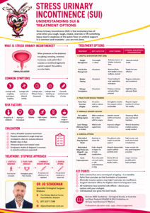
Stress urinary incontinence (SUI) is one of the most common forms of urinary incontinence in women. It occurs when urine leaks because pressure inside the abdomen rises faster than the urethra and pelvic floor can close.

Coughing, sneezing, laughing, lifting, running, jumping and exercise can all trigger leakage. For some women the leakage is only a few drops; for others it can be substantial enough to interfere with exercise, work, intimacy and everyday life.

The good news is that SUI is treatable, and treatment can be tailored to the severity of symptoms, the woman’s lifestyle and preferences, previous surgery, pelvic floor function and other bladder symptoms.

What are the symptoms and signs of stress urinary incontinence?

The classic symptom is involunta

ry urine leakage with physical exertion.

Common triggers include:

  • Coughing or sneezing
  • Laughing
  • Getting up from a chair
  • Lifting children or heavy objects
  • Running or jumping
  • Exercise, particularly high-impact exercise
  • Sexual intercourse
  • Changes in position
  • Sports such as tennis, running or aerobics

Some women notice only occasional leakage, while others need pads every day.

Stress incontinence versus urgency incontinence

It is important to distinguish SUI from urgency urinary incontinence, where urine leakage follows a sudden, difficult-to-defer urge to urinate.

Many women have mixed urinary incontinence, with both stress and urgency leakage.

This distinction matters because the treatment strategy can be quite different. Treating the wrong component first can produce disappointing results.


How is stress urinary incontinence diagnosed?

For many women, the diagnosis can be made from the history and examination without an extensive battery of tests.

A specialist assessment usually includes:

1. Medical history

Your doctor will ask about:

  • The nature and severity of leakage
  • What activities trigger leakage
  • Urinary urgency and frequency
  • Night-time urination
  • Recurrent urinary tract infections
  • Previous pregnancies and childbirth
  • Menopause and hormonal symptoms
  • Previous pelvic or continence surgery
  • Pelvic-organ prolapse
  • Neurological conditions
  • Medications
  • Impact on exercise, work, social activities and sexual function

2. Bladder or voiding diary

A bladder diary records fluid intake, urinary frequency, urine volumes, urgency episodes and leakage over several days.

It can be surprisingly revealing. It helps determine whether the problem is predominantly stress leakage, urgency, excessive fluid intake, frequent small-volume voiding or a combination of these.

3. Physical examination

A pelvic examination can assess:

  • Pelvic organ prolapse
  • Pelvic floor muscle strength
  • Urethral mobility
  • Vaginal and vulval health
  • Atrophic changes associated with menopause
  • Evidence of previous surgery

A cough stress test may be performed with a comfortably full bladder. Leakage of urine through the urethra during coughing supports the diagnosis of stress urinary incontinence.

4. Urine testing

A urine test is often appropriate, particularly when there are symptoms suggesting infection, blood in the urine or other urinary abnormalities.

5. Measurement of residual urine

A bladder ultrasound can measure the amount of urine left after voiding. This is particularly useful if there are symptoms suggesting incomplete bladder emptying or voiding dysfunction.


Do I need urodynamic studies?

This is one of the most common questions asked by women considering treatment.

Not every woman with straightforward stress urinary incontinence requires urodynamics.

Urodynamic testing measures how the bladder, urethra and pelvic floor behave while the bladder fills and empties. It can determine whether leakage is associated with increased abdominal pressure, whether involuntary bladder contractions occur and whether there is evidence of obstruction or impaired bladder emptying.

Contemporary guidelines generally do not recommend routine urodynamics before surgery in women with uncomplicated, clinically demonstrable SUI because it has not been shown to improve surgical outcomes.

Urodynamics becomes particularly useful when:

  • The diagnosis is uncertain
  • There are significant urgency or other storage symptoms
  • Mixed urinary incontinence is present
  • Voiding dysfunction is suspected
  • There is significant pelvic organ prolapse
  • Previous continence surgery has failed
  • The symptoms and examination findings do not match
  • There is suspected intrinsic sphincter deficiency
  • The results may change the choice of treatment

In these situations, urodynamics can turn a clinical puzzle into a much clearer picture.


Treatment of stress urinary incontinence

Treatment is usually progressive, beginning with conservative measures and moving towards minimally invasive or surgical treatment when necessary.

The appropriate treatment depends on the severity of the leakage and, importantly, how much it bothers you.

1. Pelvic floor muscle training

Pelvic floor muscle training (PFMT) is generally the first-line treatment for SUI.

The pelvic floor muscles provide support to the bladder neck and urethra. Strengthening and correctly coordinating these muscles can significantly reduce leakage.

A structured program with a pelvic floor physiotherapist is preferable to simply being told to “do your Kegels”.

A good program may include:

  • Identification of the correct muscles
  • Strength training
  • Endurance training
  • Rapid contractions for coughs and sneezes
  • Coordination of the pelvic floor with movement
  • Techniques for managing leakage during exercise

Guidelines recommend supervised pelvic floor muscle training for at least three months as initial therapy for SUI.

The advantage is obvious: there is no incision, no anaesthetic and essentially no procedural risk.

The disadvantage is that it requires commitment, correct technique and time.


2. Lifestyle measures

Lifestyle modifications can make a meaningful difference, particularly when combined with pelvic floor rehabilitation.

These may include:

  • Weight reduction where appropriate
  • Optimizing fluid intake
  • Reducing excessive caffeine intake
  • Treating constipation
  • Addressing chronic coughing
  • Smoking cessation
  • Modifying high-impact exercise temporarily while rehabilitation is undertaken

These measures rarely “cure” significant anatomical SUI on their own, but they can reduce symptoms and improve the results of other treatments.


3. Continence pessary

A vaginal continence pessary can provide mechanical support to the urethra and bladder neck.

It may be particularly useful for women who:

  • Want to avoid surgery
  • Have leakage predominantly during exercise
  • Are awaiting surgery
  • Have coexisting pelvic organ prolapse
  • Want an additional treatment alongside pelvic floor rehabilitation

A pessary is fitted inside the vagina and can sometimes be removed and inserted by the patient.

Possible problems include vaginal irritation, discharge, discomfort and difficulty with insertion or removal. Regular review is important.


4. Vaginal oestrogen

For postmenopausal women with vaginal or genitourinary symptoms, local vaginal oestrogen may improve vaginal tissue health and urinary symptoms.

It is not a replacement for continence surgery when significant anatomical SUI is present, but it can be a useful component of treatment in appropriately selected women.


5. Urethral bulking injections

Urethral bulking involves injecting a material around the urethra to improve its ability to remain closed.

One example used clinically is polyacrylamide hydrogel.

The procedure is minimally invasive and can be attractive to women who:

  • Prefer to avoid sling surgery
  • Are poor surgical candidates
  • Have recurrent SUI after previous surgery
  • Want a relatively quick procedure

The trade-off is durability.

Bulking agents are generally less effective than sling procedures and repeat injections may be necessary. Their effectiveness can diminish with time.

Potential side effects include:

  • Temporary urinary urgency
  • Urinary tract infection
  • Difficulty emptying the bladder
  • Blood in the urine
  • Discomfort
  • Recurrent leakage
  • Need for repeat treatment

6. Mid-urethral sling

For women with bothersome SUI who have not obtained sufficient improvement from conservative treatment, a mid-urethral sling (MUS) is one of the most established surgical treatments.

The sling sits beneath the middle portion of the urethra and provides support when abdominal pressure rises.

Two principal approaches have been used:

Retropubic mid-urethral sling

The tape passes behind the pubic bone.

Mid-urethral slings have substantially reduced the morbidity and complexity of traditional continence surgery, with high rates of improvement and continence. Contemporary evidence suggests broadly similar effectiveness between retropubic and transobturator approaches, although their complication profiles differ.

Importantly, mid-urethral sling surgery is not the same as the transvaginal mesh products that were used for pelvic organ prolapse. The Australian regulatory position is different. Mid-urethral slings for SUI remain approved products in Australia, with approved devices subject to Class III medical-device requirements.


What are the risks of a mid-urethral sling?

Although most women do well, sling surgery is not risk-free.

Potential complications include:

  • Bleeding or haematoma
  • Infection
  • Bladder or urethral injury during insertion
  • Temporary or persistent difficulty passing urine
  • Urinary retention
  • New or worsening urgency
  • Recurrent stress incontinence
  • Pelvic, groin or suprapubic pain
  • Pain during sexual intercourse
  • Mesh exposure or erosion
  • Rarely, chronic pain or other mesh-related complications
  • Need for further surgery

The TGA specifically identifies complications including bladder or urethral injury, mesh exposure or erosion, voiding dysfunction, urinary retention, urgency and acute or chronic pain among the potential adverse outcomes associated with urogynaecological mesh.

The decision to use a sling should therefore involve an individual discussion about benefits, alternatives and risks rather than treating the operation as a one-size-fits-all solution.


7. Autologous fascial sling

For women who wish to avoid synthetic mesh, another established surgical option is an autologous fascial sling.

A strip of the woman’s own tissue, usually rectus fascia from the lower abdominal wall, is fashioned into a sling and placed beneath the bladder neck/urethra.

This avoids a synthetic implant.

It can be particularly useful in selected women with:

  • Severe stress incontinence
  • Intrinsic sphincter deficiency
  • Previous failed sling surgery
  • Previous mesh complications
  • A strong preference to avoid synthetic mesh

The disadvantages are that it is generally a more extensive operation, requires harvesting tissue and can have a higher risk of postoperative voiding dysfunction. Fascial harvest can also cause abdominal wound pain, infection, seroma or, rarely, hernia.


What about an allograft mid-urethral sling?

This is an important distinction.

An allograft is tissue obtained from another human donor, rather than tissue harvested from the woman herself. Historically, cadaveric fascia lata has been used to construct pubovaginal slings.

The attraction is obvious: it avoids harvesting the woman’s own rectus fascia and therefore avoids the additional abdominal wound associated with an autologous sling.

However, there is an important catch.

The evidence for allograft slings is considerably weaker than for modern mid-urethral synthetic slings or autologous fascial slings. This based on older studies and different mechanisms to prepare the graft.

Older studies of cadaveric fascia lata reported encouraging short-term outcomes, including reduced postoperative pain compared with harvesting autologous fascia. However, other studies reported substantial recurrence of stress incontinence, raising concerns about long-term durability.

Systematic evidence reviews have also concluded that the evidence supporting biological/allograft slings is limited and of low or very low certainty. In comparisons with synthetic slings, there has not been convincing evidence that cadaveric fascia lata provides a superior outcome.

For this reason, an allograft sling should not simply be regarded as a “mesh sling without mesh.”

Its role is much more selective.

Where available, it may be considered in carefully selected women who want to avoid synthetic mesh and would otherwise be candidates for a fascial sling, but the surgeon should explain the limitations of the evidence and uncertainty around long-term durability.

In Australia, the availability and regulatory status of any particular allograft product or technique should be confirmed with the treating surgeon and relevant institution. The TGA’s current approved urogynaecological mesh list specifically identifies approved devices for SUI, while not all historical mesh or biological products remain available.


8. Burch colposuspension

Burch colposuspension is a traditional operation in which sutures are placed to support the tissues around the bladder neck and urethra.

It can be performed through an abdominal approach, including laparoscopically or robotically in appropriately experienced centres.

It remains an important non-mesh surgical option, although it is more invasive than a mid-urethral sling.

Potential complications include:

  • Bleeding
  • Infection
  • Bladder injury
  • Postoperative urinary retention or voiding dysfunction
  • New urgency
  • Recurrent SUI
  • Pelvic or abdominal pain
  • Development or worsening of pelvic organ prolapse

Choosing the right operation

There is no single “best” operation for every woman.

The decision should take into account:

Consideration Why it matters
Severity of SUI Determines how much treatment is justified
Previous surgery May alter the choice of procedure
Intrinsic sphincter deficiency May favour a different surgical approach
Pelvic organ prolapse May require combined treatment
Urgency/mixed incontinence May affect expected outcome
Desire to avoid synthetic mesh Makes fascial procedures more relevant
Previous mesh complications May favour non-mesh options
General health Influences operative risk
Future pregnancies May influence timing and choice of surgery
Patient preference An essential part of shared decision-making

The aim is not simply to make the pad dry. The aim is to find the treatment that gives the woman the best balance between continence, bladder function, quality of life and procedural risk.


What happens after treatment?

Following continence surgery, women are generally reviewed to assess:

  • Continence
  • Urgency and frequency
  • Bladder emptying
  • Residual urine
  • Pelvic pain
  • Sexual function
  • Vaginal healing
  • Mesh exposure where a mesh sling has been used

If symptoms persist or recur, further investigation may be necessary. In women with failed previous SUI surgery, repeat urodynamics and specialist assessment can be particularly valuable.

Australia also has the Australasian Pelvic Floor Procedures Registry, which collects information about stress incontinence and pelvic floor procedures with the goal of improving quality and long-term safety.


The bottom line

Stress urinary incontinence is common, but it is not something women simply have to put up with.

Treatment ranges from pelvic floor physiotherapy and lifestyle modification through to pessaries, urethral bulking injections and surgery.

For women requiring surgery, options in Australia include mid-urethral sling procedures, autologous fascial sling surgery and colposuspension, with the appropriate choice depending on individual circumstances.

An allograft fascial sling is a more specialised option. Although it avoids harvesting the patient’s own fascia, the evidence base is considerably less robust than that for established mid-urethral sling and autologous fascial sling procedures. It should therefore be discussed as a selective option rather than assumed to be a routine alternative to synthetic mesh.

Most importantly, treatment should be individualised. A woman should understand not only the likelihood of becoming dry, but also the possibility of urgency, voiding problems, pain, recurrent incontinence and other procedure-specific complications before making a decision.

If urinary leakage is interfering with exercise, work, relationships or everyday life, an assessment by a GP, urogynaecologist, urologist or pelvic-floor physiotherapist is a good place to start.

This information is intended for education and does not replace an individual medical assessment. Treatment availability, device approvals and clinical recommendations can change, so patients should discuss the current options with their treating clinician.

Don’t suffer in silence. Come see what your Brisbane Urologist Uro-Jo can do for you.

Post-Prostatectomy Urinary Incontinence

Sources

Cadaveric Fascial Sling – Fascia Lata

Why is it done?

  • Stress incontinence
  • A combination of stress incontinence and detrusor over-activity of which DO the lesser
  • Involuntary urine leakage with any exertion, coughing or sneezing
  • Risk factors
    • More than 2 pregnancies, big babies, complicated deliveries, episiotomy
    • Smokers
    • Being overweigh
  • Where Intrinsic Sphincter Deficiency has been proved due to a failed previous sling
  • Failed previous incontinence procedures


How is it done?

  • This procedure is done under a spinal / general anaesthetic, as decided by the anaesthetist.
  • The legs will be elevated into the lithotomy position.
  • A cadaveric fascia-lata will be used
  • A small incision is made in the vagina.
  • The sling is placed behind the pubic bone and brought to the skin above the pubic bone, through the incision.
  • The sling is placed with some tension.
  • The bladder will be inspected with a Cystoscopy to exclude any injuries to the bladder wall.
  • The wounds are closed with dissolvable sutures and/or skin glue.
  • A local anaesthetic is given for pain relief.
  • A urinary catheter is placed for 24hrs.
  • A vaginal plug will also be placed.
  • The catheter and plug will be removed early the next morning.
  • The patient’s urine output will be measured each time they urinate, and the residual will be measured. (Patients will be required to do this up to 3 times.)
  • If the residual amount of urine is more than 1/3 of the total bladder capacity, the patient may have to self-catheterize, until the residual volume is acceptable.
  • Prophylactic antibiotics will be given to prevent infection.


What to expect after the procedure?

  • Any anaesthetic has its risks, and the anaesthetist will explain all such risks.
  • Complications:
    • hemorrhaging, requiring blood transfusion <1%;
    • bladder perforation, requiring an open repair <1%.
  • Patients will wake up with a catheter in the urethra and bladder. This will remain in the bladder for at least 24 hrs.
  • Wound discomfort/pain will persist for a few days but this will subside / settle.
  • You may be required to self catheterize for a week or two.
  • If there is no improvement the sling may be cut, to allow spontaneous urination
  • NB! Each person is unique and for this reason  symptoms may vary!


What next?

  • Patients will have a trial of void without catheter the next day.
  • Patients will be discharged as soon as they can completely empty the bladder.
  • Patients may be required to self catheterize for a week or two.
  • Patients may initially suffer from urge incontinence but this will improve within the next 6 weeks.
  • Allow 6 weeks for symptoms to stabilise.
  • May also have abdominal pain with coughing and sneezing due to tension on rectus muscle
  • There may be some blood in the urine. This can be remedied by drinking plenty of fluids until it clears.
  • On discharge a prescription may be issued for patients to collect.
  • Patients are to schedule a follow-up appointment in 6 weeks.
  • Please direct all queries to Dr Schoeman’s rooms.
  • PLEASE CONTACT THE HOPSITAL DIRECT WITH ANY POST-OPERATIVE CONCERNS AND RETURN TO THE   HOSPITAL IMMEDIATELY SHOULD THERE BE ANY SIGNS OF SEPSIS.

Wes Sling Rectus Fascial CADAVERIC

Sacro Neuromodulation – Removal of Device

Why is it done?

  • To remove a SNM device:
    • Failed to alter bladder and bowel incontinence


How is this done?

  • A sedation / Local Anaesthetic administered
  • You will be placed prone (on your stomach) with lower back and buttocks exposed
  • An incision made over the old scars.
  • The lead is removed from the sacrum
  • The Battery removed from its pouch.
  • Wounds irrigated with Betadine and closed


What to expect

  • Wound healing takes 10 days
  • Keep dressings X 3-5 days
  • Sutures dissolve and is not required to be removed

Wes Sacro Neuro Modulation-Removal Leads