PUJ obstruction: when urine cannot drain freely from the kidney
Understanding pelvi-ureteric junction obstruction, its symptoms, investigations and treatment, including robotic-assisted pyeloplasty.
The kidneys produce urine, which collects in a funnel-shaped area called the renal pelvis before travelling down the ureter to the bladder. The pelvi-ureteric junction (PUJ) is where this funnel joins the ureter.
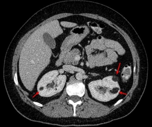
PUJ obstruction means urine does not pass freely through this junction. It is also called ureteropelvic junction obstruction, or UPJ obstruction. The collecting system can enlarge, a finding called hydronephrosis, and significant obstruction may eventually damage the affected kidney. However, a dilated collecting system does not automatically mean surgery is needed. Assessment must establish whether drainage is impaired and whether kidney function is at risk. [1,2]
What causes PUJ obstruction?
Some people are born with a short segment that is narrow or does not propel urine normally. Other anatomical causes include kinking or a ureter that joins the renal pelvis unusually high up. A blood vessel supplying the lower part of the kidney may cross the junction and contribute to compression. Seeing a crossing vessel on a scan does not, by itself, prove that it is causing obstruction.
A congenital problem may first become apparent in adulthood. Acquired narrowing can follow scarring, inflammation or previous surgery. Stones and other causes of upper urinary tract blockage must also be considered during investigation. [2,12]
How does it present?
Symptoms range from none at all to episodes of significant pain. Possible presentations include:
- Aching or intermittent pain in the flank, the side of the back below the ribs.
- Episodes of more severe pain, sometimes with nausea or vomiting.
- Pain after a large fluid intake, when urine production increases.
- Recurrent urinary infections, including infection of the kidney.
- Blood in the urine or kidney stones.
- An incidental finding of hydronephrosis on a scan.
Intermittent painful distension is sometimes called a Dietl’s crisis. These symptoms can also have other causes, so pain or blood in the urine should not simply be attributed to a known PUJ problem. [3,12]
Children may be diagnosed following an antenatal ultrasound. Childhood assessment and treatment decisions differ from those in adults and should involve a paediatric urology team. [3]
Which investigations are useful?
The investigation plan answers three questions: What does the anatomy look like? How well does each kidney function? Is urine drainage genuinely obstructed?
Urine and blood tests
Urine testing looks for blood and evidence of infection; a urine culture is useful when infection is suspected. Blood tests include creatinine and estimated glomerular filtration rate (eGFR). These assess overall kidney function, but can remain normal when the other kidney compensates for an affected kidney. [2,10]
Ultrasound
Ultrasound shows collecting-system dilatation and assesses the thickness of the kidney tissue. It is useful for follow-up because it does not involve radiation. Ultrasound alone cannot reliably establish the functional significance of a PUJ narrowing. [2,3]
CT or MR urography
CT urography can define the junction, identify stones and assess surrounding structures, including crossing vessels. The use of intravenous contrast depends on kidney function and other clinical factors.
MR urography is an alternative in selected patients, particularly when avoiding radiation is desirable. The choice of scan is individualised. [1,2]
MAG3 diuretic renogram
This nuclear medicine scan follows a small injected tracer through the kidneys. A diuretic, usually furosemide, helps assess drainage.
It measures:
- Differential or split renal function: the proportion contributed by each kidney.
- Drainage: how effectively tracer leaves the collecting system.
Interpretation considers the images, drainage curves, kidney function, hydration and bladder emptying. A slow drainage number alone is insufficient to diagnose clinically significant obstruction: a large collecting system or poorly functioning kidney can make interpretation difficult. [4]
Further tests in selected cases
A retrograde pyelogram introduces contrast into the ureter during a telescopic bladder examination. It can clarify anatomy when other imaging is incomplete. It is not routinely needed in every patient. [2]
Does every PUJ obstruction need treatment?
No. Observation can be appropriate when symptoms are absent or minor, kidney function is stable, and assessment does not suggest harmful obstruction. Monitoring may include symptoms, ultrasound and repeat functional imaging when indicated. The interval depends on the findings. [5]
Intervention is more likely to be recommended for persistent or troublesome symptoms, recurrent kidney infections, associated stones, or deteriorating function. Decisions use the overall clinical picture rather than the ultrasound appearance alone. [2]
What are the treatment options?
| Option | When it may be considered | Main limitation |
|---|---|---|
| Observation | Stable function with few symptoms | Requires planned follow-up |
| Ureteric stent or nephrostomy | Urgent drainage or temporary management | Usually does not permanently correct the narrowing |
| Endopyelotomy or balloon dilatation | Selected narrowings or recurrent obstruction | Generally less durable than reconstructive repair |
| Pyeloplasty | A significant obstruction requiring reconstruction | Requires an operation and recovery |
Temporary or emergency drainage
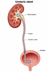
A ureteric stent passes internally from the kidney to the bladder. A nephrostomy drains the kidney through a tube placed through the skin of the back. These can relieve obstruction while infection is treated or definitive surgery is planned. [5,9]
An infected obstructed kidney is an emergency. Antibiotics may be needed together with urgent drainage. Definitive reconstruction is usually considered after the acute problem has been controlled. [9,10]
Endoscopic treatment
Endopyelotomy opens the narrowed segment from inside the urinary tract; balloon dilatation stretches it. Suitability depends on the anatomy, kidney function and previous treatment.
A large observational study found more repeat procedures after endopyelotomy than after pyeloplasty. Because patients were not randomly assigned to treatment, these results inform discussion but cannot predict an individual outcome. [1,6]
Robotic-assisted pyeloplasty: how is the repair performed?
Pyeloplasty reconstructs the junction to improve drainage. It can be performed through an open incision, conventional laparoscopic keyhole surgery, or robotic-assisted keyhole surgery.
The commonly used Anderson–Hynes dismembered pyeloplasty removes the narrowed segment. The surgeon opens the ureter more widely and stitches it back to the renal pelvis, creating a broader connection. Where a crossing vessel contributes to obstruction, the reconstruction can be positioned in front of it while preserving the vessel. [3]
During robotic-assisted surgery, the surgeon controls the instruments from a console. Magnified three-dimensional vision and articulated instruments assist dissection and suturing. The robot does not operate independently. [7]
A temporary internal stent is commonly placed to support healing. A bladder catheter and, sometimes, a drain may also be used initially. [5,7]
How successful is pyeloplasty, and is robotic surgery better?
Published literature commonly reports success above 90% for primary pyeloplasty, including open, laparoscopic and robotic approaches. Success may mean symptom relief, improved drainage or freedom from further surgery; definitions and follow-up differ between studies. These figures are not a guarantee. [1]
Robotic surgery is an established option, but it should not be presented as necessary or universally superior. Comparative research supports good outcomes with both conventional laparoscopic and robotic repair. Much of the evidence is observational, with differences in patient selection, surgical experience and follow-up. The choice should consider anatomy, surgeon expertise, availability and cost. [8]
The goals are to improve drainage, relieve attributable symptoms and preserve function. Previously lost kidney function may not fully recover, even after a technically successful operation. [11]
What are the risks and recovery expectations?
Risks include bleeding, infection, urine leakage, injury to nearby structures, recurrent narrowing, persistent pain and the need for another procedure. Anaesthetic complications and blood clots are also possible. Occasionally, conversion to open surgery is required. [5,7]
Stents can cause urinary frequency, urgency, blood in the urine and discomfort. Their removal must be arranged. Many patients leave hospital after approximately one to three days following uncomplicated keyhole repair, but recovery varies. Stents are often removed after four to six weeks. [5,7]
Follow-up may include ultrasound and a repeat drainage scan. Residual dilatation does not automatically mean the repair has failed; symptoms, drainage and function are assessed together. [4]
What if the kidney already functions poorly?
A low split-function result does not automatically mean the kidney must be removed. Selected patients may benefit from reconstruction or temporary drainage followed by reassessment. A systematic review found that pyeloplasty may relieve symptoms and stabilise function in poorly functioning kidneys, but the evidence was limited and recovery was unpredictable. [11]
Nephrectomy may be considered if the kidney has negligible useful function and causes ongoing pain or infection. This requires an individual discussion, including the health of the other kidney.
When should I seek urgent help?
Seek urgent medical assessment for fever or shaking chills with flank pain, severe uncontrolled pain, persistent vomiting, markedly reduced urine output, or feeling seriously unwell. The threshold for urgent assessment is particularly low if you have a single functioning kidney. [9,10]
This page provides general education and does not replace an individual assessment. The most suitable investigation and treatment depend on symptoms, anatomy, kidney function and overall health.
References
- Khan F, Ahmed K, Lee N, et al. Management of ureteropelvic junction obstruction in adults. Nature Reviews Urology. 2014;11:629–638. doi:10.1038/nrurol.2014.240.
- Borin JF. Ureteropelvic Junction Obstruction in Adults. Reviews in Urology. 2017;19(4):261–264. Full text.
- Oxford University Hospitals NHS Foundation Trust. PUJ Obstruction and Pyeloplasty: information for parents and carers. 2025. Patient leaflet. Used for congenital presentation and reconstruction principles; adult management differs.
- SNMMI/EANM. Procedure Standard/Practice Guideline for Diuretic Renal Scintigraphy in Adults With Suspected Upper Urinary Tract Obstruction 1.0. 2018. Guideline.
- British Association of Urological Surgeons. Keyhole reconstruction of the kidney pelvis: laparoscopic pyeloplasty. 2023. Patient leaflet.
- Jacobs BL, Lai JC, Seelam R, et al. The comparative effectiveness of treatments for ureteropelvic junction obstruction. Urology. 2018;111:72–77. doi:10.1016/j.urology.2017.09.002.
- Kingston and Richmond NHS Foundation Trust. Kidney obstruction removal with robot. Patient information.
- Uhlig A, Uhlig J, Trojan L, et al. Surgical approaches for treatment of ureteropelvic junction obstruction—a systematic review and network meta-analysis. BMC Urology. 2019;19:112. doi:10.1186/s12894-019-0544-7.
- Guy’s and St Thomas’ NHS Foundation Trust. Nephrostomy catheter to drain urine from the kidney. Patient information.
- NHS. Hydronephrosis. Patient information.
- Pyeloplasty in Adults With Ureteropelvic Junction Obstruction in Poorly Functioning Kidneys: A Systematic Review. 2021. PubMed.
- Cleveland Clinic. Ureteropelvic Junction Obstruction. Reviewed 2024. Patient information.
Prepared October 2026.