
Kidney Stones: Symptoms, Causes and Treatment Options Explained
Kidney stones: symptoms, treatment and prevention
Kidney stones can cause sudden, intense pain, but some are discovered by chance and cause no symptoms. The right treatment depends on where a stone sits, its size, whether urine can drain, whether infection is present and how well the kidneys are working. If you have suspected stone pain, an assessment can establish which approach is safest for you.
What is a kidney stone?
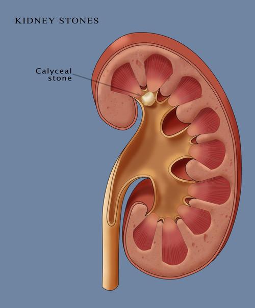
A kidney stone forms when substances in urine crystallise and grow. Calcium-based stones are common; other types include uric acid, infection-related (struvite) and cystine stones. A stone may remain in the kidney or move into the ureter, the tube carrying urine to the bladder. A ureteric stone can obstruct urine flow and cause renal colic.
Symptoms to recognise
- Severe pain in the side or back that may travel towards the lower abdomen or groin, often in waves
- Blood in the urine, which may be visible or detected on a test
- Nausea or vomiting
- Urgency, frequent urination or discomfort passing urine, particularly when a stone is near the bladder
Cloudy urine, fever or chills may indicate infection. These symptoms also have other possible causes, so imaging and tests are important.
Seek urgent care at an emergency department if you have fever or chills with suspected stone pain, cannot pass urine, cannot keep fluids down, have pain that cannot be controlled, or have a single functioning kidney or known kidney disease with possible obstruction. An infected, obstructed kidney is a urological emergency: antibiotics and urgent drainage with a ureteric stent or nephrostomy may be needed before definitive stone treatment.
Why do stones form?
Low fluid intake, heavy sweating, a high-salt diet, previous stones and family history can increase risk. Other contributors include certain medicines and conditions such as gout, bowel disease or recurrent infection. The causes differ by stone type. Finding the type matters when planning prevention.
How are kidney stones diagnosed?
Assessment usually includes your symptoms and medical history, urine testing for blood and infection, and blood tests to check kidney function when appropriate. A low-dose CT scan without contrast is often used for adults with suspected renal colic because it shows the size and position of a stone and signs of obstruction. Ultrasound may be preferred first in pregnancy and some other circumstances. If you pass or have a stone removed, laboratory analysis can guide future prevention.
What are the treatment options?
Observation and pain relief. Many small ureteric stones pass without a procedure. This is reasonable when pain is controlled, there is no infection or concerning obstruction, and kidney function is safe. Your clinician will arrange follow-up to confirm that the stone has passed or the obstruction has resolved. Pain relief is tailored to your health; anti-inflammatory medicines may be useful but are unsuitable for some people, including those with impaired kidney function. Drink normally and avoid dehydration; forcing large amounts of water during acute colic does not push a blocked stone through.
Medication to help passage. An alpha blocker may improve passage for selected stones in the lower ureter, especially those around 5–10 mm. This use is off-label and should be discussed alongside possible adverse effects, such as dizziness. It is not suitable as a substitute for urgent treatment if infection, uncontrolled pain or worsening kidney function develops.
Shock wave lithotripsy (SWL or ESWL). Shock waves delivered from outside the body break a suitable stone into fragments. Its success depends on factors including stone size, position and density; further treatment can sometimes be needed.
Ureteroscopy and laser treatment. A fine telescope passes through the urethra and bladder into the ureter or kidney. The stone can be removed or fragmented with a laser. A temporary ureteric stent is sometimes placed and can cause urinary discomfort until removal.
Percutaneous nephrolithotomy (PCNL). For some large or complex kidney stones, instruments are passed through a small opening in the back to remove the stone. This is more invasive than ureteroscopy or shock wave treatment and has its own bleeding and infection risks.
Your urologist will discuss the expected chance of clearing the stone, anaesthetic needs, recovery, possible stenting and risks of each appropriate option.
Reducing the chance of another stone
For most people, drinking enough fluid to keep urine dilute is a useful starting point, particularly in Queensland heat. If you have heart failure or kidney disease, follow your treating team’s fluid advice instead of increasing intake on your own. Reduce excess salt, maintain a balanced diet and avoid unnecessarily restricting normal dietary calcium. Further dietary advice or preventive medication should be based on the stone’s composition and, for recurrent or higher-risk stones, blood and 24-hour urine testing. Follow-up imaging may be recommended to check for residual or new stones.
When should you see a urologist in Brisbane?
Arrange an assessment if you have a diagnosed stone that has not passed, recurrent stones, persistent pain, blood in the urine, or concerns about kidney function. If you are acutely unwell or have fever with stone symptoms, go to an emergency department rather than waiting for a routine appointment.
This article is general information and does not replace individual medical advice.
References
- European Association of Urology. EAU Guidelines on Urolithiasis (2026).
- National Institute for Health and Care Excellence. Renal and ureteric stones: assessment and management (NG118).
- Healthdirect Australia. Kidney stones: symptoms, diagnosis and treatment.
- Kidney Health Australia. Kidney stones.


Leave a Reply
Want to join the discussion?Feel free to contribute!