
Blood in the Urine After Radiotherapy: Understanding Radiation Cystitis
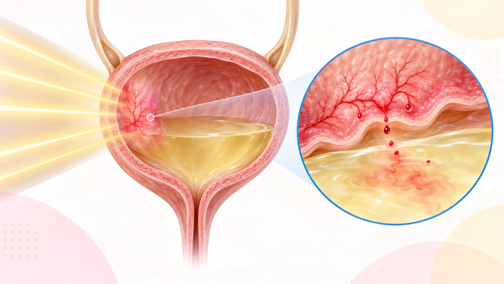
Pelvic radiotherapy is an important and often highly effective treatment for prostate, bladder, rectal and gynaecological cancers. However, radiation can leave the small blood vessels and lining of the bladder fragile. Months or even many years later, this may cause urinary symptoms or bleeding known as radiation cystitis. When bleeding is prominent, the condition is also called radiation-induced haemorrhagic cystitis.
Most episodes can be controlled, but visible blood in the urine must never simply be attributed to previous radiotherapy. Infection, urinary stones, recurrent cancer and a new bladder or upper urinary tract cancer must first be considered.
Seek urgent medical care if you cannot pass urine, are passing large clots, feel faint or short of breath, develop fever or severe pain, or have heavy ongoing bleeding.
What causes radiation cystitis?
Radiotherapy damages cancer cells, but the bladder may receive some radiation because it lies close to the treatment area. Early inflammation can make the bladder lining swollen and irritable. Late injury is different: progressive damage to small blood vessels causes reduced oxygen supply, scarring and fragile abnormal vessels called telangiectasia. These vessels may bleed with little provocation.
Radiation damage can also reduce bladder capacity and elasticity. In severe cases, ulceration, fibrosis, fistula formation or obstruction may occur.
When does it present?
Radiation-related bladder problems have two broad patterns:
- Acute radiation cystitis occurs during radiotherapy or within the first few weeks or months. Frequency, urgency, burning and pelvic discomfort are common; substantial bleeding is less usual. Symptoms often settle after treatment finishes.
- Late radiation cystitis generally begins more than six months after radiotherapy and may appear years or even decades later. The Canadian Urological Association review notes pathological vascular changes from about 6–12 months, with new symptoms reported as long as 20 years after treatment.
The risk varies with radiation dose and field, treatment technique, previous pelvic surgery, smoking, vascular disease, diabetes and combined cancer treatments.
How can it present?
Presentation ranges from microscopic blood found on a urine test to recurrent heavy bleeding with clots. Symptoms may include:
- pink, red or cola-coloured urine;
- small or large blood clots;
- urinary frequency, urgency, burning or bladder pain;
- interrupted flow or complete retention when clots obstruct the outlet;
- tiredness, dizziness or breathlessness from anaemia; and
- a small, painful or poorly compliant bladder in advanced disease.
Bleeding may be intermittent. A clear urine sample between episodes does not exclude radiation cystitis.
Does anticoagulant or antiplatelet therapy matter?
Anticoagulants such as warfarin, apixaban, rivaroxaban or dabigatran, and antiplatelet drugs such as aspirin or clopidogrel, do not create radiation cystitis. They can, however, make bleeding from its fragile vessels more prolonged or severe. Excess anticoagulation, impaired kidney function, drug interactions and combined anticoagulant–antiplatelet therapy can further increase bleeding risk.
These medicines may be preventing a stroke, heart attack, pulmonary embolus or thrombosis of a coronary stent. Do not stop them yourself. During significant bleeding, the urologist, emergency team and the clinician responsible for the medication should jointly balance:
- the severity of bleeding and haemoglobin fall;
- the reason for treatment and the risk of thrombosis if it is interrupted;
- the last dose, kidney function and, for warfarin, the INR;
- whether a temporary hold, reversal or dose adjustment is justified; and
- when and how treatment should safely restart.
Importantly, anticoagulant or antiplatelet use does not remove the need to investigate haematuria. It may reveal bleeding from an otherwise silent bladder or upper-tract tumour.
How is haematuria investigated after radiotherapy?
Assessment is tailored to the patient and severity, but commonly includes:
- History and examination: radiation site, dose and timing; cancer history; smoking; infection symptoms; bleeding pattern; and all medicines.
- Urine testing: urinalysis and culture. Urine cytology may be appropriate in selected patients, but it does not replace cystoscopy.
- Blood tests: full blood count, kidney function and coagulation studies. Severe or ongoing bleeding may require group-and-screen or crossmatch.
- Imaging of the upper urinary tracts: usually CT urography when appropriate; ultrasound or alternative imaging may be chosen when contrast or radiation exposure is unsuitable.
- Cystoscopy: inspection of the urethra and bladder to identify typical diffuse telangiectasia, exclude a tumour, evacuate clots and sometimes cauterise bleeding vessels. Suspicious areas require biopsy, performed carefully because irradiated tissue heals poorly.
A stepwise approach to treatment
Treatment depends on the rate of bleeding, clot retention, anaemia, bladder function, medical fitness and local expertise. No single treatment suits every patient.
1. Stabilisation and bladder drainage
Heavy bleeding may require hospital admission, intravenous fluids, correction of anaemia or clotting abnormalities, and blood transfusion when clinically necessary. A large three-way catheter permits manual clot washout and continuous bladder irrigation with saline. Persistent clots may require cystoscopic evacuation under anaesthesia.
Treat a proven urinary infection, but antibiotics do not treat sterile radiation injury. Medication contributing to bleeding should be reviewed collaboratively rather than stopped automatically.
2. Cystoscopy and endoscopic haemostasis
Cystoscopy can confirm the diagnosis and exclude malignancy. Focal bleeding may be treated with diathermy, laser or another endoscopic coagulation technique. This is often effective initially, although diffuse disease may recur and repeated aggressive cautery can worsen scarring or perforation risk.
3. Intravesical and systemic options
Options used for persistent or recurrent bleeding include:
- Alum bladder irrigation: may control bleeding relatively quickly, but recurrence is possible. It requires caution in substantial kidney impairment because aluminium toxicity can occur.
- Hyaluronic acid, sometimes combined with chondroitin sulphate: aims to restore the bladder’s protective lining. Evidence suggests benefit for haematuria and urinary symptoms, but treatment is gradual and is not suitable for an unstable major bleed.
- Oral sodium pentosan polysulphate: has limited, slower-onset evidence. Long-term exposure also requires discussion of pigmentary maculopathy and eye monitoring.
- Other agents have been reported, but supporting evidence is generally limited.
Formalin can rapidly seal bleeding vessels but may cause severe pain, bladder contraction, reflux, ureteric damage, fistula or systemic complications. It is therefore reserved for life-threatening or otherwise uncontrollable bleeding, used at the lowest effective concentration by experienced teams after the upper tracts have been assessed and protected.
4. Hyperbaric oxygen therapy
Hyperbaric oxygen therapy (HBOT) is one of the best-studied treatments for persistent late radiation cystitis. The patient breathes 100% oxygen in a pressurised chamber. This increases tissue oxygen levels and encourages new blood-vessel growth and healing in chronically oxygen-deprived bladder tissue; it is not simply a short-lived attempt to “oxygenate the blood.”
How effective is it?
The evidence is encouraging, although success definitions and patient populations vary:
- A meta-analysis cited by the Canadian Urological Association included 602 patients with at least one year of follow-up; 84% achieved partial or complete resolution of haematuria.
- The multicentre randomised RICH-ART trial found a clinically meaningful improvement in patient-reported urinary symptoms after HBOT compared with standard care. At five years, 48 of 70 followed patients (68.6%) met the study’s responder definition, and the mean improvement among responders remained substantial. This supports durability for many—but not all—patients.
- HBOT is not guaranteed. Some patients have incomplete improvement, relapse, or still require endoscopic or more invasive treatment. Earlier referral after recurrent bleeding may be preferable to waiting until the bladder is severely fibrotic or the patient has needed repeated transfusions.
How long does it take?
A usual course is 30–40 weekday sessions, sometimes more. Each treatment commonly involves approximately 80–90 minutes breathing oxygen at pressure, although total chamber time is longer. In practical terms, treatment usually takes six to eight weeks. Benefit may develop during the course and continue over subsequent weeks or months as tissue healing progresses.
HBOT is unsuitable or requires specialist assessment in some circumstances. An untreated pneumothorax is an absolute contraindication. Ear or sinus pressure injury, temporary visual change, claustrophobia and, rarely, oxygen-related seizure can occur. Lung disease, certain chemotherapy drugs, implanted devices and difficulty equalising ear pressure require individual review. Availability and daily travel are practical limitations.
5. Arterial embolisation
For ongoing significant bleeding despite less invasive measures, selective or super-selective embolisation can block the bleeding arterial supply. Modern targeted techniques reduce, but do not eliminate, risks such as pelvic pain, tissue ischaemia and non-target embolisation. It can be valuable in frail patients who are poor candidates for major surgery.
When should urinary diversion be considered?
Urinary diversion is a last-resort, potentially life-saving strategy, not an early treatment for uncomplicated bleeding. It should be discussed in a multidisciplinary setting at an experienced centre when there is:
- life-threatening, transfusion-dependent or recurrent clotting haematuria despite endoscopic treatment, HBOT, appropriate intravesical therapy and/or embolisation;
- a severely contracted, painful, non-functional bladder with intolerable frequency or poor storage;
- fistula, necrosis, major outlet or ureteric damage, or progressive upper-tract deterioration;
- repeated admissions and unacceptable loss of quality of life; or
- inability to control bleeding safely by less invasive means.
Options include nephrostomy tubes or ureteric occlusion as temporary or palliative measures; cutaneous ureterostomy; or an ileal conduit. Diversion without removing the bladder may be considered in a very high-risk patient, but the retained irradiated bladder can continue to bleed, become infected or painful, and may later require surgery. Cystectomy with diversion provides definitive removal of the diseased bladder but is a major operation. Previous radiation makes tissue planes, healing and bowel surgery more difficult, so complication and mortality rates are substantially higher than for routine cystectomy.
The decision should incorporate the patient’s cancer status, cardiovascular and respiratory fitness, frailty, kidney and bowel function, previous operations, goals of care and willingness to manage a stoma or external drainage.
The practical message
Radiation cystitis may appear long after the original cancer treatment and can range from mild intermittent haematuria to a medical emergency. Blood-thinning medication may worsen the episode, but it should neither be blamed as the sole cause nor stopped without a coordinated medical plan. A careful evaluation to exclude malignancy and other treatable causes comes first.
Management is progressive: stabilisation and irrigation, cystoscopic treatment, selected bladder therapies, HBOT and embolisation before major diversion surgery. HBOT offers worthwhile, durable improvement for many appropriately selected patients, but requires a substantial weekday treatment commitment. Diversion is reserved for a devastated bladder or bleeding that remains dangerous despite comprehensive treatment.
References
- Goucher G, Saad F, Lukka H, Kapoor A. Canadian Urological Association Best Practice Report: Diagnosis and management of radiation-induced hemorrhagic cystitis. Can Urol Assoc J. 2019;13(2):15–23. doi:10.5489/cuaj.5788
- Oscarsson N, Müller B, Rosén A, et al. Radiation-induced cystitis treated with hyperbaric oxygen therapy (RICH-ART): a randomised, controlled, phase 2–3 trial. Lancet Oncology. 2019;20(11):1602–1614. doi:10.1016/S1470-2045(19)30494-2
- Abramowitz DJ, Warner JN. Clinical management of radiation cystitis: a narrative review. AME Med J. 2021;6:9. doi:10.21037/amj-20-62
- Liem X, Saad F, Delouya G. A practical approach to the management of radiation-induced hemorrhagic cystitis. Drugs. 2015;75:1471–1482. doi:10.1007/s40265-015-0443-5
- Smit SG, Heyns CF. Management of radiation cystitis. Nat Rev Urol. 2010;7:206–214. doi:10.1038/nrurol.2010.23
- AUA/SUFU. Microhematuria Guideline (2020; amended 2025). American Urological Association. AUA guideline
- Oscarsson N, Rosén A, Müller B, et al. Radiation-induced cystitis treated with hyperbaric oxygen therapy (RICH-ART): long-term follow-up of a randomised controlled, phase 2–3 trial. EClinicalMedicine. 2025;83:103214. doi:10.1016/j.eclinm.2025.103214
- Yang TK, Wang YJ, Li HJ, et al. Efficacy and safety of hyperbaric oxygen therapy for radiation-induced hemorrhagic cystitis: a systematic review and meta-analysis. J Clin Med. 2024;13(16):4724. doi:10.3390/jcm13164724
This article provides general information and does not replace individual medical advice. Treatment availability and suitability vary. Visible haematuria, particularly with clots or difficulty passing urine, requires prompt medical assessment.







Leave a Reply
Want to join the discussion?Feel free to contribute!