
Nephroptosis: is a “floating kidney” really a thing?
Can a kidney move enough to cause pain? Yes. Nephroptosis is a recognised anatomical condition, and in selected people it can cause symptoms. However, finding a mobile kidney does not automatically explain abdominal or flank pain. Establishing that connection is the central challenge, and the reason this condition has attracted controversy for more than a century.[1,2]
What is nephroptosis?
The kidneys normally sit towards the back of the abdomen, supported by surrounding fat and connective tissues. Some movement with breathing and changes in posture is normal.
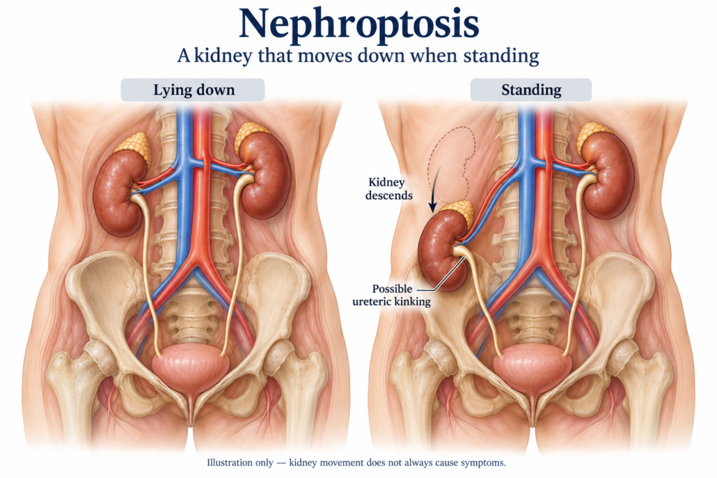
Nephroptosis, also called a floating or mobile kidney, is conventionally defined as downward movement of more than 5 cm, or approximately two vertebral body heights, when a person moves from lying down to standing. A kidney can also rotate or move towards the midline.[3,4]
This differs from a congenital pelvic kidney, which developed in a lower position and stays there.
Published reports describe it more often in slim women and more often on the right, although men and people with other body types can be affected. Reduced support from surrounding fat is a proposed contributor. The true prevalence of symptomatic nephroptosis is uncertain; historical figures from people undergoing particular imaging tests should not be treated as estimates for the general population.[4]
How does it present?
Many mobile kidneys cause no symptoms. When symptoms occur, a suggestive pattern is:
- An ache, dragging sensation or intermittent pain in the flank, the side between the lower ribs and hip, or abdomen.
- Pain that becomes worse with standing, prolonged activity or particular positions.
- Improvement when lying down.
- Occasionally, a feeling of something moving within the abdomen.
Some patients experience more severe episodes with nausea or vomiting. Positional pain is a useful clue, but it does not establish the diagnosis.[2,5]
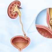
Possible mechanisms include intermittent kinking of the ureter, which drains urine to the bladder; stretching or twisting of the kidney’s blood vessels; or traction on nerves around the kidney. These mechanisms may differ between patients. Severe episodic pain from intermittent upper urinary tract obstruction is sometimes called a Dietl’s crisis, but that term is not specific to nephroptosis.[4,5]
Blood in the urine or urinary infections require investigation in their own right and should not simply be attributed to a mobile kidney.
How is the diagnosis made?
There are two separate questions: Does the kidney move excessively? And is that movement responsible for symptoms or impaired function?
1. Establish the symptom pattern and assess other causes
A consultation should explore where the pain occurs, its relationship to posture and activity, what relieves it, and associated urinary or gastrointestinal symptoms. Examination, urine testing and blood tests for kidney function help guide the investigation.
Other explanations include kidney stones, infection, obstruction at the junction between the kidney and ureter, musculoskeletal pain, and gastrointestinal or gynaecological conditions. Depending on the presentation, these may be more likely. A mobile kidney can coexist with an unrelated cause of pain.[2,3]
2. Compare imaging in different positions
A normal scan performed lying down may not exclude a problem that occurs when upright. If the clinical pattern raises suspicion, the imaging request should make the positional question explicit.[3,5]
| Investigation | What it can contribute | Main limitation |
|---|---|---|
| Ultrasound lying down and upright | Demonstrates movement and checks for changes in collecting-system dilatation | Operator dependent; requires a deliberate positional examination |
| Doppler ultrasound in different positions | May identify posture-related changes in renal blood flow | An adjunct rather than a definitive test of the cause of pain |
| Positional nuclear medicine renal scan | Assesses drainage and each kidney’s contribution to function; may reveal changes when upright | Needs an appropriate protocol and careful interpretation |
| Intravenous urography with supine and erect images | Historically used to measure kidney descent | Used less often today; involves radiation and contrast |
| Selected positional CT urography | Can clarify anatomy, rotation or medial movement | Routine supine CT may miss positional mobility; repeat imaging adds radiation |
The choice should be agreed with radiology or nuclear medicine. Not every centre routinely performs these protocols, and not every patient needs every test.[3,5,6]
3. Link the findings to the symptoms
Diagnostic confidence is stronger when the side and timing of the pain match the demonstrated movement, particularly if there is associated impaired drainage or blood flow. Nevertheless, some symptomatic patients have no demonstrable obstruction. The absence of obstruction does not conclusively exclude symptomatic nephroptosis, but it makes careful exclusion of other causes especially important.[3,4,6]
Normal overall kidney function does not, by itself, exclude an intermittent positional problem. Equally, descent beyond a measurement threshold is not sufficient justification for surgery.[2,5]
What are the treatment options?
Observation and symptom management
An incidentally discovered mobile kidney without symptoms or functional consequences generally does not require surgery.[5]
For mild symptoms, an individual plan may include modifying provoking activities, appropriate pain relief and reviewing other possible contributors. Abdominal supports, exercises and nutritional measures have been used historically, but convincing evidence that they reliably correct symptomatic nephroptosis is limited. They should not be presented as proven cures. Weight gain is not a universal recommendation.[1,2]
Nephropexy: fixing the kidney in position
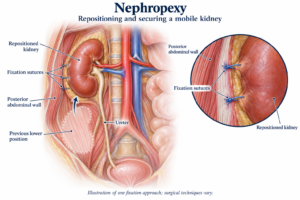
Nephropexy repositions the kidney and secures it to surrounding tissues to limit excessive movement. It aims to preserve the kidney and relieve symptoms or positional impairment.
It may be considered when symptoms substantially affect daily life, nephroptosis has been demonstrated with appropriate imaging, other explanations have been reasonably excluded, and the evidence suggests a meaningful relationship between movement and symptoms. Documented positional obstruction or impaired perfusion strengthens the rationale.[2,3,5]
Modern surgery is usually laparoscopic, through the abdomen or behind it. Robotic assistance has also been described, but there is no convincing comparative evidence that it produces better outcomes than conventional laparoscopy for this condition. Fixation techniques vary, and some published methods use mesh while others use sutures.[3]
Open surgery is less commonly used. Percutaneous techniques, intended to stabilise the kidney through a tract and subsequent scarring, have also been reported. A recent report involved only four patients: such evidence is too limited to establish equivalence with laparoscopic surgery or support routine use.[7]
How effective is surgery, and what are the risks?
Small surgical series describe substantial improvement in selected patients. For example, a long-term study reported 13 patients assessed at a median of 5.9 years: 11 were completely satisfied and two moderately satisfied.[8]
That is encouraging, but it is not a reliable prediction for every patient. Small numbers, selection of patients, loss to follow-up and the absence of a non-operative comparison group limit what can be concluded. Fixing the kidney successfully does not guarantee that pain will disappear.[2,8]
Potential risks include bleeding, infection, injury to nearby organs, vessels or the urinary tract, anaesthetic complications, persistent or new pain, and recurrent kidney movement. If mesh is proposed, its specific benefits and risks should be discussed. Individual risks and expected recovery depend on the technique and the patient’s circumstances.
Why is nephroptosis controversial?
The controversy concerns which patients have symptoms caused by kidney mobility, and which benefit from treatment.
Historically, nephroptosis was blamed for a wide range of nonspecific symptoms, and nephropexy was performed much more frequently than it is today. Recognition that many mobile kidneys were harmless, together with inconsistent surgical results, led to scepticism and a marked decline in treatment.[1]
Modern positional imaging and minimally invasive surgery have renewed interest. However, the evidence remains dominated by small observational series and case reports rather than robust randomised comparisons. There is also no universally accepted combination of symptoms and test findings that predicts success for an individual.[2,6,8]
The appropriate response is to take the symptoms seriously while investigating the cause carefully. A normal routine scan does not mean pain is imagined. A mobile kidney does not mean an operation will solve it.
When should you seek urgent care?
Severe or escalating flank pain, fever or rigors, persistent vomiting, visible blood in the urine, or a marked reduction in urine output warrant prompt medical assessment. These symptoms can signal infection, obstruction or another problem requiring urgent treatment, regardless of whether nephroptosis is present.
This article provides general education. Diagnosis and treatment require an individual clinical assessment.
Patient information by Dr Jo Schoeman | Evidence reviewed October 2026
References
- Moss SW. Floating kidneys: a century of nephroptosis and nephropexy. Journal of Urology. 1997;158(3 Pt 1):699–702. doi:10.1016/S0022-5347(01)64296-4.
- Barber NJ, Thompson PM. Nephroptosis and nephropexy—hung up on the past? European Urology. 2004;46(4):428–433. doi:10.1016/j.eururo.2004.03.023.
- Mogorovich A, Selli C, De Maria M, et al. Clinical reappraisal and state of the art of nephropexy. Urologia. 2018;85(4). doi:10.1177/0391560317749191.
- Senaratne R, Anderson S, Kelly C, et al. Rolling in the deep: an illustrative report of symptomatic nephroptosis. Radiology Case Reports. 2024;19(9):4044–4048. doi:10.1016/j.radcr.2024.06.047. Case report.
- Murari SB, Gadepalli T, Rao VP, Ram R. Renal scintigraphy in diagnosis and management of nephroptosis. Indian Journal of Nuclear Medicine. 2012;27(1):52–54. doi:10.4103/0972-3919.108874. Case report.
- Strohmeyer DM, Peschel R, Effert P, et al. Changes of renal blood flow in nephroptosis: assessment by color Doppler imaging, isotope renography and correlation with clinical outcome after laparoscopic nephropexy. European Urology. 2004;45:790–793. PubMed.
- Liu H, Gao W, Zhang Z, et al. Modified percutaneous nephropexy for nephroptosis: technical description and short-term results. Current Urology. 2025;19(3):168–172. doi:10.1097/CU9.0000000000000238. Small technical series; four patients.
- Plas E, Daha K, Riedl CR, Hübner WA, Pflüger H. Long-term followup after laparoscopic nephropexy for symptomatic nephroptosis. Journal of Urology. 2001;166(2):449–452. PubMed.




Leave a Reply
Want to join the discussion?Feel free to contribute!