
Conservative Management of a Distal Ureteric Stone: When Can You Wait and When Is Surgery Needed?
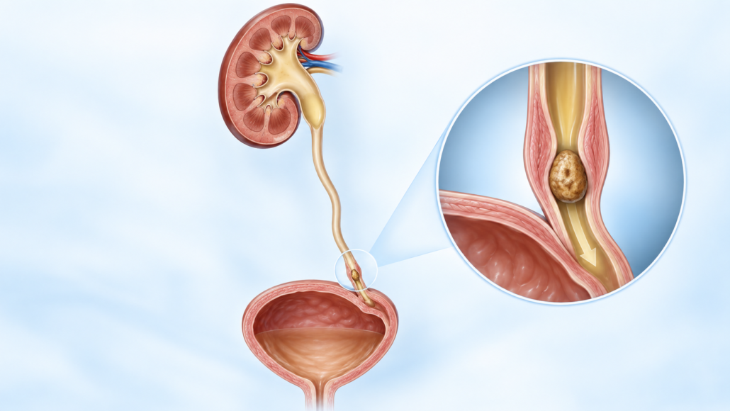
A distal ureteric calculus is a stone located in the lower part of the ureter—the tube carrying urine from the kidney to the bladder. These stones are often close to the ureterovesical junction, where the ureter enters the bladder.
As the stone moves towards the bladder, it can cause severe intermittent pain known as renal colic. Pain commonly begins in the side or back and travels into the lower abdomen, groin, testicle or labia. Blood in the urine, nausea, vomiting, urinary urgency and frequency may also occur.
Many small distal ureteric stones pass naturally. However, conservative management is only safe when pain is controlled, infection is absent, kidney function is satisfactory and appropriate follow-up is arranged.
When is a ureteric stone an emergency?
A stone obstructing an infected kidney is a urological emergency. Antibiotics alone may be insufficient because infected urine cannot drain normally.
Seek urgent medical assessment if stone symptoms are accompanied by:
- Fever or shaking chills
- Feeling severely unwell, weak, confused or drowsy
- Persistent vomiting or inability to drink
- Pain that remains severe despite medication
- Passing very little or no urine
- Known poor kidney function
- A solitary functioning kidney
- Pregnancy with significant pain or fever
The obstructed kidney may need urgent drainage with a ureteric stent or nephrostomy tube. Definitive stone treatment is usually delayed until the infection has been controlled. The European Association of Urology recommends immediate antibiotics and urgent drainage for sepsis associated with an obstructing stone. EAU Guidelines on Urolithiasis
Australian Healthdirect similarly advises patients with renal colic and fever to attend a doctor or hospital emergency department promptly. Healthdirect Australia—Kidney stones
What determines whether a stone will pass naturally?
The likelihood of spontaneous passage depends mainly on:
- Stone size
- Position within the ureter
- Degree of obstruction
- Ureteric anatomy
- Whether the stone is impacted
- Previous stone history
- Associated swelling and inflammation
- The duration for which the stone has remained in the same position
Smaller stones located close to the bladder have the greatest likelihood of passing.
The EAU reports that distal ureteric stones collectively have a spontaneous passage rate of approximately 68–83%. A distal ureteric stone smaller than 5 mm has an estimated passage rate of approximately 89%. The likelihood decreases as stone size increases.
These percentages describe groups of patients and cannot predict exactly what will happen to an individual stone. A 4 mm stone may occasionally become impacted, while a larger stone may sometimes pass without surgery.
The average reported passage time is approximately 17 days, although passage can occur sooner or take several weeks. EAU Guidelines on Urolithiasis
Who may be suitable for conservative management?
Conservative management, also called observation, expectant management or a trial of passage, may be appropriate when:
- The stone is small enough to have a reasonable chance of passing
- The stone is in the distal ureter
- Pain can be controlled with oral medication
- There is no fever or evidence of urinary infection
- Kidney function is stable
- The patient can drink and keep medication down
- There is no complete obstruction threatening kidney function
- The patient is passing urine normally
- There is no high-risk situation involving a solitary kidney or bilateral obstruction
- The patient understands the warning symptoms
- Follow-up and repeat imaging can be arranged
- The patient is comfortable waiting for the stone to pass
Stones of 5 mm or less are particularly suitable for observation when no complications are present.
Selected distal stones between 5 and 10 mm may also be managed conservatively. These stones are less likely to pass than smaller stones, but some patients may avoid surgery with careful observation and medical expulsive therapy.
Stones larger than 10 mm are substantially less likely to pass naturally and are more commonly treated with an intervention.
What assessment is required?
Before recommending conservative management, the diagnosis and severity of obstruction should be established.
Assessment may include:
- A detailed history and physical examination
- Urine dipstick testing
- Urine culture when infection is suspected
- Kidney-function blood tests
- Full blood count and inflammatory markers
- Non-contrast CT scan of the kidneys, ureters and bladder
- Ultrasound in selected patients
- A plain abdominal X-ray when the stone is visible on X-ray
- Pregnancy testing when clinically relevant
A low-dose non-contrast CT scan is often the most accurate test for determining the stone’s size and location. It can also assess hydronephrosis, the swelling of the kidney caused by obstruction and identify alternative causes of pain.
Ultrasound is particularly useful when radiation should be avoided, including during pregnancy, although it may not show every ureteric stone.
What does conservative management involve?
Pain relief
Non-steroidal anti-inflammatory medicines, NSAIDs, are often the most effective initial treatment for renal colic when they are medically safe.
Depending on the patient, treatment may include:
- Ibuprofen
- Diclofenac
- Paracetamol
- A prescribed opioid for breakthrough pain
- Medication for nausea or vomiting
NSAIDs may be unsuitable for people with:
- Reduced kidney function
- Previous stomach ulcers or gastrointestinal bleeding
- Certain cardiovascular conditions
- Anticoagulant treatment
- NSAID allergy
- Some stages of pregnancy
Pain medication should be selected according to the patient’s medical history rather than taken indiscriminately.
Hydration
Patients should generally remain normally hydrated and avoid becoming dehydrated.
Drinking excessive volumes of water during an episode of acute obstruction does not necessarily force the stone through and may increase discomfort. The aim is steady, sensible fluid intake unless a doctor has advised otherwise.
Straining the urine
Passing urine through a stone strainer can help recover the calculus. A captured stone can be sent for laboratory analysis, which may guide future prevention.
Pain disappearing does not always prove that the stone has passed. Occasionally a stone stops causing pain while obstruction remains, so follow-up imaging may still be required.
Activity
Normal gentle activity is usually reasonable if the patient feels well. Some patients find walking helpful, although exercise cannot guarantee stone passage.
Driving, remote travel and hazardous work may be unsafe while unpredictable severe pain or medication-related drowsiness remains possible.
Medical expulsive therapy
Medical expulsive therapy—usually abbreviated to MET, uses medication to help a ureteric stone pass.
The most frequently used medicines are alpha-blockers, including:
- Tamsulosin
- Silodosin
- Alfuzosin
Tamsulosin is commonly selected because it is widely available and familiar to urologists.
Alpha-blockers are primarily used to improve urinary symptoms caused by prostate enlargement. Their use for ureteric stone passage is generally off-label, meaning that stone passage is not necessarily the indication listed in the medicine’s formal registration.
Off-label use does not mean that treatment is experimental or prohibited. It means that the potential benefits, limitations and side effects should be discussed before prescribing it.
How do alpha-blockers affect the distal ureter?
The ureter contains smooth muscle and alpha-adrenergic receptors. These receptors are particularly relevant in the distal ureter.
When a stone enters the ureter, the surrounding muscle can contract and spasm. Swelling may develop around the stone, increasing resistance to its movement.
Alpha-blockers may:
- Relax distal ureteric smooth muscle
- Reduce ureteric spasm
- Reduce pressure below and around the stone
- Increase the ureter’s ability to accommodate the calculus
- Improve the likelihood of stone passage
- Shorten passage time in some patients
- Reduce episodes of renal colic and analgesic requirements in selected cases
An alpha-blocker does not:
- Dissolve most stones
- Make the stone physically smaller
- Treat a urinary infection
- Remove a completely impacted stone
- Protect a kidney from prolonged significant obstruction
- Replace drainage of an infected obstructed kidney
How effective are alpha-blockers?
Research into alpha-blockers has produced mixed results.
When all ureteric stones are grouped together, some large studies have shown little or no overall benefit. This may be because very small stones frequently pass without medication and very large or impacted stones remain unlikely to pass despite treatment.
The clearest benefit appears to be in patients with distal ureteric stones between approximately 5 and 10 mm.
The EAU recommends offering an alpha-blocker as one treatment option for conservatively managed distal ureteric stones in this size range, while explaining that treatment is off-label. EAU Guidelines on Urolithiasis
For a stone smaller than 5 mm, the natural passage rate is already high, so an alpha-blocker may add relatively little benefit. Treatment decisions should therefore be individualised.
Side effects and precautions with alpha-blockers
Possible side effects include:
- Dizziness
- Light-headedness on standing
- Low blood pressure
- Weakness or fatigue
- Headache
- Nasal congestion
- Palpitations
- Reduced semen volume
- Failure of ejaculation or retrograde ejaculation
Extra caution is required in patients who:
- Already have low blood pressure
- Have a history of fainting or falls
- Take several blood-pressure medicines
- Use medication for erectile dysfunction
- Have significant cardiovascular disease
- Are planning cataract or glaucoma surgery
Tamsulosin has been associated with intraoperative floppy iris syndrome during cataract surgery. Patients should tell their ophthalmologist about current or previous use.
Alpha-blocker treatment should be stopped and medical advice obtained if infection, uncontrollable pain or deterioration in kidney function develops.
How long can conservative management continue?
There is no single safe waiting period for every patient. The decision depends on symptoms, obstruction, kidney function, stone movement and the likelihood of passage.
Many stones that pass naturally do so within two to four weeks. A monitored trial of passage may sometimes continue for up to approximately four to six weeks, provided that:
- Pain remains manageable
- Infection does not develop
- Kidney function remains stable
- Obstruction is not causing progressive harm
- Follow-up imaging is performed
- The patient still prefers conservative treatment
Observation should not become open-ended. A painless obstructing stone can still impair kidney function.
Follow-up may involve an X-ray, ultrasound or low-dose CT scan. The most suitable test depends on whether the stone was visible on the original X-ray and whether ongoing obstruction needs to be assessed.
When has conservative management failed?
Conservative management should be reconsidered when:
- The stone has not passed within an appropriate observation period
- Repeat imaging shows that the stone has not moved
- Pain remains severe despite adequate medication
- Repeated emergency presentations are required
- Nausea or vomiting prevents oral fluids or medication
- Fever or urinary infection develops
- Kidney function deteriorates
- Hydronephrosis persists or worsens
- Urine output falls significantly
- The stone has a low likelihood of spontaneous passage
- The patient has a solitary kidney or bilateral obstruction
- The patient cannot safely continue waiting
- Work, caring responsibilities or travel make unpredictable colic unacceptable
- The patient prefers definitive removal
The EAU identifies persistent pain, persistent obstruction, impaired kidney function and a low likelihood of spontaneous passage as indications for active stone removal. EAU Guidelines on Urolithiasis
Failure of conservative management is not a personal failure. It usually means that the stone is too large, impacted, anatomically trapped or producing complications that make further waiting unsafe.
Surgical and procedural treatment
The two main definitive treatments for a distal ureteric calculus are:
- Ureteroscopy with laser lithotripsy or stone extraction
- Shock wave lithotripsy
The most appropriate option depends on stone size, density and position, as well as the patient’s anatomy, medical health, preferences and treatment availability.
Ureteroscopy and laser lithotripsy
Ureteroscopy is commonly used for distal ureteric stones and generally provides the best chance of becoming stone-free after one procedure.
Under anaesthesia, a small telescope is passed through:
- The urethra
- The bladder
- The opening of the ureter
- The ureter to the level of the stone
The stone may be removed intact with a small basket or fragmented using a holmium or thulium laser. The pieces are then extracted or allowed to pass naturally.
No external incision is usually required.
Is a ureteric stent required?
A temporary ureteric stent may be inserted when:
- The ureter is swollen
- Access was difficult
- Infection is a concern
- Stone fragments remain
- There was ureteric trauma
- Reliable drainage is required
- Kidney function is vulnerable
A stent extends from the kidney to the bladder. It can cause:
- Urinary frequency and urgency
- Bladder discomfort
- Pain in the kidney during urination
- Blood in the urine
- Discomfort during activity
- A sensation of incomplete emptying
Stents must be removed or exchanged at the planned time. A forgotten stent can become encrusted and cause serious complications.
Risks of ureteroscopy
Possible complications include:
- Urinary infection or sepsis
- Bleeding
- Failure to reach or remove the stone
- Residual fragments
- Ureteric perforation
- Ureteric narrowing or stricture
- Need for a further procedure
- Anaesthetic complications
Major ureteric injury is uncommon but can require additional surgery.
Compared with shock wave treatment, ureteroscopy generally offers a higher chance of clearing the stone in one procedure, although it is more invasive and has a higher complication rate. EAU Guidelines on Urolithiasis
Shock wave lithotripsy
Shock wave lithotripsy, SWL or ESWL, uses externally generated shock waves focused onto the stone. These waves fragment the calculus into smaller pieces that can pass down the ureter.
Advantages may include:
- No telescope passing up the ureter
- No surgical incision
- Lower procedural morbidity
- Day-treatment suitability in many patients
Limitations include:
- Lower single-treatment stone-free rates than ureteroscopy
- Possible need for repeat treatment
- Pain while fragments pass
- Residual fragments
- Difficulty targeting some distal stones
- Reduced success with dense or impacted stones
- Reduced effectiveness with greater skin-to-stone distance
- Possible need for subsequent ureteroscopy
SWL may not be suitable during pregnancy, with untreated infection, uncorrected bleeding disorders, an aneurysm near the treatment field or an anatomical obstruction below the stone.
The American Urological Association recognises both ureteroscopy and shock wave lithotripsy as options when active treatment is required for a distal ureteric stone. AUA Surgical Management of Kidney and Ureteral Stones Guideline
Emergency drainage with a ureteric stent
A ureteric stent may be inserted urgently to bypass an obstructing stone and allow urine to drain from the kidney.
This is particularly important when there is:
- Infection or sepsis
- Deteriorating kidney function
- A solitary obstructed kidney
- Bilateral obstruction
- Uncontrollable pain
- Severe obstruction where immediate stone removal is unsuitable
Emergency stenting relieves the obstruction but does not always remove the stone. Definitive ureteroscopy or SWL may be scheduled later.
Percutaneous nephrostomy
A nephrostomy tube is inserted through the skin of the back directly into the kidney under imaging guidance.
It may be used when:
- Rapid drainage of an infected kidney is required
- A ureteric stent cannot be inserted
- The patient is too unwell for a longer procedure
- Ureteric anatomy prevents retrograde access
Both ureteric stenting and nephrostomy provide effective emergency drainage. The choice depends on clinical circumstances, local expertise and the patient’s condition.
Open, laparoscopic or robotic stone removal
Open or keyhole ureterolithotomy is now rarely required for an isolated distal ureteric stone.
It may occasionally be considered for:
- A very large impacted stone
- Unusual urinary anatomy
- Failure of less invasive treatments
- A stone requiring treatment during another planned reconstruction
Most distal stones can be treated successfully with ureteroscopy or SWL.
Which treatment is best?
There is no single procedure that is best for every patient.
Ureteroscopy may be preferred when:
- Rapid and reliable stone clearance is important
- The stone is impacted
- The stone is dense or unlikely to fragment with SWL
- Previous SWL has failed
- The patient has significant obesity
- The stone is difficult to target externally
- The patient wishes to minimise the likelihood of repeat treatment
SWL may be preferred when:
- The stone can be targeted clearly
- The stone has favourable size and density
- The patient wishes to avoid ureteroscopy
- Anaesthetic considerations favour a less invasive approach
- The patient accepts the possibility of repeat treatment
The decision should include a discussion of success rates, anaesthesia, stent requirements, recovery, complications and the possible need for another procedure.
Preventing another stone
Once the acute episode has resolved, prevention becomes important.
General measures may include:
- Drinking enough fluid to produce at least 2–2.5 litres of urine daily, unless medically restricted
- Reducing excessive salt intake
- Maintaining normal dietary calcium rather than eliminating calcium
- Moderating excessive animal-protein intake
- Maintaining a healthy body weight
- Avoiding recurrent dehydration
- Capturing the stone for analysis
- Completing blood and urine testing when indicated
Patients with recurrent stones, a solitary kidney, childhood stone disease, unusual stone types or a strong family history may require a formal metabolic evaluation, including 24-hour urine testing.
Prevention should be tailored to the stone composition and the patient’s metabolic risk factors.
The bottom line
Many small distal ureteric stones can be managed safely without surgery. Stones smaller than 5 mm near the bladder have the greatest likelihood of passing naturally.
Conservative treatment usually involves appropriate pain relief, sensible hydration, urine straining, follow-up imaging and—in selected patients—an alpha-blocker such as tamsulosin.
Alpha-blockers relax the smooth muscle of the distal ureter. Their greatest likely benefit is for distal stones between approximately 5 and 10 mm. They do not dissolve the stone and should not delay treatment when infection, uncontrolled pain, persistent obstruction or declining kidney function develops.
Ureteroscopy with laser treatment provides the most reliable single-procedure clearance for many distal stones. Shock wave lithotripsy is less invasive but may require repeat treatment. An infected obstructed kidney requires urgent drainage rather than continued observation.
This article provides general information and does not replace individual medical assessment. Anyone with renal colic and fever, inability to pass urine, persistent vomiting or uncontrollable pain should seek urgent medical care.
Need help deciding whether your ureteric stone can safely pass?
If you have been diagnosed with a distal ureteric calculus, appropriate management depends on more than its size alone. Stone position, pain, infection, kidney function and the degree of obstruction must all be considered.
Your local Brisbane urologist, Dr Jo Schoeman, can assess whether observation, medical expulsive therapy, ureteroscopy or shock wave treatment is the most appropriate option for you.



Leave a Reply
Want to join the discussion?Feel free to contribute!