
Calcium Oxalate Kidney Stones: Treatment, Surgery and Prevention
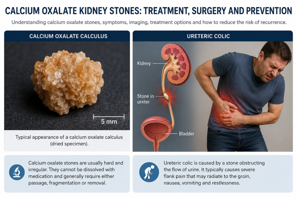
Calcium oxalate stones are the most common type of urinary tract stone. Although the name may suggest that eating too much calcium is the problem, the biology is considerably more complicated. Stone formation reflects the interaction between urine volume, calcium, oxalate, citrate, sodium, dietary factors, genetics and, in some patients, underlying metabolic or gastrointestinal conditions.
The encouraging news is that most calcium oxalate stones can be treated effectively, and the risk of forming further stones can often be substantially reduced with appropriate investigation and prevention.
This guide explains conservative management, imaging, surgical treatment, ureteric pre-stenting, laser fragmentation, recovery and long-term prevention.
What is a calcium oxalate stone?
Calcium oxalate crystals form when urine becomes sufficiently concentrated with calcium and oxalate for crystals to develop and grow.
Two principal forms occur:
- Calcium oxalate monohydrate, which tends to be harder and more resistant to shock-wave fragmentation.
- Calcium oxalate dihydrate, which is generally more readily fragmented.
Importantly, calcium oxalate stones cannot usually be dissolved with medication. This differs from uric acid stones, which can sometimes be dissolved by alkalinising the urine.
Treatment therefore involves either allowing a suitable stone to pass naturally, monitoring an asymptomatic stone, or physically removing or fragmenting it when intervention is required.
How do calcium oxalate stones present?
A stone sitting quietly inside the kidney may produce no symptoms at all and may be discovered incidentally during imaging performed for another reason.
When a stone moves into the ureter, symptoms can be dramatic and may include:
- Severe loin or flank pain
- Pain radiating towards the groin or testicle/labia
- Nausea and vomiting
- Blood in the urine
- Urinary frequency or urgency
- Burning with urination
- Restlessness during an episode of renal colic
Fever or chills associated with an obstructing stone require urgent medical assessment. An infected obstructed kidney is a urological emergency and may require immediate drainage with a ureteric stent or nephrostomy rather than definitive stone treatment at that time.
Imaging calcium oxalate stones
CT scan
A non-contrast CT of the kidneys, ureters and bladder (CT KUB) is generally the most accurate investigation for suspected urinary tract calculi.
CT provides valuable information about:
- Stone size
- Exact location
- Number of stones
- Degree of urinary obstruction
- Hydronephrosis
- Stone density
- Anatomy relevant to treatment planning
Stone density is measured in Hounsfield units (HU) and can sometimes help predict how readily a stone may fragment with shock-wave treatment.
The major disadvantage of CT is radiation exposure, although modern low-dose CT protocols can substantially reduce the radiation dose.
Ultrasound
Ultrasound avoids radiation and is particularly useful for:
- Follow-up
- Monitoring known renal stones
- Detecting hydronephrosis
- Pregnancy
- Patients requiring repeated imaging
Its disadvantage is that small ureteric stones can be missed, and measurement of stone size is generally less accurate than CT.
Plain X-ray – KUB
Most calcium oxalate stones are radiopaque and therefore visible on a plain abdominal X-ray.
KUB imaging can consequently be useful for monitoring selected stones, particularly following treatment or when considering shock-wave lithotripsy.
Does every calcium oxalate stone require surgery?
No.
A small stone without infection, significant obstruction or uncontrolled pain can frequently be managed conservatively.
Small asymptomatic stones within the kidney may also simply be monitored.
Observation is reasonable when the potential risks and inconvenience of treatment exceed the likely benefit.
Current European guidelines recognise active surveillance as an option for selected asymptomatic renal stones, although stone growth, development of symptoms or obstruction may eventually prompt intervention.
Conservative management
For a small ureteric stone that is likely to pass spontaneously, treatment may include:
Hydration
Maintain normal good hydration. Trying to “flush” a painful obstructing stone through by drinking enormous quantities of water is generally unnecessary and can actually worsen discomfort.
Pain relief
Anti-inflammatory medication is frequently particularly effective for renal colic, provided there are no medical contraindications.
Additional analgesia or anti-nausea medication may occasionally be required.
Medical expulsive therapy
An alpha-blocker such as tamsulosin may be prescribed in selected patients to facilitate passage of a ureteric stone.
The greatest benefit appears to occur with distal ureteric stones larger than approximately 5 mm, although its use for stone passage is off-label in many jurisdictions.
Straining the urine
Catching the stone is surprisingly useful. Sending a retrieved stone for laboratory analysis confirms its composition and helps direct prevention.
The small pebble that caused an astonishing amount of trouble can therefore become an important diagnostic specimen.
When should a stone be removed?
Intervention may be recommended when there is:
- Persistent or recurrent pain
- Failure of the stone to progress
- Significant urinary obstruction
- Deterioration in renal function
- Recurrent urinary infection
- Increasing stone size
- A low likelihood of spontaneous passage
- A large renal stone burden
- Occupational or lifestyle reasons where an unpredictable episode of renal colic would be problematic
Treatment is individualised according to stone size, location, density, renal anatomy, symptoms and patient preference.
Surgical treatment options
1. Extracorporeal Shock-Wave Lithotripsy – ESWL/SWL
Shock-wave lithotripsy focuses externally generated shock waves onto the stone, breaking it into smaller fragments that can subsequently pass through the urinary tract.
Advantages
- No incision
- Usually relatively rapid recovery
- May avoid ureteroscopy
- Useful for appropriately selected renal and ureteric stones
Disadvantages
- Not all calcium oxalate stones fragment equally well
- Hard calcium oxalate monohydrate stones may be resistant
- Several treatment sessions may be required
- Fragments still need to pass down the ureter
- Residual fragments can remain
- Temporary renal colic can occur
- Less effective for some lower-pole renal stones and larger stones
The success of SWL is strongly influenced by stone size, location, composition, patient anatomy and stone hardness.
2. Ureteroscopy and laser lithotripsy
Ureteroscopy has transformed modern stone treatment.
A fine telescope is passed through the natural urinary tract:
urethra → bladder → ureter → kidney
There is therefore no external surgical incision.
Rigid or semi-rigid ureteroscopy can be used for ureteric stones, while flexible ureteroscopy allows access to stones within the kidney.
Laser fragmentation
Once the stone is identified, laser energy is delivered through an extremely fine fibre passed through the ureteroscope.
Modern systems include:
- Holmium laser
- Thulium fibre laser (TFL)
Both are highly effective technologies for flexible ureteroscopic stone treatment, and contemporary European guidelines recommend Ho or TFL for ureteroscopic laser lithotripsy.
The stone can be treated using several strategies.
Fragmentation and extraction
The stone is divided into several pieces, which are removed using a small basket.
Dusting
The laser progressively converts the stone into extremely small particles or “dust”, allowing much of the material to pass spontaneously.
Pop-dusting or further fragmentation
Larger fragments can be further reduced until they are sufficiently small to pass or be extracted.
Advantages of ureteroscopy and laser treatment
Ureteroscopy provides:
- Direct visualisation of the stone
- High stone-clearance rates
- Treatment of stones resistant to shock-wave lithotripsy
- Access to most areas of the ureter and kidney
- Immediate fragmentation
- Ability to retrieve fragments for stone analysis
- No external incision
- Usually short hospitalisation
Compared with shock-wave lithotripsy, ureteroscopy generally offers a greater likelihood of becoming stone-free after a single procedure, although this comes at the price of greater invasiveness and a somewhat higher complication rate.
Possible complications of ureteroscopy
Although generally safe, complications can include:
- Blood in the urine
- Urinary infection
- Pain
- Ureteric swelling
- Temporary difficulty passing urine
- Residual stone fragments
- Need for repeat ureteroscopy
- Ureteric perforation
- Ureteric stricture
- Sepsis
Major ureteric injury is uncommon.
The EAU reports overall ureteroscopy complication rates of approximately 4–25%, with most complications being minor. Urosepsis is an important but uncommon serious complication.
Pre-operative urine testing and treatment of urinary infection are therefore important components of safe stone surgery.
What is pre-stenting?
A ureteric stent is a thin flexible tube extending from the kidney to the bladder.
Sometimes the ureter is too narrow to safely introduce the instruments required for flexible ureteroscopy.
Instead of forcing access, a temporary stent can be inserted.
The stent gently allows passive dilatation of the ureter before definitive surgery.
Ureteroscopy is then performed at a later date, commonly after the ureter has had time to accommodate the stent.
Does everyone need pre-stenting?
No.
Routine pre-stenting before ureteroscopy is not necessary.
However, pre-stenting can be useful when:
- The ureter is particularly narrow
- Previous access has been unsuccessful
- A large renal stone burden is anticipated
- An access sheath is likely to be required
- Staged ureteroscopy is planned
- Emergency drainage was previously required because of infection or obstruction
Evidence suggests that pre-stenting can improve access and may improve stone-free outcomes for renal stones, although the benefit is less clear for ureteric stones.
The price of this convenience is that the patient has to live temporarily with a ureteric stent.
Ureteric stent side effects
Stents are useful pieces of equipment, but they rarely win popularity contests.
Possible symptoms include:
- Urinary frequency
- Urgency
- Bladder discomfort
- Burning during urination
- Blood in the urine
- Flank discomfort during urination
- Pelvic or groin discomfort
Symptoms disappear after the stent is removed.
Alpha-blocker medication can reduce stent-related symptoms in selected patients.
Is a stent required after laser treatment?
Not always.
After straightforward uncomplicated ureteroscopy with complete stone clearance and no ureteric trauma, a postoperative stent may not be necessary.
A stent is more likely to be placed following:
- Difficult ureteric access
- Significant ureteric swelling
- Ureteric trauma
- Large stone burden
- Residual fragments
- Bleeding
- Infection concerns
- Prolonged surgery
- Staged treatment
Current evidence supports avoiding routine postoperative stenting following uncomplicated ureteroscopy.
3. Percutaneous nephrolithotomy – PCNL
Very large renal stones are usually better approached directly through the back rather than attempting to remove the entire stone burden through the ureter.
PCNL involves creating a small tract through the skin into the kidney, through which instruments can fragment and extract the stone.
PCNL remains the standard treatment for large renal calculi, particularly stones greater than approximately 2 cm and complex or staghorn stone burdens.
Advantages
- Excellent clearance of large stone burdens
- Large fragments can be removed directly
- Often more efficient than multiple ureteroscopies for large stones
Disadvantages
- More invasive
- Bleeding risk
- Infection/sepsis risk
- Longer recovery than routine ureteroscopy
- Potential need for nephrostomy drainage
- Rare injury to surrounding structures
Recovery after ureteroscopy and laser lithotripsy
Most patients recover relatively quickly.
It is common to experience:
- Mild burning when passing urine
- Pink or blood-stained urine
- Urinary frequency
- Mild flank discomfort
- Stent-related symptoms
Many patients return to light activities within several days, although recovery depends on the extent of the procedure and whether a stent remains.
Heavy physical activity may need to be avoided for a short period.
Patients should seek medical attention for:
- Fever or chills
- Increasing severe pain
- Persistent vomiting
- Inability to pass urine
- Heavy persistent bleeding
- Feeling systemically unwell
Have we finished once the stone has gone?
Not quite.
Removing a stone treats today’s stone.
Preventing another one requires identifying why it formed.
This distinction is important because calcium oxalate stone disease can recur.
Current EAU data suggest approximately 26% of first-time stone formers experience recurrence within five years, while a smaller group develops highly recurrent disease.
Patients with recurrent stones, multiple stones, bilateral stones, young-onset stone disease or particular metabolic risk factors deserve more detailed investigation.
Metabolic investigation
Depending on the clinical situation, evaluation may include:
Blood tests
- Calcium
- Creatinine and renal function
- Electrolytes
- Uric acid
- Bicarbonate
- Parathyroid hormone when indicated
Stone analysis
Any retrieved stone should ideally be sent for formal analysis.
24-hour urine collection
This can measure:
- Total urine volume
- Calcium
- Oxalate
- Citrate
- Sodium
- Uric acid
- Urinary pH
- Other relevant metabolic parameters
The results allow prevention to be targeted rather than relying on a generic “kidney stone diet”.
Preventing calcium oxalate stones
1. Drink more fluid
For many patients, the single most important intervention is increasing urine volume.
Rather than concentrating stone-forming chemicals into a small volume of urine, additional fluid keeps them diluted.
Water should generally form the majority of fluid intake.
The required intake varies with climate, exercise, occupation and perspiration. Someone working outdoors during an Australian summer may require considerably more fluid than someone sitting in an air-conditioned office.
2. Do not automatically restrict calcium
This is one of the most common misconceptions about calcium oxalate stones.
Calcium in the stone does not mean calcium should disappear from the diet.
Normal dietary calcium is generally desirable because calcium within the intestine binds dietary oxalate. This reduces oxalate absorption and consequently reduces the amount reaching the urine.
The EAU specifically advises that dietary calcium should generally not be restricted unless there is a particular reason to do so.
3. Reduce excessive salt intake
A high sodium intake increases urinary calcium excretion.
Reducing dietary salt can therefore help reduce urinary calcium and forms an important part of recurrence prevention.
Pay particular attention to hidden salt in:
- Processed foods
- Takeaway meals
- Processed meats
- Sauces
- Packaged snacks
- Some breads and prepared foods
4. What about oxalate?
Traditional advice often involved handing patients an intimidating list of foods containing oxalate.
Contemporary Australian CARI guidance takes a more nuanced approach and recommends against a blanket low-oxalate diet for calcium oxalate stone prevention. Instead, maintaining appropriate dietary calcium and addressing excessive intake in patients with hyperoxaluria may be more useful.
Patients with documented high urinary oxalate may need individualised dietary advice.
5. Avoid excessive vitamin C supplementation
Vitamin C can be metabolised to oxalate.
For recurrent calcium oxalate stone formers, particularly those with elevated urinary oxalate, very high-dose vitamin C supplements should generally be avoided unless there is a specific medical indication.
6. Moderate excessive animal protein
Large amounts of animal protein can alter urinary chemistry in ways that encourage stone formation, including reducing urinary citrate and increasing acid and uric acid loads.
The aim is generally moderation rather than elimination.
7. What about cola and other soft drinks?
Patients frequently ask whether cola-type soft drinks contribute to kidney stones.
The answer is more nuanced than simply blaming carbonation. The bubbles themselves are not the problem. Of greater relevance are the acid composition, sugar content and the fact that regular soft-drink consumption can displace water and other more favourable fluids from the diet.
Cola-type drinks
Many dark cola-style beverages contain phosphoric acid. High consumption of these beverages has been associated in some studies with an increased risk of stone formation or recurrence.
One clinical trial examining patients with recurrent stones found that reducing soft-drink consumption lowered recurrence, with the benefit appearing particularly relevant among people whose preferred beverages were acidified with phosphoric acid.
This does not mean that an occasional cola-type drink will automatically produce a kidney stone. The concern is frequent or high-volume consumption, particularly in someone already predisposed to recurrent stones.
Sugar-sweetened soft drinks
Regular consumption of sugar-sweetened beverages may also be undesirable for stone prevention.
Large amounts of sugar, particularly fructose-containing sweeteners, may alter urinary chemistry and have been associated with a greater risk of kidney stone formation in observational studies.
There is another very practical issue: every large glass of soft drink may be replacing a glass of water.
For a recurrent stone former, that is not an especially favourable trade.
Are sugar-free versions better?
Removing sugar eliminates one potential problem, but it does not necessarily make a cola-type beverage ideal for someone with recurrent stones.
Sugar-free varieties may still contain phosphoric acid, and frequent consumption may still replace water in the daily fluid intake.
What should I drink instead?
For most calcium oxalate stone formers:
Water remains the preferred everyday drink.
Citrus-containing fluids may also be useful because citrate is a natural inhibitor of calcium stone formation, although the citrate content and sugar load of different beverages vary considerably.
The practical message is therefore not that a patient can never have another soft drink. Rather:
make water the routine drink and soft drinks the occasional one.
The kidneys are generally more interested in what happens every day than what happens at the occasional barbecue.
Preventative medication
Medication is not required for every patient.
Treatment should ideally be directed by stone analysis, metabolic assessment and 24-hour urine results.
Potassium citrate
Potassium citrate increases urinary citrate.
Citrate is helpful because it binds calcium and inhibits calcium crystal formation.
It can be particularly useful in patients with hypocitraturia.
Potential side effects include:
- Gastrointestinal discomfort
- Nausea
- Diarrhoea
- Elevated blood potassium in susceptible patients
It requires particular caution in patients with impaired renal function or medications that increase serum potassium.
Current Australian CARI guidance considers potassium citrate an important pharmacological option for prevention of recurrent stones when appropriately indicated.
Thiazide and thiazide-like diuretics
These medications reduce urinary calcium excretion and may be considered in patients with persistent hypercalciuria despite appropriate dietary measures.
Potential side effects include:
- Low blood pressure
- Dizziness
- Low sodium
- Low potassium
- Increased uric acid
- Changes in glucose metabolism
The evidence surrounding thiazides has become more nuanced following recent clinical trials, and treatment should therefore be individualised rather than automatically prescribed to every recurrent calcium stone former.
Allopurinol
Allopurinol is not a routine treatment for every calcium oxalate stone former.
It may have a role in selected patients with hyperuricosuria or other specific metabolic abnormalities.
Treatment should be guided by appropriate biochemical evaluation rather than simply by the presence of a calcium oxalate stone.
The pros and cons of the main approaches
| Treatment | Advantages | Disadvantages |
|---|---|---|
| Observation | No surgery or anaesthetic | Stone may grow, move or cause future symptoms |
| Medical expulsive therapy | May help selected ureteric stones pass | Not suitable for infection, significant obstruction or large stones |
| Shock-wave lithotripsy | Non-invasive, relatively quick recovery | May require repeat treatment; fragments must pass; harder stones may resist fragmentation |
| Ureteroscopy + laser | High clearance rate, direct visual treatment, no skin incision | Anaesthetic, possible stent, infection and ureteric injury risks |
| PCNL | Excellent treatment for large renal stone burdens | More invasive with greater bleeding and recovery considerations |
| Preventative medication | Can significantly alter relevant urinary risk factors | Requires correct patient selection, monitoring and long-term adherence |
How effective is treatment?
There is no single “best” treatment for every calcium oxalate stone.
A 5 mm distal ureteric stone, a 12 mm lower-pole renal stone and a 30 mm renal pelvic stone may all be made from exactly the same material, yet require completely different management.
The objective is therefore not simply to treat calcium oxalate, but to treat:
the right stone, in the right patient, with the least invasive treatment likely to achieve reliable clearance.
For appropriately selected ureteric and renal stones, modern flexible ureteroscopy and laser lithotripsy provide excellent clearance with rapid recovery. Larger stone burdens may be better managed with PCNL, while smaller asymptomatic stones may need nothing more than surveillance.
Reducing the risk of another stone
For most calcium oxalate stone formers, prevention revolves around a few principles:
- Maintain a high urine volume.
- Keep normal dietary calcium rather than unnecessarily restricting it.
- Reduce excessive dietary sodium.
- Avoid excessive animal protein and high-dose vitamin C supplementation.
- Investigate recurrent stone formers metabolically.
- Use potassium citrate, thiazide therapy or other preventative medication when a specific metabolic indication exists.
- Continue appropriate imaging surveillance.
Australian CARI guidelines emphasise nutrition therapy before pharmacological treatment for many stone formers, with earlier medication appropriate for selected high-risk metabolic abnormalities or patients with a high symptom burden.
The bottom line
Calcium oxalate stones are extremely common, but their treatment has become increasingly precise.
Small uncomplicated stones can often be observed or allowed to pass naturally. Stones requiring treatment can be managed with shock-wave lithotripsy, ureteroscopy with laser fragmentation, or PCNL, depending primarily on their size and location.
Ureteroscopic laser treatment offers excellent access to the urinary tract without an external incision, and modern holmium and thulium fibre lasers can fragment even very hard calcium oxalate calculi. Pre-stenting is useful in selected patients but is not routinely necessary.
Perhaps the most important message comes after the operation: removing the stone is only half the job.
Stone analysis, appropriate imaging and metabolic evaluation can identify why stones are forming. Increased fluid intake, sensible dietary modification and targeted preventative medication can then reduce the likelihood that another small crystal grows into the next large problem.
This information is intended for general patient education and does not replace individual assessment by a urologist. Management should be tailored to stone size and location, kidney function, infection risk, medical history and individual metabolic findings.






Leave a Reply
Want to join the discussion?Feel free to contribute!