
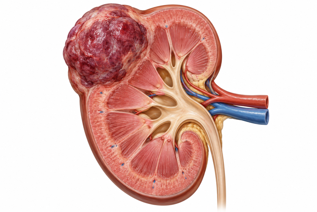
A kidney tumour larger than 3 cm: what does it mean?
By Dr Jo Schoeman | Patient information | September 2026
Finding a kidney mass can be frightening. The first question is whether it is cancer; the next is whether it is confined to the kidney. A mass measuring 3.1 cm is not automatically advanced cancer. If a renal cell carcinoma (RCC) is no more than 4 cm and remains within the kidney, it is classified as T1a. A tumour over 4 cm but no more than 7 cm, still confined to the kidney, is T1b. Lymph-node and distant spread are assessed separately. Some kidney masses are benign, and imaging alone cannot always tell us which. [1–3]
How might it present?
Many tumours are discovered by chance during an ultrasound or CT scan for another problem. Others cause blood in the urine, persistent pain in the side or back, or occasionally a palpable lump. Unexplained weight loss, fatigue, anaemia or fever warrant assessment, although they have many possible causes. The combination of blood in the urine, pain and a palpable mass is uncommon. Visible blood in the urine should always be investigated; a kidney mass does not rule out another source of bleeding. [2]
What investigations are needed?
- Dedicated imaging: A multiphase contrast CT of the kidneys and abdomen shows whether the lesion enhances, its exact position, its relationship to major blood vessels and the collecting system, and whether there are enlarged nodes or signs of spread. Chest imaging completes staging; its extent is tailored to the tumour and clinical circumstances. MRI may clarify an uncertain lesion or suspected tumour extension into a vein, or be useful when CT contrast is unsuitable. A complex kidney cyst needs its own imaging assessment. [2]
- Blood and urine tests: Kidney function (creatinine and eGFR), a full blood count and other tests guided by the clinical picture help assess fitness for treatment and preserve kidney function. Urine testing may identify infection or another cause of bleeding. [2]
- Biopsy: A needle biopsy can be useful if its result will change management, particularly before ablation or drug treatment without prior tissue diagnosis. A typical surgically removable solid mass does not always need a biopsy beforehand. A biopsy can occasionally be inconclusive. [2]
- Further staging: Bone or brain scans are generally reserved for relevant symptoms or suspected advanced disease. Routine PET scans and bone scans are not recommended for initial RCC staging. [2]
The team should review the images, kidney function, other medical conditions and the patient’s priorities together. An indeterminate mass should not be labelled definitively as RCC before appropriate assessment.
A note about my practice: I no longer perform nephrectomies. I can assess your kidney mass, discuss the findings and treatment options with you, and, if an operation is appropriate, refer you to a colleague who performs kidney surgery. We can also arrange input from a multidisciplinary team where needed.
Treatment when the tumour is confined to the kidney
Partial nephrectomy removes the tumour while preserving the rest of the kidney. This is generally preferred for T1 cancers when technically feasible, particularly if kidney function is reduced or there is only one functioning kidney. It can be performed by robotic, laparoscopic or open surgery according to tumour complexity and surgical expertise. Risks include bleeding, urine leak, infection, reduced kidney function and, occasionally, the need to remove the whole kidney. [1]
Radical nephrectomy removes the kidney and may be appropriate when a safe partial nephrectomy is not feasible, particularly with a larger or more complex tumour. Robotic or laparoscopic surgery offers an approach to the operation; it does not change the underlying choice between partial and radical removal. [1]
Active surveillance may suit selected people with a small, slow-growing mass, competing health risks or a strong preference to defer treatment. It involves planned scans and a clear trigger to reconsider treatment. A tumour just above 3 cm needs a particularly careful discussion because the room for further growth before 4 cm is limited. Thermal ablation, such as cryoablation or radiofrequency or microwave ablation, may suit selected small tumours, especially when surgery is unsuitable; a biopsy is recommended before ablation. Local recurrence and the possible need for repeat treatment should be discussed. [1, 2]
Stereotactic ablative radiotherapy (SABR) is also being used in specialist centres for selected patients with localised RCC who cannot undergo surgery. It is a distinct option from routine radiation after an operation, and its long-term comparative evidence continues to mature. [1]
What if lymph nodes look enlarged?
An enlarged node on a scan is suspicious, not proof of cancer; inflammation can also enlarge nodes. The team considers the appearance and site of the nodes, whether there is disease elsewhere and whether tissue confirmation will change treatment. If nephrectomy is planned, visibly enlarged regional nodes may be removed for diagnosis and staging. Removing normal-looking nodes routinely has not been shown to improve survival for organ-confined RCC. The survival benefit of removing involved nodes is uncertain. [1]
Confirmed regional node involvement is a substantial change in risk even if the kidney tumour is small. It calls for multidisciplinary discussion about whether all visible disease can be removed, whether postoperative drug treatment is appropriate, and how closely to monitor. Nodes beyond the regional area, or spread to organs such as the lung or bone, may represent metastatic disease and require a different plan. Surgery on the kidney is not automatically the best first step when cancer is widespread. [1, 3]
When does immunotherapy help?
For selected patients with clear-cell RCC at higher risk of recurrence after complete surgery, a course of pembrolizumab may lower recurrence risk and has shown an overall survival benefit in the KEYNOTE-564 study. The eligible groups include some patients with confirmed regional node involvement and no distant metastases. A 3–4 cm node-negative tumour confined to the kidney would not usually qualify based on size alone. The final pathology, stage and overall health matter. [1, 4]
If cancer has spread and cannot all be removed, initial treatment for clear-cell RCC often combines immune checkpoint drugs with each other (nivolumab plus ipilimumab) or combines immunotherapy with an oral targeted drug (for example, pembrolizumab plus axitinib, pembrolizumab plus lenvatinib, or nivolumab plus cabozantinib). The best regimen depends on risk group, other illnesses, kidney function, previous treatment and the specific cancer subtype. Papillary and other non-clear-cell cancers may need different approaches. Availability and subsidy in Australia should be checked for the individual indication. [1]
Immunotherapy can cause serious inflammation of the bowel, lungs, liver, kidneys or hormone-producing glands; targeted drugs can cause high blood pressure, diarrhoea, fatigue and other adverse effects. Some immune effects can persist after treatment ends. Benefits and risks need an individual discussion with a medical oncologist. [1, 4]
Is chemotherapy useful?
Conventional cytotoxic chemotherapy is generally ineffective for the common types of RCC and is not standard treatment for ordinary clear-cell RCC. Rare subtypes, including collecting duct and renal medullary carcinoma, are exceptions in which chemotherapy may have a role. A cancer arising from the lining of the renal pelvis (upper-tract urothelial carcinoma) is a different disease with different chemotherapy options; establishing the diagnosis matters. [1]
Does radiation offer a benefit?
Radiotherapy is not routinely given after nephrectomy to prevent recurrence of localised RCC. Its clearest established role is treating specific problems from spread, such as painful bone lesions, brain metastases or symptoms from another site. Precisely targeted radiation may also control selected limited metastases or treat a primary tumour in someone unsuitable for surgery. Whether it adds benefit to systemic therapy or surgery in an individual case depends on the site, number and behaviour of lesions and is best decided with radiation oncology input. It should not be presented as a standard substitute for an operable partial nephrectomy. [1, 5]
Questions worth asking at your consultation
- Is this definitely a solid enhancing kidney tumour, and what is its exact size and location?
- Is the disease confined to the kidney? Are any nodes merely enlarged, or proven to contain cancer?
- Can we safely preserve part of the kidney? What is my current kidney function?
- Would a biopsy change the plan?
- If the cancer is clear-cell and higher risk, would adjuvant pembrolizumab be suitable?
- Would drug treatment or targeted radiation offer a benefit in my particular situation?
Seek prompt medical attention for heavy bleeding with clots, inability to pass urine, severe persistent pain, or sudden neurological symptoms. This article is general information and cannot replace review of your scans and pathology at a multidisciplinary meeting.
References
- European Association of Urology. EAU Guidelines on Renal Cell Carcinoma: Disease Management. 2026.
- European Association of Urology. EAU Guidelines on Renal Cell Carcinoma: Diagnostic Evaluation. 2026.
- European Association of Urology. EAU Guidelines on Renal Cell Carcinoma: Staging and Classification Systems. 2026.
- National Cancer Institute. Adjuvant Keytruda improves kidney cancer survival, reporting updated KEYNOTE-564 outcomes. 2024.
- National Cancer Institute. Renal Cell Cancer Treatment (PDQ), Health Professional Version. Accessed September 2026.





Leave a Reply
Want to join the discussion?Feel free to contribute!