New Premises

From an Empty Shell to a Fresh New Practice at The Wesley Hospital

It is remarkable how quickly an empty shell can become a welcoming new medical practice.

After months of planning, construction and careful attention to detail, the renovation of our new rooms at The Wesley Hospital was completed in December 2025. The finished practice is modern, fresh and thoughtfully designed to provide a comfortable, calm and professional environment for our patients.

The new rooms officially opened on 12 January 2026.

Here is a look back at the transformation—from the first bare walls to the completed rooms ready to welcome patients.

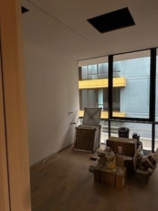
7 October 2025: The empty shell

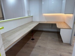
Every renovation has to begin somewhere. In early October, the new space was still an open shell: bare, unfinished and difficult to imagine as a functioning medical practice.

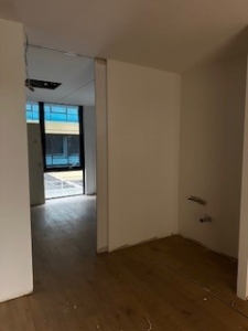
29 October 2025: The walls go up

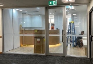
By the end of October, the internal walls were in place and the layout of the practice was beginning to emerge. For the first time, it was possible to see the consulting rooms, reception areas and shared spaces taking shape.

8 November 2025: Flooring installed

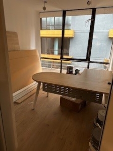
The installation of the flooring brought warmth and character to the space. What had looked like a building site only weeks earlier was starting to feel like a real practice.

14 November 2025: Paint, windows and lighting

Fresh paint, finished windows and new lighting transformed the atmosphere of the rooms. The design began to feel bright, clean and contemporary, while still maintaining the warmth and privacy that patients expect from a medical practice.

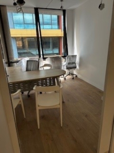
26 November 2025: The furniture arrives

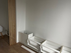
The arrival of the furniture was another important milestone. Consulting rooms, workspaces and patient areas could finally be arranged as they had been envisioned. The practice was no longer simply a completed fit-out—it was beginning to feel ready for people.

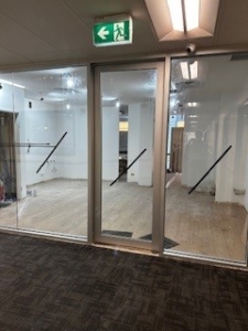
9 December 2025: One day before sign-off

With only the final checks and finishing touches remaining, the long-planned vision had become a reality. The rooms were clean, organised and almost ready for handover.

10 December 2025: Hours before handover

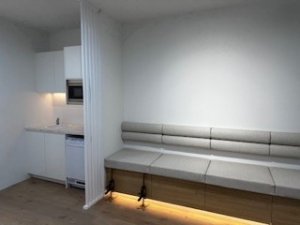
On handover day, the completed lighting and final finishes brought everything together. After progressing from a bare shell through framing, flooring, painting and furnishing, the new practice was finally complete.

Opening the doors

Following the December handover and preparations over the holiday period, we officially opened the doors to our new rooms on 12 January 2026.

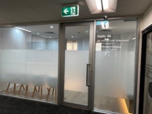
The completed practice offers a modern and fresh setting at The Wesley Hospital, designed around patient comfort, privacy and efficient care. We are delighted with the result and grateful to everyone whose planning, skill and hard work helped bring the project to life.

Most importantly, thank you to our patients and referring colleagues for their patience and support throughout the transition. We look forward to welcoming you to the new rooms.

Renovations have been completed. This promises to be modern and fresh.

Living with an indwelling urethral catheter

An indwelling urethral catheter is a soft tube passed through the urethra into the bladder. A small balloon holds it in place and urine drains into a bag. It may be needed for a few days after an operation or for longer when the bladder cannot empty safely. The reason for the catheter and a plan for review should be clear to the patient and the people helping with their care.[1, 2]

Why might a catheter be needed?

Common reasons include acute urinary retention, an obstruction to urine flow, temporary drainage after surgery, and selected cases of chronic retention when other options are unsuitable. In hospital, a catheter may be used to monitor urine output in a critically ill person or manage bleeding and clots. It can occasionally support comfort at the end of life. A urethral catheter is generally not the first response to urinary leakage alone.[1]

When practical, the team should discuss whether the catheter can be removed after a trial of void, whether intermittent self-catheterisation is possible, or whether a suprapubic catheter may be more suitable for longer-term drainage. The best choice depends on bladder function, dexterity, the underlying condition and the person’s wishes.[1, 3]

What might it feel like, and what can go wrong?

Some people notice discomfort or a sense of needing to pass urine after insertion. Bladder spasms can cause cramping or urine to leak around the tube. Leakage can also mean that the catheter is kinked or blocked, so it should be checked rather than simply treated as incontinence. Other possible problems include blood in the urine after insertion or a change, skin irritation, accidental pulling or displacement, blockage from debris or encrustation, and infection.[2, 3]

A long-term urethral catheter can also cause pressure or trauma at the urethral opening; persistent soreness or a change in its appearance deserves review. Bladder stones and repeated blockages are further reasons to reassess the drainage plan. The longer a catheter remains, the more opportunity there is for bacteria to colonise it, so it should stay in place only while needed.[3, 4]

How often should a long-term catheter be changed?

There is no single safe change interval for every catheter and every patient. The plan depends on the catheter material and manufacturer’s instructions, local nursing policy, comfort, drainage, and whether it repeatedly blocks or becomes encrusted. Some community protocols plan changes approximately every four to eight weeks, but an individual plan may differ. The interval should not exceed the relevant product’s recommended duration.[1, 3]

Changing a catheter more frequently just to prevent infection has not been shown to help. A catheter may instead need an earlier change if it is blocked, damaged, displaced, causing problems, or as part of managing a symptomatic infection. People with a history of difficult insertion, urethral injury or bleeding should have a specific plan for who can safely perform the change.[1, 3]

At each review, it is worth asking: Is the catheter still necessary? Could a trial without it or another drainage method be considered?[1]

Day-to-day care at home

Keep the drainage bag below bladder level, avoid kinks in the tubing, and secure the catheter so it does not pull. Wash your hands before and after handling the bag. Follow the nurse’s instructions for emptying it and for cleaning the area where the catheter enters the body. Ordinary hygiene is usually enough; do not disconnect the closed drainage system, flush the catheter or take preventive antibiotics unless your treating team has given a specific instruction.[3, 5]

Drink according to your usual health advice. If you have a fluid restriction for heart or kidney disease, follow that plan rather than trying to drink extra to “flush” the catheter. Ask your nurse which supplies to keep at home and whom to call after hours if drainage stops.[2]

Bacteria in urine: when are antibiotics needed?

With a catheter in place, bacteria commonly grow on the catheter surface. After a month, bacteriuria is found in nearly everyone with a long-term catheter. A positive urine culture without symptoms usually reflects colonisation, not an infection requiring antibiotics. Routine screening or treatment of asymptomatic bacteriuria is generally discouraged because antibiotics can cause side effects and encourage resistant bacteria. Exceptions include pregnancy and certain invasive urological procedures, where the treating team will advise on testing and treatment.[4, 6]

Seek clinical assessment for possible infection if you develop fever, rigors, new pelvic or flank pain, feel distinctly unwell, or have other new symptoms that could indicate infection. In a frail person, a new change in function or confusion warrants assessment of several possible causes rather than automatically assuming a UTI. Cloudy or strong-smelling urine, sediment, or a positive dipstick on its own does not usually justify antibiotics.[4, 6, 7]

When a symptomatic catheter-associated UTI is suspected, a clinician should assess you and arrange a properly collected urine specimen if indicated—not from the drainage bag. Treatment is chosen in light of symptoms, culture results, allergies and local guidance. If the catheter has been in place for more than two weeks and still needs to remain, changing it as part of treatment is commonly recommended. Routine antibiotics at every scheduled catheter change are not recommended.[7, 8]

When to get help promptly

Contact your nurse or doctor promptly if urine stops draining, the catheter falls out, you have significant pain or new bleeding, or urine is leaking around the catheter with little in the bag. Check for a kink or a full bag, but do not forcefully flush or reinsert a catheter yourself unless specifically trained and instructed. Seek urgent medical care if drainage has stopped and you have a painful or swollen lower abdomen, or if you have fever, shaking chills or feel seriously unwell.[2]

Catheter support in Bundaberg

Some people can have catheter assessment and planned changes at home through a community nursing service. Ozcare and BlueCare provide home nursing in the Bundaberg area; whether a nurse can provide catheter care for an individual depends on referral, staffing, clinical needs, service area and funding arrangements. Ask the provider directly about availability and costs before relying on a visit.[9, 10]

For my Bundaberg patients, Sandra Ilett, a continence nurse with Community Nurse Service, is another local contact for bladder and catheter-related care. Sandra and her colleague Carla Kerr have also seen patients at the nurse-led clinic at The Friendlies Medical Suites. Patients can discuss a suitable referral and whether a home visit or clinic appointment is available. Community Nurse Service: (07) 4126 2002.[11] This mention recognises local nursing support; it is not a claim that one service is preferable for every patient.

A written catheter plan should record the reason for drainage, catheter details, the planned review or change, who will provide care, and whom to call if problems arise. Community nurses, the GP and urology team can then coordinate care across visits.

This article provides general information. Follow your individual catheter plan and seek clinical advice for new symptoms or a catheter that is not draining.

References

  1. Queensland Health. Urinary catheter insertion or change: indications and review.
  2. Healthdirect Australia. Catheter problems.
  3. Queensland Spinal Cord Injuries Service. Indwelling catheters.
  4. US Centers for Disease Control and Prevention. Indwelling urinary catheter culture stewardship.
  5. US Centers for Disease Control and Prevention. CAUTI prevention: summary of recommendations.
  6. Infectious Diseases Society of America. Management of asymptomatic bacteriuria.
  7. Queensland Spinal Cord Injuries Service. Management of urinary tract infection.
  8. US Centers for Disease Control and Prevention. Catheter urine culture collection guidance.
  9. Ozcare Bundaberg: home nursing and service area.
  10. BlueCare: community nursing at home; Bundaberg community service listing.
  11. Dr Jo Schoeman. Nurse-led urology clinic in Bundaberg: Sandra Ilett and Carla Kerr.

Renal angiomyolipoma (AML): when to watch, when to treat and when to seek urgent help

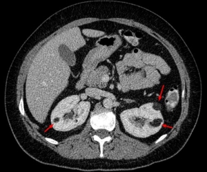
An angiomyolipoma, usually shortened to AML, is a growth in the kidney made up of blood vessels, muscle-like tissue and fat. Most are found by chance on a scan and never cause trouble. The main concern is that some AMLs can bleed, occasionally quite severely. The right plan depends on the diagnosis, the growth’s features and your circumstances, not its diameter alone. [1,2]

In this article, “AML” means angiomyolipoma, not acute myeloid leukaemia.

Is an AML cancer?

A classic renal AML is generally a benign tumour. Most occur on their own (sporadic AML). Some occur with tuberous sclerosis complex (TSC) or, less commonly, lymphangioleiomyomatosis (LAM); these can be multiple, affect both kidneys and require a different surveillance and treatment approach. A rare epithelioid AML can behave aggressively and needs specialist assessment. Not every solid renal mass presumed to be an AML can be confidently identified on a scan, particularly when little visible fat is present. [1,3,4]

What symptoms can it cause?

Many AMLs have no symptoms. Possible symptoms include aching or pain in the side, visible blood in the urine, or, less commonly, a palpable swelling. A sudden bleed into or around the kidney may cause abrupt, severe flank or abdominal pain, sometimes with dizziness, fainting, weakness or a racing heart. Blood in the urine and flank pain can also have other causes, including stones or cancer, so they should be assessed rather than attributed automatically to a known AML. [1–3]

Emergency warning: If you develop sudden severe side or abdominal pain, especially with faintness, collapse, a racing heart or visible bleeding, call 000 in Australia or attend an emergency department immediately. A bleeding AML can cause major blood loss even when blood is not visible in the urine. Do not wait for a routine appointment. [1,3]

How is it diagnosed?

An AML may first appear on an ultrasound performed for another reason. CT or MRI helps confirm whether it contains fat and assess its size, location, blood vessels and any evidence of bleeding. Contrast-enhanced imaging may be needed for an indeterminate lesion or procedural planning, with the choice adapted to kidney function, pregnancy and other factors. A classic fat-rich AML is often diagnosed on imaging alone. A fat-poor AML may resemble kidney cancer; specialist review, further imaging or a biopsy may be appropriate if the result would change management. Biopsy has limitations and is chosen case by case. [1,3,4]

The assessment may also include blood pressure, kidney function and a blood count if bleeding is suspected. Multiple or bilateral AMLs, especially in a younger person, should prompt consideration of TSC or LAM and appropriate specialist input. [3,5]

Is there a critical size that must be treated?

There is no single diameter at which every AML must be removed. The traditional rule to treat an AML once it reaches 4 cm is no longer considered an automatic trigger. The European Association of Urology (EAU) states that the relationship between size and bleeding risk is uncertain and that no universal intervention threshold exists. Some larger AMLs can still be watched safely; a smaller one may need treatment if it bleeds or causes persistent symptoms. [1,2]

Size remains one part of the discussion. The team may also consider growth, vascular features or aneurysms seen on imaging, prior bleeding, pain, kidney function, pregnancy plans, blood-thinning medicines and access to urgent care. An aneurysm measurement such as 5 mm has been proposed as a risk marker, but recent evidence questions how reliably it predicts bleeding on its own. These findings guide an individual discussion; they do not supply a stand-alone rule. [1,3,6]

Management options

Active surveillance

For a confidently diagnosed, symptom-free AML, monitoring is often appropriate. It avoids exposing people with stable lesions to procedure risks. A surveillance plan records the starting size and imaging characteristics, checks for new symptoms and uses ultrasound, MRI or CT when appropriate to look for change. There is no single imaging interval suitable for all sporadic AMLs. A small, stable lesion may need little or no ongoing imaging after specialist review; a larger or changing lesion usually needs closer follow-up. Agree on the next scan, who will review it and what changes should prompt earlier contact. [1–3]

Selective arterial embolisation

An interventional radiologist passes a catheter into the blood vessels feeding the AML and blocks selected branches. Embolisation can control active bleeding and can be considered in advance for selected higher-risk or symptomatic tumours, while preserving kidney tissue. Pain, fever and nausea can occur afterwards; infection, non-target embolisation and repeat treatment are possible. In the EAU systematic review, around 30% of embolised cases required a further intervention, although individual outcomes vary. [1,2]

Kidney-sparing surgery

A partial nephrectomy removes the AML while aiming to preserve the rest of the kidney. Open, laparoscopic or robotic approaches may be considered depending on anatomy and surgical expertise. Surgery may be useful when imaging is uncertain, symptoms persist, embolisation is unsuitable or a definitive removal is preferred. It carries anaesthetic, bleeding, urine leak and kidney-function risks. Removal of the whole kidney is generally reserved for situations where kidney-sparing care is not feasible, especially in an emergency. [1–3]

Ablation and medicines

Thermal ablation is used less often for AML, and the evidence is more limited than for surveillance, embolisation and surgery. For TSC-associated AML, specialist teams may use an mTOR inhibitor such as everolimus for an asymptomatic but growing lesion over 3 cm when treatment is appropriate. This TSC recommendation should not be applied automatically to an isolated sporadic AML. Medicines require monitoring for adverse effects and interactions. [1,2,5]

What happens if an AML bleeds?

In hospital, the immediate priorities are resuscitation, blood tests and imaging to locate the bleed. Treatment may include intravenous fluids, blood transfusion and urgent selective arterial embolisation to stop the bleeding. Surgery is considered if embolisation is unavailable, unsuccessful or otherwise unsuitable. After recovery, follow-up checks the AML, remaining kidney function and whether further treatment is needed. [1,3]

How is surveillance different with tuberous sclerosis?

TSC-related AMLs require a coordinated long-term plan because lesions may occur in both kidneys and kidney function needs protection over a lifetime. The 2021 international TSC recommendations advise abdominal MRI every 1–3 years, with blood pressure, kidney function and urine protein checked at least yearly; the interval is tailored to individual risk. Growing AMLs greater than 3 cm may merit first-line mTOR treatment when intervention is indicated, while acute bleeding is generally managed with embolisation. A TSC specialist team can help balance treatment against the need to preserve kidney tissue. [5]

Questions to bring to your appointment

  • Is the imaging diagnosis of a classic AML secure, or is more investigation needed?
  • What are its size, growth pattern and vascular features?
  • What is my personal risk of bleeding, and how does kidney function affect my options?
  • If we monitor it, when is my next scan and what result would change the plan?
  • If treatment is recommended, why is embolisation, surgery or another option best for me?

This article provides general information and does not replace individual assessment or advice.

References

  1. European Association of Urology. EAU Guidelines on Renal Cell Carcinoma, section on other renal tumours and AML. Accessed September 2026.
  2. Fernández-Pello S, et al. Management of sporadic renal angiomyolipomas: a systematic review to guide EAU recommendations. European Urology Oncology. 2020;3:57–72. doi:10.1016/j.euo.2019.04.005.
  3. Guo Y, et al. Canadian Urological Association best practice report: Diagnosis and management of sporadic angiomyolipomas. Canadian Urological Association Journal. 2020;14–E536. doi:10.5489/cuaj.6942.
  4. Wilson MP, et al. Diagnostic performance of MRI in detection of renal lipid-poor angiomyolipomas: systematic review and meta-analysis. Radiology. 2020;296:511–520.
  5. Northrup H, et al. Updated international TSC diagnostic criteria and surveillance and management recommendations. Pediatric Neurology. 2021;123:50–66. See also Tuberous Sclerosis Australia: Kidneys.
  6. Swärd J, et al. Renal angiomyolipoma: investigating radiological signs indicative of bleeding risk. Insights into Imaging. 2025.

Urethral Meatal Stenosis: Congenital, Acquired and Correct Management

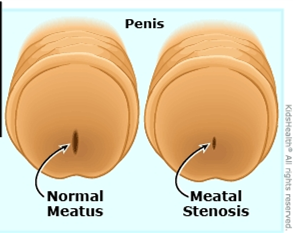
The urethral meatus is the opening through which urine leaves the body. Meatal stenosis means that this opening has become abnormally narrow and is interfering with urinary flow. It is most often discussed in boys and men, although narrowing of the female urethral opening can also occur.

An important point is that a meatus can look small without causing obstruction. Treatment should therefore be based on the complete picture, symptoms, the appearance of the opening, the urinary stream and, where appropriate, objective testing, not appearance alone.
What symptoms can it cause?

Typical symptoms include:

  • a thin, forceful or upward-deflected stream;
  • spraying or difficulty aiming the urine;
  • taking longer to pass urine or needing to strain;
  • burning or discomfort during urination;
  • a small spot of blood at the meatus;
  • urinary frequency, urgency or incomplete emptying; and
  • recurrent urinary infection in selected patients.

In toilet-trained boys, an abnormal stream is often the most useful clue. Published patient-reported data show that improvement after meatotomy is most predictable when the preoperative problem is an abnormal or deflected stream. Frequency, urgency, wetting or dysuria may have another cause and should not automatically be attributed to a narrow-looking meatus.

Complete inability to pass urine is uncommon but requires urgent medical attention.

Congenital meatal stenosis

Congenital meatal stenosis is present from birth. True isolated congenital narrowing is uncommon and should be distinguished from normal variation in meatal size. It may also occur as part of another developmental urethral condition, including hypospadias, or following congenital urethral reconstruction.

In a baby or young child, symptoms can be difficult to recognise. The diagnosis becomes more apparent after toilet training, when a persistently narrow, spraying or markedly deflected stream can be observed.

Not every anatomically small meatus needs surgery. An asymptomatic child with a satisfactory stream, no urinary infections and normal bladder emptying can often be observed. Symptomatic obstruction, however, should be assessed by a paediatric urologist.

Acquired meatal stenosis

Acquired stenosis develops after birth. Its causes vary with age.

In boys

Meatal stenosis is recognised after circumcision, although published estimates vary substantially because studies use different definitions and methods of examination. Proposed mechanisms include irritation and inflammation of the exposed meatus, contact with wet nappies, meatal ulceration and subsequent scar formation. It often becomes clinically obvious between early childhood and school age rather than immediately after circumcision.

Other causes include inflammation, trauma, catheterisation and previous surgery, particularly repair of hypospadias. Following hypospadias surgery, narrowing may involve more than the external opening and must be assessed in the context of the reconstructed urethra.

In adolescents and adults

Important causes include:

  • lichen sclerosus (also called balanitis xerotica obliterans or BXO), which can scar the foreskin, glans, meatus and more proximal urethra;
  • repeated urethral instrumentation, catheterisation or endoscopic surgery;
  • prior hypospadias repair or other penile surgery;
  • trauma, infection or chronic inflammation; and
  • previous radiotherapy or treatment affecting the urethra.

In adults, it is essential to determine whether narrowing is confined to the meatus or extends into the fossa navicularis or penile urethra. Treating only the visible opening will fail if more extensive scar disease has been overlooked.

Female meatal or urethral stenosis is uncommon. Symptoms may resemble recurrent urinary infection or other causes of bladder-outlet obstruction. Diagnosis should be made carefully, as urinary symptoms alone do not prove that the urethra is narrowed.

How is it diagnosed?

Assessment may include:

  1. A detailed history: including the direction and calibre of the stream, spraying, pain, infections, prior circumcision, catheterisation, surgery, trauma and skin disease.
  2. Examination: assessing the meatus and surrounding skin for scarring, pallor, inflammation, lichen sclerosus, hypospadias or surgical change.
  3. Observation of the urinary stream, particularly in a toilet-trained child.
  4. Uroflowmetry and post-void residual ultrasound when symptoms are unclear, the patient is older, or more extensive obstruction is suspected.
  5. Urinalysis or urine culture if pain, blood or infection is suspected.
  6. Urethral calibration, cystoscopy or urethrography selectively, especially in adults, recurrent disease, previous urethral surgery or suspected extension beyond the meatus.

Kidney and bladder ultrasound is not required for every straightforward case, but may be appropriate when there are recurrent infections, incomplete emptying, significant obstruction or concern about the upper urinary tract.

What is the correct management?

Management must match the patient’s symptoms, cause, age and extent of narrowing.

1. Observation

Observation is reasonable when the meatus is merely small in appearance but the patient has no relevant symptoms, passes a satisfactory stream and empties the bladder normally. Treating an incidental finding is unlikely to improve unrelated urgency, frequency or wetting.

2. Treat active skin or inflammatory disease

When lichen sclerosus or another inflammatory disorder is present, the underlying disease must be treated as well as the narrowing. Potent topical corticosteroid treatment is commonly used for genital lichen sclerosus under medical supervision. Circumcision may be indicated when the foreskin is affected, but established meatal or urethral scar may also require surgery.

Persistent or suspicious penile lesions may need biopsy. Long-term review can be appropriate because lichen sclerosus can recur, extend into the urethra and is associated with a small but important risk of penile malignancy.

3. Meatotomy or meatoplasty

For a short, symptomatic stenosis confined to the meatus, meatotomy or meatoplasty is usually the definitive treatment.

  • A meatotomy enlarges the opening with a controlled incision.
  • A meatoplasty reconstructs and sutures the edges to create a durable, appropriately shaped opening.

Both can provide excellent relief in appropriately selected children. A large paediatric series reported that meatotomy required more early manual spreading and had a higher reoperation rate than meatoplasty; technique and postoperative care therefore matter. In another study, 95% of families reported their child was at least somewhat improved after meatotomy, with the strongest benefit in boys treated for an abnormal stream.

The operation may be performed under local anaesthesia in selected cooperative patients or under general anaesthesia, particularly in younger children. Expected short-term effects include stinging, minor spotting of blood and temporary spraying while swelling settles. The surgeon may advise ointment and gentle separation of the meatal edges during early healing; instructions vary according to the procedure used.

4. Dilatation

Repeated blind dilatation is generally not a durable solution for dense scar-related meatal stenosis. It may cause further tearing and scarring and can commit a patient to repeated procedures. Carefully selected dilation or self-dilatation may have a role as temporary or palliative management, or as part of a specialist regimen for inflammatory disease, but it should not replace an appropriate reconstructive assessment in recurrent or complex disease.

5. Recurrent, adult or extended distal disease

If narrowing recurs, is associated with lichen sclerosus, or extends into the fossa navicularis/distal urethra, a simple repeat incision may not be sufficient. Options include formal meatoplasty or distal urethroplasty using an oral mucosal graft. The European Association of Urology recommends offering open meatoplasty or distal urethroplasty for meatal and distal urethral strictures; the precise operation should be individualised to stricture length, tissue quality and the patient’s priorities.

In lichen-sclerosus-related urethral disease, genital skin should not be used as a graft because the disease may recur in that tissue. Oral mucosa is generally preferred when graft reconstruction is required.

Follow-up and recurrence

Most patients with an isolated, properly treated meatal stenosis do well. Follow-up should assess:

  • improvement in stream direction and calibre;
  • pain, bleeding, infection or difficulty voiding;
  • bladder emptying when clinically indicated; and
  • restenosis or progression of an underlying condition such as lichen sclerosus.

Prompt reassessment is advisable if the stream narrows again, spraying persists after healing, voiding becomes painful or difficult, infections recur, or new skin changes appear.

The practical message

Meatal stenosis is not simply “a small hole.” Correct care begins by confirming that the narrowing is clinically important and identifying its cause and extent. A symptomatic, short stenosis confined to the meatus is usually treated successfully with meatotomy or meatoplasty. Recurrent stenosis, adult disease, previous hypospadias repair or lichen sclerosus requires a more detailed urethral assessment and sometimes formal reconstruction.

This article provides general educational information and does not replace an individual assessment. Seek urgent care if you or your child cannot pass urine, develops fever with urinary symptoms, or has significant bleeding or pain.

Selected references

  1. European Association of Urology. EAU Guidelines on Urethral Strictures: Disease Management in Males. 2026. https://uroweb.org/guidelines/urethral-strictures/chapter/disease-management-in-males
  2. European Association of Urology. EAU Guidelines on Urethral Strictures: Definition, Epidemiology, Aetiology and Prevention. 2026. https://uroweb.org/guidelines/urethral-strictures/chapter/definition-epidemiology-aetiology-and-prevention
  3. Wessells H, et al. Urethral Stricture Disease Guideline Amendment (2023). Journal of Urology. 2023. doi:10.1097/JU.0000000000003482.
  4. Dothan D, et al. Surgical Treatment of Meatal Stenosis: Lessons Learned from the Pediatric Urology Practice. Urology. 2023;171:220–224. PMID: 35981660.
  5. Varda BK, et al. Minor procedure, major impact: patient-reported outcomes following urethral meatotomy. Journal of Pediatric Urology. 2018;14(2):165.e1–165.e5. doi:10.1016/j.jpurol.2017.11.018.
  6. Godley SP, et al. Meatal stenosis: a retrospective analysis of over 4000 patients. Journal of Pediatric Urology. 2015;11(1):38.e1–38.e6. doi:10.1016/j.jpurol.2014.09.016.
  7. Morris BJ, Krieger JN. Does circumcision increase meatal stenosis risk? A systematic review and meta-analysis. Urology. 2017;110:16–26. doi:10.1016/j.urology.2017.07.027.
  8. Wang MH. Surgical management of meatal stenosis with meatoplasty. Journal of Visualized Experiments. 2010;(45):2213. doi:10.3791/2213.

Prepared for patient education.

Pelvic floor rehabilitation post prostatectomy

Pelvic floor physiotherapy after a prostatectomy helps regain bladder control and addresses erectile dysfunction through exercises and other therapies. A physiotherapist can create a personalized plan, which may include pre- and post-operative pelvic floor muscle training (Kegels), bladder retraining, and, in some cases, treatments like focused shockwave therapy for erectile dysfunction. 

What pelvic floor physiotherapy involves

  • Pelvic floor muscle training: A physiotherapist will guide you on how to perform exercises (like Kegels) to strengthen the pelvic floor muscles. This can significantly improve urinary incontinence and support bladder control.
  • Bladder retraining: This involves a combination of exercises, tracking bladder use, and scheduling toilet breaks to improve bladder control and reduce urgency or leakage.
  • Individualized assessment: Each person’s recovery is unique, so a physiotherapist will conduct a comprehensive assessment to create a treatment plan tailored to your specific symptoms and needs.

When to start

  • Pelvic floor exercises can be started before surgery or soon after the catheter is removed.
  • Starting pelvic floor rehabilitation before surgery is recommended by some organizations, as it can help with recovery. 

Why it’s important

  • The prostate gland sits below the bladder, and its removal can weaken the surrounding pelvic floor muscles and surrounding structures.
  • This can lead to issues like urinary incontinence (leaking), urgency, and erectile dysfunction.
  • Pelvic floor physiotherapy helps by strengthening the muscles that support bladder and sexual function. 

Getting started

  • It is highly beneficial to see a physiotherapist, as they can provide guidance and ensure you are performing exercises correctly.
  • You can start with basic exercises on your own, but a physiotherapist can help you get more benefit from your training. 


I use 3 groups of physiotherapy practices depending on your location:

1. Wesley Hospital Physiotherapy

Women and men’s physiotherapy | Allied health | The Wesley Hospital

2. Bodyworks Physiotherapy in Kallangur – Rashiq Patel

BODYWORKS PHYSIOTHERAPY

3. Coral Coast Physiotherapy in Bargara, Bundaberg – Reuben Wharerau

Bargara Physiotherapy | Coral Coast Physiotherapy Bargara


Dr Jo Schoeman, Urologist
Suite 46, Level 4
The Wesley Medical Centre
Wesley Hospital
Auchenflower Brisbane

Richard Marsden

Many of my longstanding patients in Bundaberg will remember Richard Marsden, a highly respected urologist who served the Bundaberg community for many years.

For much of that time, Richard was Bundaberg’s only urologist. He was deeply committed to regional Queensland and to ensuring that people living outside the major cities could receive high-quality specialist care close to home. This remarkable man passed away on 5 November.

I owe my own life-changing move from South Africa to Australia, in no small part, to Richard.

We first met in 2005 at the biennial UROSA conference, the Urological Association of South Africa meeting held at Sun City. We struck up a conversation, and by the end of it Richard had handed me his business card. He told me that he was preparing to retire and invited me to contact him if I might be interested in taking over his urology practice in Bundaberg.

At the time, I did not fully appreciate the significance of his offer. It took several weeks for me to realise what an extraordinary opportunity he had placed before me.

A few months later, my wife, Natasha, and I travelled to Bundaberg for what South Africans often call an “LSD trip” a “look, see and decide” visit undertaken before making the enormous decision to uproot a family and step into the unknown.

Australia represented the promise of safety, freedom and a hopeful future for our daughters. From the moment we arrived in Bundaberg, we felt an immediate connection with the town. Its ocean, sugarcane fields and humid subtropical climate reminded us of the east coast of South Africa and the former province of Natal.

Richard and his wife, Carol, welcomed us warmly. Richard generously shared his daily routine with me and introduced me to the operating theatre and ward staff. He gave us an honest and personal insight into both the practice and the community we were considering making our home.

We also discovered that Richard understood the immigrant journey firsthand. Born in the United Kingdom, he spent part of his childhood during the Second World War in Johannesburg, South Africa. After the war, he joined his parents in Melbourne before the family later returned to the United Kingdom. Richard went on to study medicine and establish his medical career in London.

Years later, he made his own courageous decision to relocate to Australia with his two daughters. In many ways, his journey mirrored the one Natasha and I were contemplating. His story, his generosity and the welcome extended by Richard and Carol helped us make one of the most difficult and ultimately most important decisions of our lives: Bundaberg was where we wanted to raise our girls.

Richard did far more than offer me a professional opportunity. He opened a door that changed the course of my career and transformed the future of my family.

I will always be grateful for his kindness, his trust and his commitment to the people of regional Queensland.

Thank you, Richard. Your legacy lives on in the community you served, the colleagues you inspired and the lives you changed—including mine.

Mid-Urethral Slings for Stress Urinary Incontinence: What Is the Current Australian Position?

Patient information for Australian women | Reviewed 22 September 2026

The word “mesh” can understandably cause concern. Public discussion has often grouped together several very different products and operations. A mid-urethral sling used to treat female stress urinary incontinence is not the same operation as transvaginal mesh used to repair pelvic organ prolapse, and it is also different from a single-incision mini-sling.

The Urological Society of Australia and New Zealand (USANZ) supports the continued availability of mid-urethral slings in Australia as one treatment option for appropriately selected women. That support is not unconditional: USANZ emphasises appropriate clinical governance, surgeon credentialing, informed consent, multidisciplinary review where appropriate, and monitoring of patient outcomes.

What is stress urinary incontinence?

Stress urinary incontinence, or SUI, is leakage caused by an increase in abdominal pressure, for example when coughing, sneezing, laughing, exercising or lifting. It is different from urge incontinence, in which leakage is associated with a sudden compelling need to pass urine.

An accurate diagnosis matters. Some women have both stress and urgency symptoms, difficulty emptying the bladder, prolapse, recurrent infections or previous pelvic surgery. A consultation may therefore include a history, examination, bladder diary, urine testing, measurement of residual urine and, in selected or complex cases, urodynamic studies or cystoscopy.

What is a mid-urethral sling?

A mid-urethral sling is a narrow strip of permanent synthetic polypropylene mesh placed beneath the middle part of the urethra. It provides support during coughing, exercise and other activities that raise abdominal pressure.

The two established approaches are:

  • Retropubic sling: the tape passes behind the pubic bone.
  • Transobturator sling: the tape passes through the obturator region towards the groin.

These approaches have different risk profiles. The retropubic route has a greater risk of bladder perforation and short-term voiding difficulty, while the transobturator route has a greater association with groin or thigh pain. The most appropriate approach depends on the woman’s anatomy, previous operations, clinical circumstances and preferences.

What does USANZ say?

USANZ states that mid-urethral slings have an established evidence base for relative safety and effectiveness and should remain available in Australia for the treatment of female SUI. In its 2023 statement, USANZ specifically linked continued use to the clinical-governance improvements introduced in Australia since 2018, including:

  • appropriate surgeon credentialing
  • careful patient selection
  • discussion through multidisciplinary processes where indicated
  • genuine informed consent
  • monitoring of outcomes through the Australasian Pelvic Floor Procedure Registry.

This position supports patient choice, not routine surgery for every woman. Conservative care should usually be considered first, and non-mesh alternatives must be discussed when surgery is being considered.

What is the TGA position?

The Therapeutic Goods Administration (TGA) regulates medical devices in Australia. Surgical mesh is classified as a Class III medical device, the highest-risk classification, requiring more stringent evidence and regulatory assessment.

Following its safety reviews, the TGA removed certain transvaginal prolapse mesh products and single-incision mini-slings from routine supply. This did not amount to a ban on established retropubic and transobturator mid-urethral slings for SUI. The TGA’s current register includes eligible Class III urogynaecological mesh devices intended for SUI; the precise indication must always be checked in the device’s approved Instructions for Use.

The TGA requires manufacturers to provide patient information leaflets and implant cards for these devices. A woman receiving a sling should know the product used and should retain her implant card.

How effective is a mid-urethral sling?

Mid-urethral sling surgery is one of the most extensively studied operations for female SUI. Australian safety and quality guidance describes it as highly effective in the short and medium term, with long-term studies showing sustained patient satisfaction for many women.

No operation can guarantee a cure. Outcomes depend on the definition of success, length of follow-up, type of sling, surgeon experience and the individual patient. Stress leakage may persist or recur, and urgency symptoms may remain, improve or occasionally develop after surgery.

What are the risks?

Most women do not develop a serious complication, but complications can be significant and may occur early or years later. Potential risks include:

  • bleeding, infection and anaesthetic complications
  • temporary or persistent difficulty emptying the bladder, sometimes requiring catheterisation or further surgery
  • bladder or urethral injury
  • urinary tract infection
  • new or worsened urinary urgency or urge incontinence
  • persistent or recurrent stress incontinence
  • vaginal mesh exposure
  • mesh erosion into the urethra or bladder
  • pelvic, vaginal, groin or thigh pain
  • pain during intercourse
  • rarely, injury to major blood vessels, bowel or other pelvic structures.

Mesh is intended to remain permanently. If a mesh complication develops, partial or complete removal may be considered, but complete removal can be technically difficult or impossible and may require more than one operation. Removal may not fully resolve pain and can cause stress incontinence to recur.

Seek medical assessment if you develop persistent pelvic or groin pain, pain during intercourse, vaginal bleeding or discharge, recurrent urinary infections, difficulty passing urine, blood in the urine, or recurrent leakage after sling surgery.

What are the alternatives?

Treatment should be individualised. Options include:

  • no active treatment or the use of continence products
  • lifestyle measures, including weight management, treatment of constipation and chronic cough, and smoking cessation
  • supervised pelvic-floor muscle training, usually for at least three months
  • a continence pessary in suitable women
  • urethral bulking injections, which are less invasive but generally less durable and may need repeating
  • an autologous fascial sling using the patient’s own tissue
  • Burch colposuspension using sutures and native tissue.

Autologous fascial sling and colposuspension avoid permanent synthetic mesh but usually involve a longer operation and recovery and have their own risks, including voiding difficulty. There is no single best operation for every woman.

Shared decision-making and informed consent

Before proceeding, a woman should have enough time and balanced information to consider:

  • whether her symptoms are predominantly stress, urgency or mixed incontinence
  • conservative, mesh and non-mesh options
  • the expected benefits and limitations of each option
  • short- and long-term risks, including mesh-specific complications
  • the surgeon’s training and experience with the proposed procedure and alternatives
  • what follow-up will occur and how complications would be managed
  • the name and TGA registration status of the proposed device.

A second opinion is reasonable, particularly if symptoms are complex, previous continence surgery has failed, chronic pelvic pain is present, or the available options remain unclear.

The balanced Australian position

Mid-urethral slings are not appropriate for every woman, but neither are they prohibited in Australia. USANZ supports their continued availability for carefully selected patients within strong clinical-governance systems. The TGA continues to regulate eligible SUI sling devices as Class III medical devices and requires enhanced patient information and traceability.

The most important principles are an accurate diagnosis, consideration of conservative care, a balanced discussion of mesh and non-mesh alternatives, surgeon credentialing, informed consent and structured follow-up.

Important: This article provides general educational information and does not replace individual medical advice. It does not claim endorsement or approval by AHPRA, USANZ or the TGA. These organisations do not pre-approve individual practitioner website articles. Treatment recommendations must be based on personal assessment and shared decision-making.

References and further reading

  1. Urological Society of Australia and New Zealand. Vaginal mesh complications: USANZ submission, position statement and patient resources.
  2. Urological Society of Australia and New Zealand. USANZ supports pause on mesh stress urinary incontinence surgery in New Zealand. 23 August 2023.
  3. Therapeutic Goods Administration. Urogynaecological (transvaginal) surgical mesh hub. Updated 28 April 2025.
  4. Therapeutic Goods Administration. Australian transvaginal surgical mesh regulatory actions.
  5. Therapeutic Goods Administration. Current status of mesh products in Australia. Updated 9 June 2026.
  6. Australian Commission on Safety and Quality in Health Care. Treatment options for stress urinary incontinence: information for consumers. 2018.
  7. Australian Commission on Safety and Quality in Health Care. Care pathway for the management of stress urinary incontinence. 2018.
  8. Australian Health Practitioner Regulation Agency. Guidelines for advertising a regulated health service.
  9. Australian Health Practitioner Regulation Agency. Summary of the advertising requirements.

Why Self-Care Is Not Selfish: It’s Your Healing Tool

Making room for yourself: self-care during stressful times

Life can become so full of work, family needs and everyday responsibilities that rest starts to feel like something we have to earn. When stress or anxiety builds, even basic things—sleeping well, eating regularly, seeing a friend—can slip down the list. Making time to care for yourself is a practical way to protect your health and stay connected to what matters to you.

Self-care does not need to be expensive, time-consuming or tied to any particular belief. For one person, it might mean prayer or time with a faith community. For another, it might be quiet reflection, a walk outside, music or a conversation with a friend. There is no single right way to rest.

What does self-care actually mean?

Self-care includes ordinary habits that support physical and emotional wellbeing: enough sleep, regular meals, movement you enjoy, time with people you trust and breaks from constant demands. It can also mean setting a boundary, asking for help, attending a medical appointment or saying no when your capacity is stretched.

It is not another task to do perfectly. When you are exhausted, a small, realistic step may be more useful than an ambitious routine that adds pressure.

1. A pause can help you reset

Stress can narrow our attention to the next problem. Taking a few minutes to breathe slowly, step outdoors or notice what is happening around you can create space to respond more calmly. The World Health Organization offers short, practical exercises for coping with stress that can be used in everyday life. You might prefer stillness, meditation, prayer or simply putting your phone away while you drink a cup of tea.

Try asking yourself: What do I need in the next ten minutes? The answer might be a glass of water, a break, company or a chance to finish one manageable task.

2. Rest supports you when demands keep growing

Self-care cannot remove every source of stress, especially when caring responsibilities, financial pressure or work conditions are involved. It can, however, help you notice when the load has become too heavy. Persistent exhaustion, irritability, poor sleep or feeling detached from things you usually value are signs to slow down and seek support.

Sometimes the most helpful step is a practical change: sharing a task, taking leave, discussing workload or arranging help at home. You do not have to solve every problem by becoming better at coping with it alone.

3. Your needs matter too

People who care for others often feel guilty about taking time for themselves. Yet your need for rest, connection and support deserves attention in its own right. A quiet hour, a hobby or an appointment with a health professional is not a reward for completing everything else.

For those who draw strength from religion or spirituality, personal practices may be part of this care. Others find meaning through nature, relationships, creativity or community. Choose what feels supportive and consistent with your own values.

4. Caring for yourself can help your relationships

Rest does not guarantee patience or solve every conflict, but it may make it easier to listen, communicate and be present. It also gives others permission to be honest about their limits. Caring for yourself and caring for others can sit alongside each other.

A simple place to start

Choose one step that fits your life this week:

  • Take a short walk or spend a few minutes outdoors.
  • Keep a regular bedtime where possible.
  • Share how you are feeling with someone you trust.
  • Make space for prayer, reflection, mindfulness or quiet, if that helps you.
  • Reduce time spent on news or social media if it leaves you feeling overwhelmed.
  • Ask for help with one specific task.

If stress or anxiety persists, affects sleep or daily life, or feels difficult to manage, speak with your GP or a mental health professional. Self-care can support recovery, but it does not have to carry the whole burden of treatment. If you feel at risk of harming your

self or need immediate help in Australia, call 000 or visit the nearest emergency department. You can also contact Lifeline on 13 11 14.

This article is general wellbeing information, not a substitute for individual medical or mental health care.

Further reading

Food and urological health: what does the evidence say?

Food and drink can influence some urinary conditions, particularly recurrent kidney stones. A balanced eating pattern also supports general health. But the evidence does not support calling individual foods “urology-friendly” treatments or claiming that a berry, seed or spice will prevent urinary tract infections (UTIs), prostate enlargement or cancer. The useful advice depends on the diagnosis and the person.[1–4]


Start with the whole diet

The Australian Dietary Guidelines recommend a variety of vegetables, fruit, wholegrain foods and suitable protein foods, while limiting foods high in salt.[1] That is a more reliable starting point than a list of purported “superfoods.” People with kidney disease, recurrent stones or other medical conditions may need more specific advice from their treating team.

1. Berries and the cranberry question

Blueberries, strawberries and raspberries can be enjoyed as fruit, but evidence about cranberry products should not be extended to all berries. A Cochrane review of 50 trials found that cranberry juice, tablets or capsules probably reduce symptomatic, culture-confirmed UTIs in women with recurrent UTIs, and found benefit in some other groups. It found little or no clear benefit in several other populations. Product preparations and doses differ, and no standard effective dose has been established.[3]

Cranberry products are a possible prevention measure for selected people, not a treatment for an infection that is already present. Recurrent urinary symptoms also warrant assessment: not every episode of burning or urgency is caused by a bacterial UTI.[3]

2. Watermelon, water and kidney stones


Watermelon contributes fluid and can be part of a varied diet, but it has no special “detoxifying” action. The kidneys filter blood continuously; eating watermelon does not flush out a diagnosed infection or stone.

For many people with recurrent stones, adequate fluid intake is one of the better supported preventive measures. European Association of Urology (EAU) guidance recommends enough fluid, preferably water, to produce more than 2.5 litres of urine in 24 hours for stone prevention.[2] This is a urine output target, not an instruction for everyone to drink a fixed amount of water. Fluid needs vary with heat, activity and health conditions. People with a prescribed fluid restriction, including some with kidney or heart disease, should follow individual advice.[2, 8]

3. Leafy greens and oxalate

Vegetables are part of a healthy eating pattern. There is no reason for everyone to avoid spinach or other leafy greens. However, spinach contains substantial oxalate, which can matter for some people with calcium oxalate stones and high urinary oxalate. In that setting, limiting excessive intake of high-oxalate foods may be useful.[2]

Stone prevention is more nuanced than cutting out one vegetable. Stone composition, blood tests and sometimes a 24-hour urine collection can guide advice. EAU guidance generally discourages restricting normal dietary calcium, since adequate calcium intake can help reduce intestinal oxalate absorption; advice on supplements is different and should be individualised.[2]

4. Fish and dietary protein

fatty fish , urology food, urology brisbane,

Fish is one protein choice within the Australian Dietary Guidelines.[1] Oily fish contains omega-3 fats, but there is insufficient clinical evidence to claim that eating it treats bladder inflammation or prevents a particular urological disorder.

For some stone formers, the overall amount of animal protein may be more relevant than selecting one type of fish. Excess animal protein can change urine chemistry in ways that favour stone formation. Any change should fit the person’s nutritional needs and stone risk profile.[2]

5. Pumpkin seeds and prostate health

Pumpkin seeds can contribute nutrients, including zinc, to a varied diet. That does not mean they prevent benign prostate enlargement or prostate cancer, or replace assessment of urinary symptoms. Cancer Council Australia concludes that evidence about specific dietary factors is insufficient to recommend a food or supplement for prostate cancer prevention.[4]

“Natural” supplements are not automatically beneficial. For example, a large prevention trial found an increased prostate cancer risk in men assigned vitamin E alone, illustrating why a plausible biological theory is not enough to justify a prevention claim.[5]

6. Turmeric: food versus supplements

urogolgy food, tumeric

Turmeric is a spice; curcumin is one of its constituents. Laboratory findings about inflammation do not demonstrate that adding turmeric to meals prevents or treats urinary disease. The US National Center for Complementary and Integrative Health says there is not enough evidence to draw firm conclusions about many proposed health benefits of oral turmeric or curcumin.[6]

Concentrated supplements are different from ordinary culinary use. Some formulations designed to increase curcumin absorption have been associated with liver injury. Tell your treating clinician about supplements, particularly before an operation or when taking regular medicines.[6]

Practical steps if you have urinary symptoms

For someone with recurrent kidney stones, the strongest dietary starting points are usually appropriate fluid intake, avoiding excess salt and adjusting diet based on stone type and urine results. EAU guidance recommends a mixed diet and generally advises against restricting normal dietary calcium.[2] For someone with recurrent UTIs, the priority is to confirm the diagnosis and consider the full range of prevention options; cranberry is one possible measure for certain groups, with limits to the evidence.[3]

Diet cannot replace assessment of blood in the urine, persistent pain, fever with urinary symptoms, difficulty passing urine or repeated infections. If you have these symptoms, speak with your GP or treating clinician. Advice can then be tailored with input from a urologist or accredited practising dietitian where needed.

This is general educational information, not an individual treatment plan. No food or supplement discussed here is presented as a cure or as a substitute for appropriate medical care.

References

  1. National Health and Medical Research Council. Australian Dietary Guidelines.
  2. European Association of Urology. Guidelines on Urolithiasis: Metabolic Evaluation and Recurrence Prevention.
  3. Williams G, et al. Cranberries for preventing urinary tract infections. Cochrane Database of Systematic Reviews, 2023.
  4. Cancer Council Australia. Prostate cancer prevention.
  5. US National Cancer Institute. Selenium and Vitamin E Cancer Prevention Trial (SELECT).
  6. US National Center for Complementary and Integrative Health. Turmeric: Usefulness and Safety.
  7. Kidney Health Australia. Kidney stones.
  8. Healthdirect Australia. Drinking water and your health.


Understanding Urology

Understanding Urology and the People Involved in Your Care

Urology is the medical and surgical specialty concerned with the urinary tract in people of all genders and the male reproductive system. A urologist may assess symptoms involving the kidneys, ureters, bladder, urethra, prostate or testes.

I’m Dr Joseph “Jo” Schoeman, a urologist practising in Brisbane. This page explains the types of concerns that may lead someone to see a urologist, what an appointment may involve, and how the wider team contributes when an operation is needed.

When might someone see a urologist?

People are referred for many reasons, including blood in the urine, recurrent urinary tract infections, kidney stones, difficulty passing urine, urinary leakage, prostate concerns or a suspected cancer of the urinary tract or male reproductive organs. These symptoms have different possible causes. An assessment helps determine what, if any, investigation or treatment is appropriate.

Urological conditions affect people of different ages and genders. Risk factors vary by condition and may include age, medical history, family history, medicines and lifestyle factors. Having a risk factor does not mean that a person will develop a particular condition.

What happens at an appointment?

I usually begin by discussing your symptoms, medical history, medicines and what matters most to you. Depending on the concern, an examination or tests such as a urine test, blood test, imaging or bladder function study may be useful. Not everyone needs every test.

Once we have enough information, we can discuss the options. These may include monitoring, changes to daily habits, medication, a procedure or referral to another clinician. The choice depends on the diagnosis, the expected benefits and downsides of each option, and your preferences. If an operation is being considered, we discuss its risks, alternatives, recovery and the possibility that symptoms may persist or recur.

The theatre team

An operation involves a team, including anaesthetists, theatre nurses and other hospital staff. Each has a role in preparation, the procedure and recovery. I regularly work alongside theatre colleagues, though the individuals involved may differ between procedures.

One informal motto among the team is “Beat the Leak.” It reflects our interest in helping people troubled by urinary leakage. It is a light-hearted phrase, not a promise of cure. Leakage can have several causes; many people can start with non-surgical care, and outcomes from any treatment vary.

If you have a urological concern, your GP can help decide whether a referral to a urologist is appropriate. For appointments with my rooms, call (07) 3371 7288. Fax: (07) 3870 5350.

Dr Jo Schoeman’s website


This page is general information. It cannot replace an assessment or predict the result of treatment for an individual.

urologist brisbane, urologist brisbane bulk billing , best urologist brisbane, urologist brisbane wesley hospital