
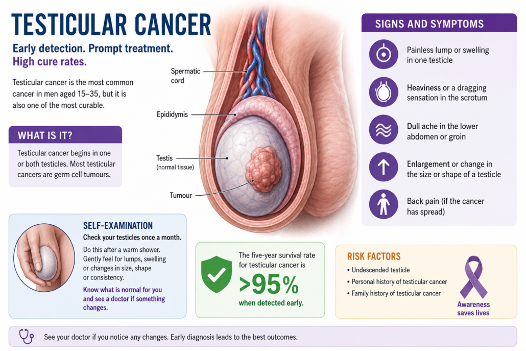
Testicular Cancer: Symptoms, Self-Examination, Diagnosis, Treatment and Prognosis
One of the most curable cancers in men
A lump in the testicle can be frightening, particularly because testicular cancer often occurs in younger men. The reassuring news is that testicular cancer is one of the most successfully treated solid cancers.
When detected early, cure rates approach 100%. Even when the cancer has spread to lymph nodes, lungs or other parts of the body, modern chemotherapy and surgery can still cure many men.
The most important message is therefore:
Know what is normal for you, and if something changes, have it checked.
Most testicular lumps are not cancer, but a new lump, enlargement, hardness or persistent change in a testicle deserves medical assessment and usually an ultrasound.
The importance of checking your testicles
There is no population screening program for testicular cancer in Australia.
This makes testicular awareness particularly important.
Cancer Council Australia recommends that men become familiar with the normal shape, size and feel of their testicles and seek medical attention if they notice a lump, swelling, heaviness, aching or another change.
This is slightly different from recommending a rigid population-wide screening program. There is currently insufficient evidence that formal scheduled self-examination reduces mortality from testicular cancer.
Nevertheless, knowing your own anatomy makes sense.
How do I check my testicles?
A convenient time is during or after a warm shower or bath when the scrotal skin is relaxed.
Gently examine one testicle at a time between your fingers and thumb.
Become familiar with:
- The usual size of each testicle
- Its shape
- Its firmness
- The fact that one testicle commonly hangs slightly lower
- The epididymis, which feels like a soft cord-like structure behind the testicle
You are not searching for microscopic abnormalities. You are simply learning what is normal for you.
Seek medical advice if you discover:
- A new hard lump
- Enlargement of one testicle
- A change in shape
- Increasing firmness
- Persistent swelling
- A heavy or dragging sensation
- Persistent testicular discomfort
- An unexplained difference from how the testicle normally feels
How often?
There is some variation between international recommendations.
The NHS in the United Kingdom advises checking the testicles regularly, approximately monthly, whereas Cancer Research UK emphasises awareness of what is normal rather than insisting upon a rigid monthly examination schedule.
In Australia, the practical message is best described as testicular awareness: become familiar with your testicles and investigate a persistent change rather than waiting to see whether it disappears.
Men at increased risk, including those with a previous undescended testicle, previous testicular cancer or significant family history, should discuss their individual surveillance with their doctor.
What is testicular cancer?
Approximately 90–95% of primary testicular cancers are germ-cell tumours.
They are divided into two broad groups:
Seminoma
Seminoma tends to behave in a relatively predictable manner and is extremely sensitive to both chemotherapy and radiotherapy.
Non-seminomatous germ-cell tumour (NSGCT)
This group includes:
- Embryonal carcinoma
- Yolk sac tumour
- Choriocarcinoma
- Teratoma
- Mixed germ-cell tumours
A tumour containing both seminoma and non-seminomatous components is treated as a non-seminomatous germ-cell tumour.
An important clinical rule is:
Pure seminoma should not produce AFP.
If AFP is significantly elevated, the tumour is managed clinically as a non-seminomatous germ-cell cancer even if seminoma is reported in the pathological specimen.
Other testicular tumours
Less common tumours include:
Leydig cell tumours
Most are benign, although malignant variants occur.
Sertoli cell tumours
Again, most are benign but malignant variants are recognised.
Testicular lymphoma
Lymphoma becomes particularly important in older men and is managed quite differently from conventional germ-cell cancer.
Occasionally another cancer can metastasise to the testicle.
How does testicular cancer present?
The classic presentation is a:
Painless testicular lump
A man may notice:
- Enlargement of one testicle
- A hard area
- A new lump
- Alteration in shape
- Increasing firmness
- Scrotal heaviness
Testicular cancer can also cause discomfort or pain.
Therefore:
Pain does not rule cancer in, and absence of pain does not rule cancer out.
Symptoms of more advanced disease
When cancer has spread beyond the testicle, symptoms can include:
- Back pain
- Abdominal discomfort
- Persistent cough
- Shortness of breath
- Chest symptoms
- Enlarged lymph nodes
- Weight loss
- Fatigue
- Breast tenderness or enlargement
Back pain can result from enlarged retroperitoneal lymph nodes behind the abdominal organs.
Diagnosis in Australia
The Australian diagnostic pathway is broadly consistent with European and British practice.
Step 1: Examination
Both testicles should be examined.
The abdomen and lymph-node regions may also be assessed.
Step 2: Testicular ultrasound
A high-resolution scrotal ultrasound with Doppler assessment is usually the first imaging investigation.
It determines whether a lesion is:
- Within the testicle
- Outside the testicle
- Solid
- Cystic
- Vascular
- Potentially malignant
A solid intratesticular mass should generally be considered malignant until proven otherwise.
Step 3: Tumour markers
Blood should ideally be collected before orchidectomy for:
AFP, Alpha-fetoprotein
May be elevated with:
- Yolk sac tumour
- Embryonal carcinoma
- Mixed germ-cell tumours
β-hCG, Beta human chorionic gonadotropin
Can be elevated in:
- Choriocarcinoma
- Embryonal carcinoma
- Mixed germ-cell tumours
- Some seminomas
LDH, Lactate dehydrogenase
LDH is less specific but provides information regarding tumour burden and prognosis.
Importantly:
Normal tumour markers do not exclude testicular cancer.
Markers are repeated after orchidectomy because the rate at which AFP and β-hCG fall provides valuable information regarding whether active cancer remains elsewhere.
Step 4: Staging
Staging commonly involves CT imaging of the:
- Chest
- Abdomen
- Pelvis
The retroperitoneal lymph nodes are particularly important because they represent the characteristic first lymphatic landing zone for many testicular cancers.
MRI can be used in selected circumstances.
PET scanning is not routinely recommended for initial staging.
FDG-PET has a specialised role after chemotherapy in selected patients with seminoma and a persistent residual mass.
Should the testicular lump be biopsied?
Usually no.
A needle biopsy through the scrotum is generally avoided when a germ-cell malignancy is suspected.
The standard procedure is:
Radical inguinal orchidectomy
The testicle and spermatic cord are removed through an incision in the groin.
This provides both treatment of the primary tumour and the tissue required for an accurate pathological diagnosis.
Fertility before treatment
This is particularly important because many patients are diagnosed while young.
The possibility of sperm banking should be discussed before chemotherapy, radiotherapy or other treatments that may impair fertility.
Ideally this conversation begins at diagnosis rather than after treatment has started.
One healthy remaining testicle will usually produce adequate testosterone and sperm, but some men with testicular cancer already have impaired sperm production before treatment.
Management of testicular cancer in Australia
Australian treatment is generally delivered through a multidisciplinary cancer team involving:
- Urologists
- Medical oncologists
- Radiation oncologists
- Radiologists
- Pathologists
- Fertility specialists when required
Complex metastatic, recurrent and post-chemotherapy disease is particularly suited to treatment through centres experienced in germ-cell cancer.
Australian practice broadly follows international evidence-based principles and is closely aligned with European and British practice, while incorporating Australian multidisciplinary cancer-care pathways.
Stage I seminoma
After radical inguinal orchidectomy, approximately 80% of men with unselected Stage I seminoma are cured by surgery alone.
For most reliable patients:
Active surveillance is generally preferred
This avoids exposing the majority of men who have already been cured to unnecessary chemotherapy or radiotherapy.
Surveillance involves scheduled:
- Clinical review
- Imaging
- Tumour markers where appropriate
If recurrence occurs, treatment is usually extremely successful.
Adjuvant carboplatin
A single cycle of carboplatin may be considered for selected patients who prefer adjuvant treatment or for whom surveillance is unsuitable.
Radiotherapy
Radiotherapy is extremely effective against seminoma but is now used much less frequently for Stage I disease because of concern regarding long-term:
- Secondary malignancies
- Cardiovascular effects
- Other radiation-related complications
Thus, in contemporary Australian practice, routine adjuvant radiotherapy for uncomplicated Stage I seminoma has largely moved into the background.
Stage I non-seminomatous germ-cell cancer
Approximately 70% of patients overall are cured by orchidectomy alone.
Management is influenced particularly by the presence or absence of lymphovascular invasion – LVI.
Without lymphovascular invasion
Surveillance is generally preferred for a reliable patient who is able to comply with follow-up.
With lymphovascular invasion
The risk of recurrence is considerably greater.
Options include:
- Surveillance
- One cycle of BEP chemotherapy
BEP consists of:
Bleomycin
Etoposide
Platinum – cisplatin
The advantages of avoiding unnecessary chemotherapy must be balanced against the increased likelihood of requiring several cycles of chemotherapy if metastatic recurrence subsequently occurs.
Retroperitoneal lymph-node dissection, RPLND
RPLND involves removal of lymph nodes from the retroperitoneum at the back of the abdomen.
It has an important but selective role.
In contemporary European and Australian-style practice, RPLND may be considered particularly for:
- Selected marker-negative Stage II NSGCT
- Residual masses after chemotherapy
- Teratoma
- Selected recurrent disease
- Particular patients in whom chemotherapy is undesirable
Modern nerve-sparing RPLND attempts to preserve the sympathetic nerves responsible for normal antegrade ejaculation.
These procedures should ideally be performed in experienced high-volume centres.
Stage II seminoma
For limited Stage IIA or IIB seminoma, treatment options can include:
- Radiotherapy
- Cisplatin-based chemotherapy
- In highly selected cases, specialist nerve-sparing RPLND
The precise choice depends upon lymph-node size, disease distribution, patient factors and the potential long-term consequences of each treatment.
For more extensive Stage IIB and Stage IIC disease, chemotherapy becomes increasingly favoured.
Metastatic seminoma and non-seminoma
Cisplatin-based combination chemotherapy transformed testicular cancer from a frequently fatal metastatic disease into one of the most curable metastatic solid cancers.
Common treatment includes:
BEP
Bleomycin + etoposide + cisplatin.
Depending upon the IGCCCG prognostic classification, treatment commonly consists of three or four cycles.
EP
Etoposide + cisplatin can be used in selected good-prognosis patients when bleomycin is unsuitable.
VIP
Etoposide + ifosfamide + cisplatin has a role in selected circumstances.
Surgery after chemotherapy
Surgery can remain crucial even after successful chemotherapy.
This is particularly important in non-seminomatous germ-cell cancer.
If tumour markers normalise but a residual retroperitoneal mass remains, surgery may reveal:
- Fibrosis or necrosis
- Mature teratoma
- Persistent viable cancer
Teratoma is particularly important because it can be relatively resistant to chemotherapy and radiotherapy.
The EAU recommends surgical resection of visible residual NSGCT masses greater than 1 cm when serum tumour markers are normal or normalising.
What about immunotherapy?
Immunotherapy has revolutionised treatment for several urological cancers.
Unfortunately, germ-cell cancer has not followed the same script.
Checkpoint inhibitors targeting PD-1, PD-L1 and related pathways have been investigated in chemotherapy-resistant germ-cell tumours, but responses have generally been disappointing.
Therefore:
Immunotherapy is not standard first-line treatment for conventional seminoma or NSGCT in Australia, Europe, Britain or the United States.
Its role is currently largely confined to highly selected refractory disease and clinical trials.
European, British, Australian and American guidelines – are they different?
The reassuring answer is:
The major principles are remarkably similar.
All emphasise:
- Prompt ultrasound of a suspicious testicular mass
- AFP, β-hCG and LDH assessment
- Radical inguinal orchidectomy
- Appropriate CT staging
- Histological distinction between seminoma and non-seminoma
- Fertility discussion and sperm banking
- Surveillance for many Stage I cancers
- Cisplatin-based chemotherapy for metastatic germ-cell cancer
- Specialist surgery for appropriate residual or retroperitoneal disease
There are, however, some interesting differences in emphasis.
European approach: EAU
The European Association of Urology guidelines strongly favour avoiding unnecessary treatment.
For Stage I seminoma, surveillance is preferred when the patient can comply with follow-up.
Routine adjuvant radiotherapy is not recommended.
For Stage I NSGCT, the EAU uses lymphovascular invasion prominently for risk-adapted counselling:
- LVI negative → surveillance generally preferred
- LVI positive → surveillance or one cycle of BEP
Primary RPLND has a relatively limited role in Stage I NSGCT.
For marker-negative Stage IIA NSGCT, however, the 2026 EAU guideline supports nerve-sparing RPLND in an experienced specialised centre.
British approach
British practice is broadly similar to European practice.
The NHS pathway centres around:
Ultrasound → tumour markers → inguinal orchidectomy → staging → multidisciplinary oncology review.
Surveillance is commonly used following orchidectomy for appropriate Stage I disease.
Carboplatin remains an option for Stage I seminoma, while BEP chemotherapy is used for appropriate non-seminomatous and metastatic disease.
Radiotherapy retains a role predominantly in selected seminoma rather than non-seminomatous cancer.
British cancer services place considerable emphasis on:
- Specialist germ-cell cancer multidisciplinary teams
- Fertility preservation
- Long-term follow-up
- Minimising unnecessary treatment toxicity
This is very similar to contemporary Australian practice.
How does the American approach differ?
American management follows the same oncological principles, but the American Urological Association – AUA – gives RPLND somewhat greater prominence as an acceptable primary treatment option in selected early-stage disease.
For Stage IA NSGCT, the AUA recommends surveillance but recognises:
- RPLND
- One cycle of BEP
as alternatives for appropriate patients who decline surveillance or may not comply reliably.
For Stage IB NSGCT, American guidance recognises:
- Surveillance
- RPLND
- One or two cycles of BEP
as options following shared decision-making.
This differs subtly from the EAU approach, where primary RPLND in Stage I NSGCT has a considerably narrower role.
Another evolving difference: RPLND for Stage II seminoma
Traditionally seminoma involving retroperitoneal lymph nodes was treated with either:
Radiotherapy or chemotherapy.
American guidelines have increasingly recognised primary RPLND as an option for carefully selected Stage IIA/IIB seminoma with limited retroperitoneal disease, particularly for patients wishing to avoid the potential long-term toxicity of chemotherapy or radiotherapy.
European recommendations are also evolving in this direction.
The 2026 EAU guideline now incorporates nerve-sparing RPLND and newer de-escalation strategies into the management discussion for selected Stage IIA/B seminoma.
This is an excellent example of why testicular cancer management continues to evolve.
Australia: where do we sit?
Australian practice sits comfortably between these international approaches.
For most Australian patients:
Stage I seminoma
Surveillance is generally preferred, with carboplatin available for selected patients.
Stage I NSGCT without LVI
Surveillance is generally preferred.
Stage I NSGCT with LVI
Surveillance or one cycle of BEP following individualised discussion.
Stage II seminoma
Radiotherapy or cisplatin-based chemotherapy depending upon disease volume, with increasingly selective consideration of surgical strategies in specialist centres.
Marker-positive metastatic disease
Cisplatin-based chemotherapy according to IGCCCG prognostic classification.
Residual NSGCT after chemotherapy
Surgical resection when indicated, ideally through an experienced germ-cell cancer service.
Australian cancer care increasingly emphasises multidisciplinary decision-making and treatment that achieves cure while reducing unnecessary long-term toxicity.
Why avoiding unnecessary treatment matters
A 25-year-old cured of testicular cancer may live another 60 years.
That changes the treatment equation.
The question is no longer simply:
“Which treatment will cure the cancer?”
It is also:
“Which treatment will cure this cancer while leaving the smallest possible footprint over the next several decades?”
This is why surveillance has become so important.
Chemotherapy and radiotherapy are extraordinarily effective, but they should be used when their benefit justifies their immediate and long-term risks.
Prognosis
The prognosis for testicular cancer is excellent.
Stage I disease has survival approaching 100%.
Even metastatic disease is frequently curable.
Prognosis depends upon:
- Seminoma versus non-seminoma
- Stage
- Tumour-marker levels
- Sites of metastatic disease
- Response to chemotherapy
- Tumour-marker decline
- Presence of residual disease
- Ability to completely resect appropriate residual masses
Importantly, doctors genuinely use the word cure when discussing metastatic testicular cancer.
After treatment: don’t forget the other testicle
Having had one testicular cancer increases the risk of developing cancer in the remaining testicle.
Men should therefore remain familiar with the remaining testicle and report any new abnormality promptly.
Long-term survivorship care may also address:
- Testosterone levels
- Fertility
- Cardiovascular health
- Kidney function
- Hearing
- Peripheral neuropathy
- Lung health following bleomycin
- Psychological wellbeing
- Sexual health
- Risk of late treatment complications
The take-home message
Testicular cancer tends to arrive at an inconvenient age, when most men are thinking about careers, relationships, families and weekend plans rather than cancer.
Fortunately, it is also one of medicine’s most impressive cancer success stories.
Know your testicles.
Become familiar with what is normal for you.
Don’t ignore a change.
A lump, enlargement, hardness, heaviness or persistent discomfort deserves examination.
Don’t be embarrassed.
Your urologist has quite literally made a career out of discussing these things.
And don’t assume a diagnosis of testicular cancer means the worst.
With modern surveillance, surgery, chemotherapy and selective radiotherapy, the overwhelming majority of men diagnosed with testicular cancer can expect to be cured.




Leave a Reply
Want to join the discussion?Feel free to contribute!