
Hydronephrosis: what it means and how it is treated
Finding out that a kidney is swollen on a scan can be worrying. The word hydronephrosis sounds like a diagnosis, but it describes an appearance: urine is collecting and stretching the drainage system of one or both kidneys. It is a sign that needs to be understood in context. It does not, by itself, tell us why it has happened, whether a cancer is present, or whether an operation is needed.
Many causes are treatable, and some cases can be monitored safely. The right approach depends on the cause, the degree and duration of blockage, kidney function, symptoms, overall health and, when cancer is involved, what matters most to the person receiving care.
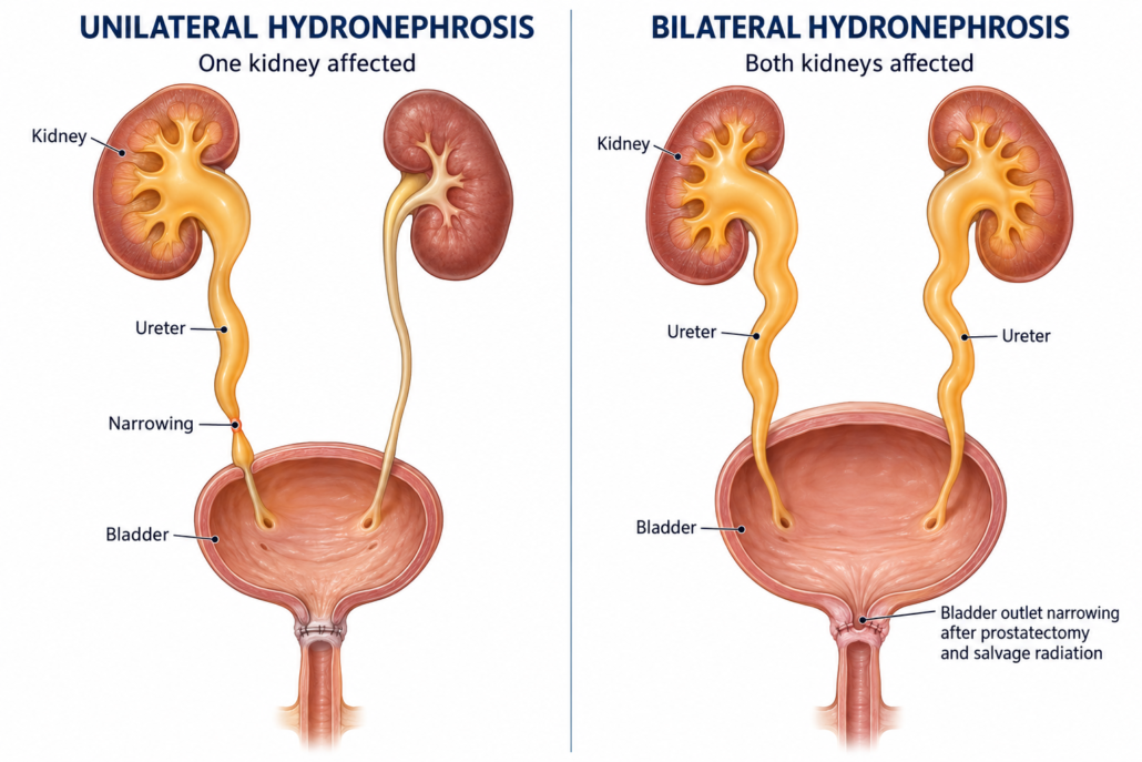
What are unilateral and bilateral hydronephrosis?
Each kidney drains urine through a tube called a ureter into the bladder. Unilateral hydronephrosis affects one kidney; bilateral hydronephrosis affects both. A blockage may be within the kidney or ureter, or lower down where it affects the bladder outlet. A full bladder or difficulty emptying can sometimes cause dilation on both sides.
One healthy kidney can often maintain overall kidney function when the other side is obstructed. This can mean that a person with a one-sided blockage has few symptoms and normal blood tests. With obstruction affecting both kidneys, or a person who has only one functioning kidney, kidney function can become impaired more quickly. The scan appearance alone does not show how well each kidney is working.
What can cause hydronephrosis?
Causes may be benign (non-cancerous) or malignant (related to cancer). They include:
- Kidney or ureteric stones, which may block urine flow and cause pain, blood in the urine or infection.
- Narrowing or scarring of the ureter (a stricture), sometimes following previous surgery, instrumentation, infection or radiation treatment.
- Bladder outlet obstruction or poor bladder emptying, including from an enlarged prostate before prostate surgery, urethral narrowing, a tight bladder-neck/anastomotic scar after surgery, or a bladder that does not contract effectively.
- Anatomical or functional conditions, such as a narrowing where the kidney joins the ureter (a pelvi-ureteric junction obstruction), reflux of urine towards the kidney, or in some circumstance, pregnancy.
- Blood clots or inflammation, which can temporarily obstruct drainage.
- Cancer, either growing in the urinary tract or pressing on a ureter from nearby tissues or lymph nodes. Possible cancers include prostate, bladder, bowel, gynaecological and other pelvic or abdominal cancers. A cancer-related cause is only one possibility; imaging and clinical assessment are needed to establish the cause.
Prostate cancer and hydronephrosis
Prostate cancer can affect urine drainage in different ways. A locally advanced tumour may involve the bladder outlet or the area where the ureters enter the bladder. Cancer in pelvic or abdominal lymph nodes can press on one or both ureters. These problems are more likely with locally advanced or recurrent disease than with an early cancer confined to the prostate, but each person’s situation is different.
Hydronephrosis in someone who has had prostate cancer does not automatically mean the cancer has returned. Stones, infection, scarring, bladder-emptying problems and unrelated conditions remain possible. The team will consider the person’s history, prostate-specific antigen (PSA) trend, symptoms, blood and urine tests, and the pattern seen on imaging. Further tests may be appropriate if the cause is not clear.
After radical prostatectomy
After prostate removal, the bladder is joined to the urethra. Scarring at this join (a vesicourethral anastomotic stenosis, sometimes called a bladder-neck contracture) can make it difficult to empty the bladder. Significant retention may raise pressure in the bladder and contribute to dilation of both upper urinary tracts. A ureteric injury or later narrowing is a less common possibility. Recurrent cancer or enlarged lymph nodes can also obstruct a ureter, but this is not the only explanation.
Assessment may include checking bladder emptying, kidney function and imaging, with a camera examination or contrast study if needed. Treatment is directed at the level and cause of the blockage: for example, bladder drainage, treatment of a narrowing, or drainage of an obstructed ureter.
After salvage radiation therapy
Radiation can cause inflammation and, less commonly, delayed scarring in urinary tissues. Narrowing of the urethra or bladder outlet can interfere with emptying. A ureteric stricture after pelvic radiation is uncommon, but may develop months or years later. Salvage radiation after prostatectomy is generally directed to the prostate bed; hydronephrosis that appears afterwards still needs a proper assessment rather than being assumed to be a radiation effect or a cancer recurrence.
If imaging shows a narrowed ureter, the team may use CT or MRI, a contrast study, or a functional kidney scan to clarify its site, length and effect on kidney drainage. Treatment can be more complex in previously irradiated tissue, so decisions are individualised and may involve a reconstructive urologist, radiation oncologist, medical oncologist or interventional radiologist.
Symptoms and when to seek urgent care
Hydronephrosis can cause no noticeable symptoms, particularly when it develops gradually or affects one kidney. When symptoms occur, they may include pain in the side, back or abdomen; nausea or vomiting; blood in the urine; difficulty passing urine; or repeated urinary infections.
Please seek urgent medical assessment for fever or shaking chills with suspected urinary blockage, severe or worsening pain, repeated vomiting, very little or no urine, confusion, or feeling suddenly very unwell. Infection behind a blockage can become serious and may need urgent antibiotics and drainage. Sudden inability to pass urine also needs prompt care.
How is it investigated?
The first aim is to understand whether there is a blockage that needs prompt relief and what is causing it. Assessment commonly includes:
- A conversation and examination, including previous cancer treatments, operations, stones, infections, urinary symptoms and medicines.
- Blood tests, especially creatinine and estimated kidney function, and urine tests for infection or blood.
- Ultrasound, which can show dilation of the kidneys and bladder and whether the bladder empties.
- CT or MRI, selected according to the suspected cause and the person’s kidney function, to look for stones, strictures, masses or enlarged lymph nodes.
- Additional tests where useful: a MAG3 renal scan can assess each kidney’s contribution and drainage; cystoscopy or contrast X-rays can clarify the bladder outlet or ureter.
Not everyone needs every test. The clinician will choose investigations that answer the question while taking account of kidney function, previous treatment and the person’s circumstances.
Management options
Treatment is tailored to the cause and the degree of risk. The goals may be to protect kidney function, treat infection or pain, restore urine flow, enable other cancer treatment, or support comfort and quality of life.
Observation and treatment of the cause
Mild, stable hydronephrosis may sometimes be monitored with repeat blood tests and imaging, particularly if there is no infection, troublesome pain or evidence of kidney function decline. A stone may pass or be treated with medication, ureteroscopy or another stone procedure. Infection is treated with antibiotics. Retention may be relieved with a bladder catheter while the cause is assessed. A benign narrowing may be treated by endoscopic incision or dilation, reconstruction, or another procedure depending on its location and the person’s health.
Draining the kidney
If drainage is needed, there are two common approaches:
- Ureteric stent (often called a JJ stent): a thin internal tube placed between the kidney and bladder, usually using a camera. It can avoid an external collection bag, but may cause urinary frequency, urgency, discomfort, blood in the urine or infection, and it usually needs planned replacement or removal. A tight narrowing or external cancer pressure can make stent placement difficult or less reliable.
- Percutaneous nephrostomy: a small tube placed through the skin of the back into the kidney, usually by an interventional radiologist, to drain urine into an external bag. It can be useful when a stent cannot be placed, has not drained adequately, or rapid reliable drainage is needed. The tube needs care and periodic changes and can affect daily activities.
The choice depends on the site and cause of obstruction, infection, anatomy, kidney function, expected duration, other treatment plans and personal preference. Sometimes a nephrostomy is used first, with an internal stent considered later. Drainage can relieve pressure and help kidney function, but it does not remove the underlying cause; further treatment or monitoring is often needed.
When might palliative care be part of the plan?
“Palliative” can sound frightening, but palliative care is focused on comfort, symptom relief, support and quality of life. It can be provided alongside active cancer treatment. It does not mean that someone is being abandoned or that all treatment has stopped.
When hydronephrosis is caused by advanced cancer, a stent or nephrostomy may help with pain, infection, kidney function, or the ability to receive cancer treatment. It can also bring burdens such as procedures, hospital visits, tube or stent symptoms, repeat exchanges and complications. In some situations, drainage may not improve symptoms or kidney function enough to outweigh those burdens. Evidence in this area is limited, and outcomes vary widely; population averages cannot predict an individual person’s future.
There is no single point at which palliation is “required.” The decision is made with the patient, their chosen supports and the treating team. It can help to discuss:
- What problem are we hoping drainage will improve?
- How likely is it to help, and how soon might we know?
- What would living with a stent or nephrostomy involve?
- Could the result allow another treatment that matters to me?
- What might happen if we monitor or manage symptoms without drainage?
- What matters most to me now: time at home, symptom relief, kidney function, further cancer treatment, or avoiding procedures?
Choosing not to have a procedure is also a valid choice when it best fits a person’s wishes and clinical circumstances. The team can still treat pain, nausea, infection and other symptoms, and provide practical and emotional support. These conversations are best held early and revisited as circumstances change.
In summary
Hydronephrosis means that urine is collecting in one or both kidneys, but it does not identify the cause on its own. Many causes are benign and treatable. Prostate cancer, previous prostate surgery and radiation can be relevant, but a careful assessment is needed before drawing conclusions. Some people need urgent drainage; others can be monitored or treated in a less invasive way. When cancer is advanced, decisions about drainage and palliative care should be compassionate, individualised and guided by what is most important to the person.
References and further reading
- National Kidney Foundation. Hydronephrosis. Updated 2023.
- Mayo Clinic. Hydronephrosis: Diagnosis and treatment. Updated 2024.
- European Association of Urology. EAU Guidelines on Urolithiasis. See section on sepsis and anuria in an obstructed kidney.
- National Institute for Health and Care Excellence. Prostate cancer: diagnosis and management (NG131). Includes recommendations on upper urinary tract decompression for obstructive uropathy secondary to hormone-relapsed prostate cancer.
- Chorbińska J, Krajewski W, Zdrojowy R. Urological complications after radiation therapy—nothing ventured, nothing gained: a narrative review. Transl Androl Urol. 2021;10(8):3553–3566. doi:10.21037/tau-20-1217.
- Carilli M, et al. Management of ureteral stricture disease after radiation therapy for pelvic malignancies. Cancers (Basel). 2024;16(21):3561. doi:10.3390/cancers16213561.
- Shah M, et al. Malignant upper urinary tract obstruction in cancer patients: a systematic review. BJUI Compass. 2024;5(11):915–925. doi:10.1002/bco2.340.
- Gauhar V, et al. Nephrostomy tube versus double J ureteral stent in patients with malignant ureteric obstruction: a systematic review and meta-analysis of comparative studies. Int Braz J Urol. 2022;48(6):903–914. doi:10.1590/S1677-5538.IBJU.2022.0225.
This page provides general information and is not a substitute for an individual assessment by a qualified health professional. If you have hydronephrosis, your clinician can explain what the scan means in your circumstances and whether further tests or treatment are needed.





Leave a Reply
Want to join the discussion?Feel free to contribute!