
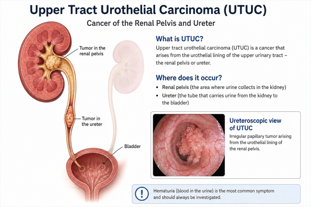
Upper Tract Urothelial Carcinoma
Cancer of the Renal Pelvis and Ureter
Urothelial carcinoma is most commonly associated with the bladder, but the same specialised lining extends all the way from the bladder up the ureters and into the collecting system of each kidney.
When a urothelial cancer develops in the renal pelvis or ureter, it is called upper tract urothelial carcinoma (UTUC).
UTUC is considerably less common than bladder cancer. Importantly, its behaviour varies enormously. Some tumours are small, superficial and relatively slow growing, while others are aggressive cancers capable of invading the kidney, surrounding tissues, lymph nodes and distant organs.
The key to successful management is therefore not simply finding the tumour, but determining how aggressive it is and how likely it is to spread.
What Is the Upper Urinary Tract?
Urine produced by the kidney drains through:
Kidney → renal calyces → renal pelvis → ureter → bladder → urethra
The inner surface of the renal pelvis, ureter and bladder is covered by urothelium, previously called transitional epithelium.
Cancer arising from these cells is called urothelial carcinoma.
UTUC can therefore occur in:
- the renal pelvis;
- one or more renal calyces;
- the upper, middle or lower ureter;
- multiple areas of the same urinary tract; or
- occasionally both upper urinary tracts.
Patients with UTUC may also develop urothelial tumours within the bladder, either at the same time or later.
What Causes Upper Tract Urothelial Cancer?
Cancer develops when genetic damage accumulates within urothelial cells, allowing them to grow and divide abnormally.
Frequently there is no single identifiable cause, but several important risk factors are recognised.
Smoking
Cigarette smoking is one of the most important preventable risk factors for urothelial carcinoma.
Carcinogenic chemicals absorbed through the lungs enter the bloodstream, are filtered by the kidneys and become concentrated in the urine. The urothelium may therefore be exposed to these substances for many years.
Stopping smoking remains important even after diagnosis because of its wider health benefits and potential relevance to future urothelial cancer risk.
Previous bladder cancer
Patients who have previously had urothelial carcinoma of the bladder have an increased risk of developing urothelial carcinoma elsewhere within the urinary tract.
Similarly, patients treated for UTUC remain at risk of subsequently developing bladder cancer.
Occupational chemical exposure
Long-term exposure to certain industrial chemicals, particularly some aromatic amines historically associated with dye, rubber, textile, leather and chemical industries, has been associated with urothelial cancer.
Lynch syndrome
A small but important proportion of UTUC occurs in people with Lynch syndrome, an inherited disorder caused by abnormalities in DNA mismatch-repair genes.
UTUC, particularly in a younger patient or someone with a strong family history of bowel, endometrial or other Lynch-associated cancers, may prompt consideration of genetic assessment.
Chronic inflammation
Long-standing urinary tract inflammation and some chronic infections may contribute to malignant change. Chronic stone disease has particularly been associated with squamous carcinoma of the renal pelvis, which is a different and much less common tumour type.
Previous analgesic exposure
Heavy historical exposure to certain analgesics, particularly phenacetin-containing preparations that are no longer routinely used in many countries, has been associated with upper tract urothelial malignancy.
What Are the Symptoms?
Blood in the urine
The most common warning sign is:
Haematuria: blood in the urine
This may be obvious, turning the urine pink, red or occasionally dark brown.
Importantly, haematuria may:
- occur only once;
- disappear for weeks or months;
- be completely painless; or
- only be detected microscopically on a urine test.
Visible blood in the urine should always be appropriately investigated, particularly in adults.
Do not assume that painless bleeding is simply due to infection, prostate enlargement or a blood-thinning medication without appropriate assessment.
Flank or loin pain
A tumour can obstruct drainage of urine from the kidney.
This may produce:
- persistent flank discomfort;
- loin pain;
- renal colic;
- hydronephrosis, where the kidney becomes dilated because urine cannot drain normally.
Blood clots passing down the ureter can occasionally produce severe colicky pain resembling a kidney stone.
Recurrent urinary symptoms
Some patients experience:
- urinary frequency;
- urgency;
- burning when passing urine;
- recurrent apparent urinary tract infections.
These symptoms are much more commonly caused by benign conditions, but persistent or unexplained symptoms may warrant further investigation.
General symptoms
More advanced disease can occasionally cause:
- unexplained weight loss;
- loss of appetite;
- fatigue;
- persistent pain;
- anaemia.
Fortunately, many tumours are detected before these symptoms develop.
How Is UTUC Diagnosed?
Investigation usually involves several complementary tests.
1. Urine testing
Urine is assessed for:
- microscopic blood;
- infection;
- renal abnormalities; and
- sometimes malignant cells.
Urine cytology
Urine cytology examines shed urinary cells under a microscope.
It is particularly useful for detecting high-grade urothelial carcinoma, although a negative cytology result does not completely exclude cancer.
Selective urine samples may sometimes be collected directly from the affected upper urinary tract.
2. CT Urography
CT urography is one of the most important imaging investigations for suspected UTUC.
Contrast-enhanced CT imaging allows assessment of:
- the kidneys;
- renal collecting systems;
- ureters;
- bladder;
- lymph nodes; and
- surrounding organs.
A tumour may appear as a filling defect, thickening of the ureter or renal pelvis, or an infiltrating mass.
CT can also identify obstruction and hydronephrosis.
3. Cystoscopy
Because urothelial cancer can occur at more than one location, the bladder should generally also be examined.
A flexible cystoscope is passed through the urethra into the bladder to look for associated bladder tumours.
Ureteroscopy and Biopsy
Sometimes imaging alone cannot provide enough information about the tumour.
A ureteroscope is a very fine telescope passed through the urethra and bladder and then into the ureter and renal collecting system.
This allows the urologist to directly inspect the tumour.
During ureteroscopy it may be possible to:
- identify the location of the tumour;
- assess whether there is one tumour or several;
- estimate its size;
- obtain selective urine cytology;
- take a biopsy; and
- in selected low-risk tumours, treat the lesion with a laser.
Biopsy is particularly helpful in determining whether the tumour is low-grade or high-grade.
An important limitation is that ureteroscopic biopsy samples are small, so determining the exact depth of invasion before definitive surgery can sometimes be difficult.
Low-Risk Versus High-Risk Disease
Modern management increasingly divides UTUC according to its risk of invasion and progression.
Factors considered include:
- tumour grade;
- appearance on CT;
- tumour size;
- number of tumours;
- presence of hydronephrosis;
- urine cytology;
- ureteroscopic appearance;
- biopsy findings;
- evidence of invasion; and
- previous urothelial cancer.
This distinction is extremely important because treatment can range from relatively conservative endoscopic therapy to removal of the entire kidney and ureter.
Treatment of Low-Risk UTUC
Selected patients with low-risk disease may be suitable for kidney-sparing treatment.
The objective is to control the cancer while preserving as much functioning kidney tissue as possible.
Ureteroscopic Laser Treatment
A ureteroscope is passed to the tumour and the lesion is treated using laser energy.
Laser treatment can:
- vaporise;
- coagulate; or
- fragment the tumour.
Modern flexible ureteroscopes allow access to much of the renal collecting system.
Advantages
Kidney-sparing treatment may:
- preserve kidney function;
- avoid major abdominal surgery;
- reduce recovery time;
- be particularly valuable in patients with impaired kidney function or a solitary kidney.
Disadvantages
The trade-off is the need for very careful surveillance.
UTUC has a tendency to recur, so patients may require repeated:
- ureteroscopy;
- urine cytology;
- CT imaging; and
- occasionally further laser treatments or biopsies.
Kidney preservation therefore does not mean that the tumour can simply be treated once and forgotten.
Segmental Ureterectomy
Some tumours confined to a relatively short section of ureter can be treated by removing only the affected portion.
This is known as segmental ureterectomy.
It is particularly useful for appropriately selected tumours of the distal ureter.
The affected segment is removed and the remaining ureter is either reconnected or reimplanted into the bladder.
This can preserve the kidney while providing a complete surgical specimen for pathological examination.
Treatment of High-Risk UTUC
For patients with high-risk localised disease, the standard definitive operation is generally:
Radical Nephroureterectomy
This involves removal of:
the kidney + entire ureter + a cuff of bladder surrounding the ureteric opening
Removing the complete ureter is important because leaving part of the affected ureter behind may allow cancer to recur within the remaining urothelium.
The procedure can often be performed using:
- robotic surgery;
- laparoscopic surgery; or
- occasionally open surgery.
The choice depends upon tumour characteristics, anatomy, previous surgery and individual circumstances.
What About the Lymph Nodes?
For selected high-risk tumours, lymph nodes draining the affected area may also be removed.
A lymph-node dissection can provide important staging information and may be incorporated into the surgical management of appropriately selected high-risk disease.
Chemotherapy
Systemic chemotherapy plays an important role in selected patients with higher-risk UTUC.
Platinum-based chemotherapy, particularly cisplatin-containing treatment, has traditionally formed an important part of systemic treatment.
Chemotherapy may be given:
Before surgery: neoadjuvant chemotherapy
One potential advantage is that both kidneys are still present, and renal function may therefore be better able to tolerate cisplatin.
After surgery: adjuvant chemotherapy
Chemotherapy may be recommended after nephroureterectomy when the final pathology demonstrates sufficiently high-risk disease.
An important consideration is that removing a kidney can reduce renal function, potentially affecting the patient’s ability to receive certain chemotherapy drugs.
Treatment decisions are therefore ideally made by a multidisciplinary uro-oncology team.
Immunotherapy and Newer Treatments
The treatment of advanced urothelial carcinoma has changed considerably in recent years.
Depending upon the stage, previous treatment and molecular characteristics of the cancer, systemic therapy may include combinations of:
- platinum-based chemotherapy;
- immune checkpoint inhibitors;
- antibody-drug conjugates; and
- targeted treatments for selected molecular abnormalities such as FGFR alterations.
These treatments are particularly relevant to patients with locally advanced, recurrent or metastatic disease.
Treatment is increasingly individualised according to the biological characteristics of the tumour and the patient’s kidney function and overall health.
Treatment Placed Directly Into the Upper Urinary Tract
For carefully selected non-invasive disease, medication may sometimes be delivered directly into the renal pelvis and ureter.
Options can include topical chemotherapy or immunotherapy in selected circumstances.
A chemoablative gel formulation of mitomycin has also been developed for selected low-grade UTUC, allowing prolonged contact between chemotherapy and the tumour.
These treatments are specialised and are not suitable for every patient.
What Happens After Treatment?
Follow-up is particularly important because urothelial carcinoma has a characteristic ability to develop at another point along the urinary tract.
After treatment, surveillance may include:
- cystoscopy;
- urine cytology;
- CT urography or other upper-tract imaging;
- blood tests to assess kidney function;
- ureteroscopy following kidney-sparing treatment.
The exact surveillance schedule depends upon whether the original tumour was low or high risk and what treatment was performed.
Can the Cancer Return in the Bladder?
Yes.
One of the distinctive features of UTUC is the significant risk of subsequently developing urothelial carcinoma within the bladder.
Published data have reported subsequent bladder tumours in a substantial proportion of patients following treatment of UTUC.
For this reason, regular cystoscopic surveillance remains important even when the original kidney or ureteric tumour has been completely removed.
What Is the Prognosis?
The outlook for UTUC depends predominantly upon:
- tumour stage;
- tumour grade;
- lymph-node involvement;
- metastatic spread;
- tumour multifocality;
- response to treatment; and
- certain pathological and molecular characteristics.
The most important question is whether the cancer remains superficial or has invaded deeply through the wall of the renal pelvis or ureter.
Superficial, localised disease
The outlook can be excellent when a tumour is identified while still superficial and confined to the upper urinary tract. The National Cancer Institute reports that more than 90% of superficial cancers confined to the renal pelvis or ureter may be curable.
Invasive disease
Once a tumour has invaded deeply into the wall or surrounding tissues, the risk of lymph-node involvement and distant spread increases substantially.
High-grade and invasive tumours therefore usually require more aggressive treatment and closer surveillance.
Metastatic disease
When UTUC has spread to distant organs, treatment is generally systemic rather than surgical alone.
Although metastatic UTUC remains a serious disease, modern systemic treatments have expanded considerably and can provide meaningful cancer control for selected patients.
UTUC and Kidney Function
Preserving kidney function is an important part of treatment planning.
Removing one kidney is usually well tolerated when the opposite kidney is healthy, but kidney function can become particularly important in patients with:
- pre-existing chronic kidney disease;
- diabetes;
- hypertension;
- a solitary kidney;
- bilateral upper-tract tumours; or
- conditions likely to affect future kidney function.
The decision between kidney-sparing treatment and radical nephroureterectomy therefore involves balancing two priorities:
adequate cancer control and preservation of renal function.
Cancer safety remains the priority, but in appropriately selected low-risk disease these objectives can often coexist.
When Should You See a Urologist?
Seek medical assessment if you develop:
- visible blood in the urine;
- persistent microscopic haematuria;
- unexplained flank or loin pain;
- recurrent unexplained urinary symptoms;
- abnormal findings on kidney imaging; or
- haematuria with a previous history of urothelial cancer.
Blood in the urine should not automatically be attributed to prostate enlargement, infection, kidney stones or blood-thinning medication without appropriate investigation.
The Bottom Line
Upper tract urothelial carcinoma is an uncommon cancer arising from the lining of the renal pelvis or ureter.
Blood in the urine is its most important warning sign.
Investigation commonly involves CT urography, cystoscopy, urine cytology and, when required, ureteroscopy with biopsy.
Treatment is increasingly tailored to the biological risk of the tumour.
Small, low-grade tumours may sometimes be managed with kidney-sparing ureteroscopic laser treatment or segmental surgery, while high-risk invasive cancers generally require radical nephroureterectomy with removal of the bladder cuff, sometimes combined with lymph-node surgery and systemic treatment.
Long-term surveillance is essential because urothelial cancer can recur elsewhere within the urinary tract, particularly the bladder.
The reassuring part is that when UTUC is detected while still superficial and localised, the likelihood of successful treatment can be very high.
A Note for Patients
Every upper tract urothelial tumour is different. Treatment depends upon the tumour’s location, size, grade, stage, number of lesions, kidney function and your general health.
Management should therefore be individualised following discussion with your urologist and, for higher-risk disease, a multidisciplinary uro-oncology team.
This information is intended for general patient education and should not replace individual medical advice, examination or treatment recommendations from your treating specialist.


Leave a Reply
Want to join the discussion?Feel free to contribute!