
Tag Archive for: urologist near me

Upper Tract Urothelial Carcinoma
Cancer of the Renal Pelvis and Ureter
Urothelial carcinoma is most commonly associated with the bladder, but the same specialised lining extends all the way from the bladder up the ureters and into the collecting system of each kidney.
When a urothelial cancer develops in the renal pelvis or ureter, it is called upper tract urothelial carcinoma (UTUC).
UTUC is considerably less common than bladder cancer. Importantly, its behaviour varies enormously. Some tumours are small, superficial and relatively slow growing, while others are aggressive cancers capable of invading the kidney, surrounding tissues, lymph nodes and distant organs.
The key to successful management is therefore not simply finding the tumour, but determining how aggressive it is and how likely it is to spread.
What Is the Upper Urinary Tract?
Urine produced by the kidney drains through:
Kidney → renal calyces → renal pelvis → ureter → bladder → urethra
The inner surface of the renal pelvis, ureter and bladder is covered by urothelium, previously called transitional epithelium.
Cancer arising from these cells is called urothelial carcinoma.
UTUC can therefore occur in:
- the renal pelvis;
- one or more renal calyces;
- the upper, middle or lower ureter;
- multiple areas of the same urinary tract; or
- occasionally both upper urinary tracts.
Patients with UTUC may also develop urothelial tumours within the bladder, either at the same time or later.
What Causes Upper Tract Urothelial Cancer?
Cancer develops when genetic damage accumulates within urothelial cells, allowing them to grow and divide abnormally.
Frequently there is no single identifiable cause, but several important risk factors are recognised.
Smoking
Cigarette smoking is one of the most important preventable risk factors for urothelial carcinoma.
Carcinogenic chemicals absorbed through the lungs enter the bloodstream, are filtered by the kidneys and become concentrated in the urine. The urothelium may therefore be exposed to these substances for many years.
Stopping smoking remains important even after diagnosis because of its wider health benefits and potential relevance to future urothelial cancer risk.
Previous bladder cancer
Patients who have previously had urothelial carcinoma of the bladder have an increased risk of developing urothelial carcinoma elsewhere within the urinary tract.
Similarly, patients treated for UTUC remain at risk of subsequently developing bladder cancer.
Occupational chemical exposure
Long-term exposure to certain industrial chemicals, particularly some aromatic amines historically associated with dye, rubber, textile, leather and chemical industries, has been associated with urothelial cancer.
Lynch syndrome
A small but important proportion of UTUC occurs in people with Lynch syndrome, an inherited disorder caused by abnormalities in DNA mismatch-repair genes.
UTUC, particularly in a younger patient or someone with a strong family history of bowel, endometrial or other Lynch-associated cancers, may prompt consideration of genetic assessment.
Chronic inflammation
Long-standing urinary tract inflammation and some chronic infections may contribute to malignant change. Chronic stone disease has particularly been associated with squamous carcinoma of the renal pelvis, which is a different and much less common tumour type.
Previous analgesic exposure
Heavy historical exposure to certain analgesics, particularly phenacetin-containing preparations that are no longer routinely used in many countries, has been associated with upper tract urothelial malignancy.
What Are the Symptoms?
Blood in the urine
The most common warning sign is:
Haematuria: blood in the urine
This may be obvious, turning the urine pink, red or occasionally dark brown.
Importantly, haematuria may:
- occur only once;
- disappear for weeks or months;
- be completely painless; or
- only be detected microscopically on a urine test.
Visible blood in the urine should always be appropriately investigated, particularly in adults.
Do not assume that painless bleeding is simply due to infection, prostate enlargement or a blood-thinning medication without appropriate assessment.
Flank or loin pain
A tumour can obstruct drainage of urine from the kidney.
This may produce:
- persistent flank discomfort;
- loin pain;
- renal colic;
- hydronephrosis, where the kidney becomes dilated because urine cannot drain normally.
Blood clots passing down the ureter can occasionally produce severe colicky pain resembling a kidney stone.
Recurrent urinary symptoms
Some patients experience:
- urinary frequency;
- urgency;
- burning when passing urine;
- recurrent apparent urinary tract infections.
These symptoms are much more commonly caused by benign conditions, but persistent or unexplained symptoms may warrant further investigation.
General symptoms
More advanced disease can occasionally cause:
- unexplained weight loss;
- loss of appetite;
- fatigue;
- persistent pain;
- anaemia.
Fortunately, many tumours are detected before these symptoms develop.
How Is UTUC Diagnosed?
Investigation usually involves several complementary tests.
1. Urine testing
Urine is assessed for:
- microscopic blood;
- infection;
- renal abnormalities; and
- sometimes malignant cells.
Urine cytology
Urine cytology examines shed urinary cells under a microscope.
It is particularly useful for detecting high-grade urothelial carcinoma, although a negative cytology result does not completely exclude cancer.
Selective urine samples may sometimes be collected directly from the affected upper urinary tract.
2. CT Urography
CT urography is one of the most important imaging investigations for suspected UTUC.
Contrast-enhanced CT imaging allows assessment of:
- the kidneys;
- renal collecting systems;
- ureters;
- bladder;
- lymph nodes; and
- surrounding organs.
A tumour may appear as a filling defect, thickening of the ureter or renal pelvis, or an infiltrating mass.
CT can also identify obstruction and hydronephrosis.
3. Cystoscopy
Because urothelial cancer can occur at more than one location, the bladder should generally also be examined.
A flexible cystoscope is passed through the urethra into the bladder to look for associated bladder tumours.
Ureteroscopy and Biopsy
Sometimes imaging alone cannot provide enough information about the tumour.
A ureteroscope is a very fine telescope passed through the urethra and bladder and then into the ureter and renal collecting system.
This allows the urologist to directly inspect the tumour.
During ureteroscopy it may be possible to:
- identify the location of the tumour;
- assess whether there is one tumour or several;
- estimate its size;
- obtain selective urine cytology;
- take a biopsy; and
- in selected low-risk tumours, treat the lesion with a laser.
Biopsy is particularly helpful in determining whether the tumour is low-grade or high-grade.
An important limitation is that ureteroscopic biopsy samples are small, so determining the exact depth of invasion before definitive surgery can sometimes be difficult.
Low-Risk Versus High-Risk Disease
Modern management increasingly divides UTUC according to its risk of invasion and progression.
Factors considered include:
- tumour grade;
- appearance on CT;
- tumour size;
- number of tumours;
- presence of hydronephrosis;
- urine cytology;
- ureteroscopic appearance;
- biopsy findings;
- evidence of invasion; and
- previous urothelial cancer.
This distinction is extremely important because treatment can range from relatively conservative endoscopic therapy to removal of the entire kidney and ureter.
Treatment of Low-Risk UTUC
Selected patients with low-risk disease may be suitable for kidney-sparing treatment.
The objective is to control the cancer while preserving as much functioning kidney tissue as possible.
Ureteroscopic Laser Treatment
A ureteroscope is passed to the tumour and the lesion is treated using laser energy.
Laser treatment can:
- vaporise;
- coagulate; or
- fragment the tumour.
Modern flexible ureteroscopes allow access to much of the renal collecting system.
Advantages
Kidney-sparing treatment may:
- preserve kidney function;
- avoid major abdominal surgery;
- reduce recovery time;
- be particularly valuable in patients with impaired kidney function or a solitary kidney.
Disadvantages
The trade-off is the need for very careful surveillance.
UTUC has a tendency to recur, so patients may require repeated:
- ureteroscopy;
- urine cytology;
- CT imaging; and
- occasionally further laser treatments or biopsies.
Kidney preservation therefore does not mean that the tumour can simply be treated once and forgotten.
Segmental Ureterectomy
Some tumours confined to a relatively short section of ureter can be treated by removing only the affected portion.
This is known as segmental ureterectomy.
It is particularly useful for appropriately selected tumours of the distal ureter.
The affected segment is removed and the remaining ureter is either reconnected or reimplanted into the bladder.
This can preserve the kidney while providing a complete surgical specimen for pathological examination.
Treatment of High-Risk UTUC
For patients with high-risk localised disease, the standard definitive operation is generally:
Radical Nephroureterectomy
This involves removal of:
the kidney + entire ureter + a cuff of bladder surrounding the ureteric opening
Removing the complete ureter is important because leaving part of the affected ureter behind may allow cancer to recur within the remaining urothelium.
The procedure can often be performed using:
- robotic surgery;
- laparoscopic surgery; or
- occasionally open surgery.
The choice depends upon tumour characteristics, anatomy, previous surgery and individual circumstances.
What About the Lymph Nodes?
For selected high-risk tumours, lymph nodes draining the affected area may also be removed.
A lymph-node dissection can provide important staging information and may be incorporated into the surgical management of appropriately selected high-risk disease.
Chemotherapy
Systemic chemotherapy plays an important role in selected patients with higher-risk UTUC.
Platinum-based chemotherapy, particularly cisplatin-containing treatment, has traditionally formed an important part of systemic treatment.
Chemotherapy may be given:
Before surgery: neoadjuvant chemotherapy
One potential advantage is that both kidneys are still present, and renal function may therefore be better able to tolerate cisplatin.
After surgery: adjuvant chemotherapy
Chemotherapy may be recommended after nephroureterectomy when the final pathology demonstrates sufficiently high-risk disease.
An important consideration is that removing a kidney can reduce renal function, potentially affecting the patient’s ability to receive certain chemotherapy drugs.
Treatment decisions are therefore ideally made by a multidisciplinary uro-oncology team.
Immunotherapy and Newer Treatments
The treatment of advanced urothelial carcinoma has changed considerably in recent years.
Depending upon the stage, previous treatment and molecular characteristics of the cancer, systemic therapy may include combinations of:
- platinum-based chemotherapy;
- immune checkpoint inhibitors;
- antibody-drug conjugates; and
- targeted treatments for selected molecular abnormalities such as FGFR alterations.
These treatments are particularly relevant to patients with locally advanced, recurrent or metastatic disease.
Treatment is increasingly individualised according to the biological characteristics of the tumour and the patient’s kidney function and overall health.
Treatment Placed Directly Into the Upper Urinary Tract
For carefully selected non-invasive disease, medication may sometimes be delivered directly into the renal pelvis and ureter.
Options can include topical chemotherapy or immunotherapy in selected circumstances.
A chemoablative gel formulation of mitomycin has also been developed for selected low-grade UTUC, allowing prolonged contact between chemotherapy and the tumour.
These treatments are specialised and are not suitable for every patient.
What Happens After Treatment?
Follow-up is particularly important because urothelial carcinoma has a characteristic ability to develop at another point along the urinary tract.
After treatment, surveillance may include:
- cystoscopy;
- urine cytology;
- CT urography or other upper-tract imaging;
- blood tests to assess kidney function;
- ureteroscopy following kidney-sparing treatment.
The exact surveillance schedule depends upon whether the original tumour was low or high risk and what treatment was performed.
Can the Cancer Return in the Bladder?
Yes.
One of the distinctive features of UTUC is the significant risk of subsequently developing urothelial carcinoma within the bladder.
Published data have reported subsequent bladder tumours in a substantial proportion of patients following treatment of UTUC.
For this reason, regular cystoscopic surveillance remains important even when the original kidney or ureteric tumour has been completely removed.
What Is the Prognosis?
The outlook for UTUC depends predominantly upon:
- tumour stage;
- tumour grade;
- lymph-node involvement;
- metastatic spread;
- tumour multifocality;
- response to treatment; and
- certain pathological and molecular characteristics.
The most important question is whether the cancer remains superficial or has invaded deeply through the wall of the renal pelvis or ureter.
Superficial, localised disease
The outlook can be excellent when a tumour is identified while still superficial and confined to the upper urinary tract. The National Cancer Institute reports that more than 90% of superficial cancers confined to the renal pelvis or ureter may be curable.
Invasive disease
Once a tumour has invaded deeply into the wall or surrounding tissues, the risk of lymph-node involvement and distant spread increases substantially.
High-grade and invasive tumours therefore usually require more aggressive treatment and closer surveillance.
Metastatic disease
When UTUC has spread to distant organs, treatment is generally systemic rather than surgical alone.
Although metastatic UTUC remains a serious disease, modern systemic treatments have expanded considerably and can provide meaningful cancer control for selected patients.
UTUC and Kidney Function
Preserving kidney function is an important part of treatment planning.
Removing one kidney is usually well tolerated when the opposite kidney is healthy, but kidney function can become particularly important in patients with:
- pre-existing chronic kidney disease;
- diabetes;
- hypertension;
- a solitary kidney;
- bilateral upper-tract tumours; or
- conditions likely to affect future kidney function.
The decision between kidney-sparing treatment and radical nephroureterectomy therefore involves balancing two priorities:
adequate cancer control and preservation of renal function.
Cancer safety remains the priority, but in appropriately selected low-risk disease these objectives can often coexist.
When Should You See a Urologist?
Seek medical assessment if you develop:
- visible blood in the urine;
- persistent microscopic haematuria;
- unexplained flank or loin pain;
- recurrent unexplained urinary symptoms;
- abnormal findings on kidney imaging; or
- haematuria with a previous history of urothelial cancer.
Blood in the urine should not automatically be attributed to prostate enlargement, infection, kidney stones or blood-thinning medication without appropriate investigation.
The Bottom Line
Upper tract urothelial carcinoma is an uncommon cancer arising from the lining of the renal pelvis or ureter.
Blood in the urine is its most important warning sign.
Investigation commonly involves CT urography, cystoscopy, urine cytology and, when required, ureteroscopy with biopsy.
Treatment is increasingly tailored to the biological risk of the tumour.
Small, low-grade tumours may sometimes be managed with kidney-sparing ureteroscopic laser treatment or segmental surgery, while high-risk invasive cancers generally require radical nephroureterectomy with removal of the bladder cuff, sometimes combined with lymph-node surgery and systemic treatment.
Long-term surveillance is essential because urothelial cancer can recur elsewhere within the urinary tract, particularly the bladder.
The reassuring part is that when UTUC is detected while still superficial and localised, the likelihood of successful treatment can be very high.
A Note for Patients
Every upper tract urothelial tumour is different. Treatment depends upon the tumour’s location, size, grade, stage, number of lesions, kidney function and your general health.
Management should therefore be individualised following discussion with your urologist and, for higher-risk disease, a multidisciplinary uro-oncology team.
This information is intended for general patient education and should not replace individual medical advice, examination or treatment recommendations from your treating specialist.
The current 2026 EAU guideline specifically incorporates updated risk stratification, kidney-sparing management, bladder-cuff and lymph-node considerations, systemic therapy, and follow-up recommendations.
Focal Therapy for Prostate Cancer: Treating the Cancer, Preserving the Prostate
For many years, treatment of localised prostate cancer largely involved choosing between active surveillance and treatment of the whole prostate gland with surgery or radiotherapy.
Modern multiparametric MRI, targeted transperineal biopsy and increasingly accurate image-guided treatment have opened a third pathway for carefully selected men: focal therapy.
Rather than treating or removing the entire prostate, focal therapy aims to identify the clinically significant cancer and destroy that area together with an appropriate safety margin, while leaving as much normal prostate tissue as possible.
A useful analogy is treating the troublesome patch rather than replacing the entire lawn.
The attraction is obvious: if the cancer can be controlled without treating the whole prostate, it may be possible to reduce the risks of urinary incontinence, erectile dysfunction and other quality-of-life effects associated with radical treatment.
However, focal therapy is not suitable for every prostate cancer, and it comes with an important trade-off: long-term cancer-control evidence is less mature than it is for radical prostatectomy and radiotherapy. Current European guidance therefore remains cautious, recommending focal therapy within clinical trials or well-designed prospective registries until stronger long-term comparative evidence becomes available.
What is focal therapy?
Focal therapy treats a selected region of the prostate containing clinically significant cancer rather than treating the entire gland.
Depending on the size and location of the tumour, treatment may involve:
- Focal ablation of an individual lesion
- Hemi-ablation, treating approximately one side of the prostate
- Quadrant or zonal ablation
- A wider “hockey-stick” ablation where disease distribution requires a larger treatment field
The treatment zone normally includes both the visible tumour and a planned margin around it.
The challenge is that prostate cancer is frequently multifocal. The largest or most biologically significant lesion is often referred to as the index lesion, but smaller cancer deposits may exist elsewhere in the gland.
For this reason, careful imaging, biopsy and follow-up are fundamental to a successful focal therapy program.
Who may be suitable for focal therapy?
The ideal candidate is generally a man with localised, clinically significant prostate cancer that can be accurately identified and safely targeted.
Potential candidates may include men with:
- Disease confined to the prostate
- A clearly identifiable lesion on multiparametric MRI
- Cancer confirmed by targeted and systematic or mapping transperineal biopsy
- Favourable intermediate-risk disease, commonly ISUP Grade Group 2 / Gleason 3+4, in an appropriate anatomical distribution
- Selected higher-volume Grade Group 1 disease where active surveillance is considered unsuitable or unacceptable
- Occasionally carefully selected Grade Group 3 disease in experienced centres, although the evidence is less established
- A lesion that can be treated with an adequate margin without unacceptable injury to the urethra, sphincter, rectum or neurovascular structures
- A strong preference to minimise the potential urinary and sexual consequences of whole-gland treatment
The decision should ideally follow review of the MRI, biopsy pathology, PSA, PSA density, prostate volume, tumour location and overall risk profile, rather than simply asking whether a particular machine can reach the tumour.
Who is generally NOT a good candidate?
Focal treatment becomes less attractive when there is:
- Extensive multifocal clinically significant cancer
- Significant bilateral disease
- High-volume high-grade cancer
- Extracapsular extension
- Seminal vesicle invasion
- Lymph-node involvement
- Metastatic disease
- Cancer that cannot be reliably seen or mapped
- Disease immediately adjacent to structures that cannot safely be included in the treatment margin
- A patient preference for the treatment with the longest-established oncological follow-up
Some men with very low-risk disease may also be better served by active surveillance rather than focal treatment, avoiding treatment altogether until there is evidence that treatment is actually necessary.
How do we determine whether focal treatment is appropriate?
Successful focal therapy begins with accurate cancer mapping.
Assessment will usually include:
Multiparametric MRI
MRI identifies suspicious lesions and helps establish their size, location and relationship to the urethra, capsule, sphincter and neurovascular bundles.
Transperineal prostate biopsy
MRI alone is not enough.
Targeted biopsy confirms the grade and extent of the MRI-visible lesion, while systematic or mapping biopsies help determine whether significant cancer exists elsewhere in the prostate.
PSA and PSA density
PSA remains useful, although interpretation after focal therapy differs from interpretation following radical prostatectomy because normal prostate tissue remains behind.
PSMA PET/CT
PSMA PET may be useful in selected patients, particularly those with higher-risk characteristics or when there is concern about disease outside the proposed treatment area.
What focal therapy options are available?
Several technologies can destroy a selected area of prostate tissue.
These include:
Irreversible Electroporation: NanoKnife
NanoKnife® is a system used to perform irreversible electroporation, or IRE.
Several fine needle electrodes are inserted through the perineum around the tumour under imaging guidance. Very short, high-voltage electrical pulses are passed between the electrodes.
Rather than primarily heating or freezing the tissue, the electrical field creates irreversible disruption of cell membranes, resulting in cell death.
IRE is therefore principally considered a non-thermal ablative technology.
Focal Laser Ablation
A laser fibre is placed directly into the target lesion and laser energy produces controlled thermal destruction of cancerous tissue.
ProFocal-Rx® is an Australian-developed focal laser technology designed specifically for targeted prostate treatment.
High-Intensity Focused Ultrasound
HIFU focuses ultrasound energy within the prostate, heating and destroying the targeted tissue without requiring needles to be placed directly throughout the treatment zone.
Cryotherapy
Needles are placed into the prostate and tissue is repeatedly frozen and thawed, producing cellular destruction.
Other technologies
Photodynamic therapy, radiofrequency ablation, focal brachytherapy and other energy-based approaches have also been investigated.
The Prostate Cancer Foundation of Australia notes that focal therapies including IRE/NanoKnife, laser ablation, HIFU and other technologies have been investigated or used in Australia, although availability varies.
NanoKnife versus ProFocal Laser Therapy
Both technologies attempt to achieve the same broad objective: destroy the cancer while preserving as much normal prostate and surrounding function as possible.
They achieve this in very different ways.
| NanoKnife / IRE | ProFocal-Rx Laser | |
|---|---|---|
| Energy | High-voltage electrical pulses | Laser energy |
| Mechanism | Irreversible electroporation | Thermal coagulative ablation |
| Thermal treatment | Principally non-thermal | Yes |
| Access | Transperineal needles/electrodes | Transperineal laser applicator |
| Treatment planning | Electrode geometry surrounds treatment zone | Laser applicator positioned within/adjacent to target |
| MRI/TRUS planning | Yes | Yes |
| Tissue effect | Cell membrane disruption | Controlled heating and tissue necrosis |
| Treatment margin | Created by electrical field between electrodes | Created by laser ablation zone |
| Near neurovascular structures | Potential theoretical advantage of non-thermal mechanism | Requires careful thermal planning |
| Anaesthesia | General anaesthesia with profound muscle relaxation generally required | General anaesthesia typically used |
| Cardiac synchronisation | Required with IRE | Not required in the same manner |
| Repeat treatment | Possible in selected cases | Potentially possible |
| Long-term oncological evidence | Growing medium-term evidence | Earlier-stage clinical evidence |
| Australian regulatory status | IRE devices are represented on the ARTG; specific device/indication should be checked | ProFocal is currently not included on the ARTG |
NanoKnife: potential advantages
The major attraction of IRE is that it does not rely primarily upon heating or freezing the prostate.
The electrical field disrupts cell membranes while potentially allowing relative preservation of extracellular structures. This makes IRE particularly interesting when treating cancers close to delicate structures.
Potential advantages include:
- Precise treatment planning
- No ionising radiation
- No prostate removal
- Preservation of untreated prostate tissue
- Low reported rates of significant urinary incontinence
- Potentially better preservation of erectile function compared with whole-gland treatment
- Ability to consider repeat focal treatment in selected patients
- Radical surgery or radiotherapy may remain possible if subsequent clinically significant cancer develops
Australian and international experience with IRE is considerably more mature than that of many newer focal technologies, although long-term comparative data against radical prostatectomy and radiotherapy are still developing. Published reviews cited by the AUA report residual or recurrent clinically significant cancer after focal ablation across all technologies, reinforcing the need for surveillance rather than considering focal therapy a “treat it and forget it” procedure.
NanoKnife: disadvantages and potential complications
IRE is still an invasive procedure.
Potential complications include:
- Temporary urinary frequency and urgency
- Dysuria
- Haematuria
- Perineal bruising or discomfort
- Urinary retention
- Temporary catheter requirement
- Urinary tract infection
- Prostatitis
- Urethral injury or stricture
- Erectile dysfunction
- Ejaculatory changes
- Rare urinary incontinence
- Incomplete tumour ablation
- Residual cancer within the treated field
- Development or recognition of cancer elsewhere in the prostate
- Need for repeat focal treatment
- Subsequent need for radical prostatectomy or radiotherapy
Because IRE uses high-voltage electrical pulses, treatment requires appropriate anaesthesia, muscle relaxation and cardiac synchronisation.
ProFocal-Rx: focal laser therapy
ProFocal-Rx is a targeted laser ablation system developed in Australia.
A treatment applicator is placed transperineally into the prostate tumour. Laser energy is then delivered into the planned treatment area, producing controlled thermal destruction.
Early Australian studies have evaluated the feasibility and safety of this approach, including clinical trials of targeted treatment for MRI-localised prostate cancer.
Potential attractions include:
- Highly localised treatment
- Direct placement of the treatment fibre into the tumour
- Relatively small treatment volumes
- Preservation of surrounding prostate tissue
- Short treatment and recovery pathways
- Potential preservation of urinary continence
- Potential preservation of erectile and ejaculatory function
However, ProFocal remains a newer technology with substantially less long-term oncological follow-up than radical prostatectomy, radiotherapy and even some other focal therapy platforms.
ProFocal: potential risks and limitations
Because laser treatment is thermal, careful treatment planning is required to prevent unintended heat injury.
Possible complications include:
- Urinary frequency or urgency
- Dysuria
- Haematuria
- Temporary urinary retention
- Infection
- Perineal discomfort
- Urethral thermal injury
- Erectile dysfunction
- Ejaculatory changes
- Rectal injury, although uncommon with appropriate treatment planning
- Incomplete ablation
- Residual clinically significant cancer
- Cancer developing or being detected elsewhere in the prostate
- Need for repeat treatment
- Need for subsequent radical prostatectomy or radiotherapy
An important additional consideration is simply the maturity of the evidence. Early results can be encouraging without necessarily predicting cancer control at 10, 15 or 20 years.
What is the TGA status in Australia?
This point deserves particular clarity.
The Australian Register of Therapeutic Goods (ARTG) is the TGA’s public register of therapeutic products that can legally be supplied in Australia, subject to applicable exemptions and special-access pathways.
IRE / NanoKnife
Irreversible electroporation technology is available in Australia and is being used clinically for selected prostate cancers. The Medical Services Advisory Committee currently has an application assessing IRE using the NanoKnife system for prostate tumour tissue, including a proposed Medicare Benefits Schedule item. That MSAC application remains under assessment rather than representing an established Medicare item.
It is important not to confuse TGA/ARTG regulatory status with Medicare funding or with endorsement of focal therapy as oncologically equivalent to prostatectomy or radiotherapy. These are separate questions.
ProFocal-Rx
As of August 2026, the manufacturer’s Australian website specifically states that:
ProFocal is not included on the TGA’s ARTG in Australia.
TGA documents also demonstrate previous Australian patient access to ProFocal-Rx through the Special Access Scheme, which is a pathway for accessing an unapproved therapeutic good in particular circumstances and is not the same as general ARTG inclusion.
This distinction is important when discussing ProFocal with Australian patients.
Regulatory status can change, so the current ARTG should always be checked when treatment is being considered.
Does focal therapy cure prostate cancer?
It can achieve local control of appropriately selected prostate cancers, but the word “cure” needs to be used carefully.
Unlike radical prostatectomy, focal therapy deliberately leaves much of the prostate behind.
There are therefore two important potential sites of future cancer:
In-field recurrence
Cancer persists or recurs within the treated area.
Out-of-field cancer
Clinically significant cancer is subsequently detected elsewhere in the untreated prostate.
Neither necessarily means that focal therapy was inappropriate, but patients need to understand from the outset that continued prostate cancer surveillance is part of the treatment strategy.
The AUA’s salvage guideline notes clinically significant cancer following focal treatment across different modalities and emphasises that recurrence remains an important consideration after focal ablation.
Follow-up after focal therapy
Focal therapy does not end prostate cancer surveillance.
Follow-up typically involves a combination of:
- Regular PSA testing
- Clinical review
- Multiparametric MRI
- Repeat targeted and systematic biopsy
- Additional imaging where clinically indicated
A common strategy is to establish a new PSA baseline after treatment and combine PSA behaviour with MRI and scheduled biopsy rather than relying on PSA alone.
This is important because the remaining normal prostate continues to produce PSA. Unlike after radical prostatectomy, the PSA is therefore not expected to become undetectable.
What happens if the cancer returns?
One of the advantages of focal treatment is that further treatment options usually remain available.
Depending upon the location, grade and extent of recurrent disease, options may include:
- Continued surveillance for insignificant disease
- Repeat focal therapy
- Radical prostatectomy
- External-beam radiotherapy
- Other appropriate salvage treatment
Patients should nevertheless understand that salvage surgery after previous focal therapy may be technically more challenging because of fibrosis and altered tissue planes.
For clinically significant recurrence following focal ablation, AUA salvage guidance recommends that men considering definitive salvage treatment be offered whole-gland treatment with radical prostatectomy or radiotherapy.
Focal therapy versus radical treatment
Focal therapy occupies an increasingly interesting middle ground.
Active surveillance aims to avoid treatment until treatment becomes necessary.
Focal therapy aims to treat the clinically significant cancer while preserving the remainder of the prostate.
Radical prostatectomy or radiotherapy aims to treat the entire prostate and therefore both known and potentially occult cancer within the gland.
There is no universally “best” choice.
The appropriate treatment depends upon:
- Cancer grade
- Cancer volume
- MRI findings
- Biopsy distribution
- PSA and PSA density
- Age and life expectancy
- Baseline urinary function
- Baseline erectile function
- Other medical conditions
- Individual attitude towards cancer risk
- Willingness to undergo continued MRI and biopsy surveillance
- Personal priorities regarding continence and sexual function
The key question: are we treating the right cancer?
The success of focal therapy depends less on the glamour of the machine and more on patient selection, accurate imaging, meticulous biopsy mapping, treatment planning and rigorous follow-up.
NanoKnife, laser, HIFU and cryotherapy are different tools. The most important step occurs before any of them are switched on: establishing exactly where the clinically significant cancer is and whether disease elsewhere in the prostate has been adequately excluded.
For the appropriately selected man, focal therapy offers an attractive possibility:
Treat the cancer that needs treatment while preserving as much of the prostate, urinary function and sexual function as possible.
For other men, active surveillance, radical prostatectomy or radiotherapy will remain the safer oncological strategy.
Important perspective
Focal therapy is an exciting and rapidly evolving field, but it should not be presented as a universally equivalent replacement for established prostate cancer treatments.
Current evidence suggests good functional outcomes in appropriately selected patients, while definitive long-term comparative oncological evidence remains incomplete. European guidelines consequently continue to recommend focal therapy within clinical trials or prospective registries.
The decision is therefore best made after a detailed discussion with a urologist experienced in prostate MRI, transperineal biopsy, focal therapy and established radical treatment options.
Australian regulatory note
At the time of writing in August 2026, ProFocal-Rx is not included on the Australian ARTG, while IRE/NanoKnife technology is available in Australia and IRE for prostate cancer is currently undergoing an MSAC assessment relating to proposed Medicare funding. Regulatory status and funding arrangements may change and should be confirmed before treatment.
This information is intended for general patient education and does not replace individual medical advice. Suitability for focal therapy requires assessment of the patient’s pathology, imaging, prostate anatomy, overall health and personal treatment priorities.
Kidney Stones: Symptoms, Causes and Treatment Options Explained
Kidney stones: symptoms, treatment and prevention
Kidney stones can cause sudden, intense pain, but some are discovered by chance and cause no symptoms. The right treatment depends on where a stone sits, its size, whether urine can drain, whether infection is present and how well the kidneys are working. If you have suspected stone pain, an assessment can establish which approach is safest for you.
What is a kidney stone?
A kidney stone forms when substances in urine crystallise and grow. Calcium-based stones are common; other types include uric acid, infection-related (struvite) and cystine stones. A stone may remain in the kidney or move into the ureter, the tube carrying urine to the bladder. A ureteric stone can obstruct urine flow and cause renal colic.
Symptoms to recognise
- Severe pain in the side or back that may travel towards the lower abdomen or groin, often in waves
- Blood in the urine, which may be visible or detected on a test
- Nausea or vomiting
- Urgency, frequent urination or discomfort passing urine, particularly when a stone is near the bladder
Cloudy urine, fever or chills may indicate infection. These symptoms also have other possible causes, so imaging and tests are important.
Seek urgent care at an emergency department if you have fever or chills with suspected stone pain, cannot pass urine, cannot keep fluids down, have pain that cannot be controlled, or have a single functioning kidney or known kidney disease with possible obstruction. An infected, obstructed kidney is a urological emergency: antibiotics and urgent drainage with a ureteric stent or nephrostomy may be needed before definitive stone treatment.
Why do stones form?
Low fluid intake, heavy sweating, a high-salt diet, previous stones and family history can increase risk. Other contributors include certain medicines and conditions such as gout, bowel disease or recurrent infection. The causes differ by stone type. Finding the type matters when planning prevention.
How are kidney stones diagnosed?
Assessment usually includes your symptoms and medical history, urine testing for blood and infection, and blood tests to check kidney function when appropriate. A low-dose CT scan without contrast is often used for adults with suspected renal colic because it shows the size and position of a stone and signs of obstruction. Ultrasound may be preferred first in pregnancy and some other circumstances. If you pass or have a stone removed, laboratory analysis can guide future prevention.
What are the treatment options?
Observation and pain relief. Many small ureteric stones pass without a procedure. This is reasonable when pain is controlled, there is no infection or concerning obstruction, and kidney function is safe. Your clinician will arrange follow-up to confirm that the stone has passed or the obstruction has resolved. Pain relief is tailored to your health; anti-inflammatory medicines may be useful but are unsuitable for some people, including those with impaired kidney function. Drink normally and avoid dehydration; forcing large amounts of water during acute colic does not push a blocked stone through.
Medication to help passage. An alpha blocker may improve passage for selected stones in the lower ureter, especially those around 5–10 mm. This use is off-label and should be discussed alongside possible adverse effects, such as dizziness. It is not suitable as a substitute for urgent treatment if infection, uncontrolled pain or worsening kidney function develops.
Shock wave lithotripsy (SWL or ESWL). Shock waves delivered from outside the body break a suitable stone into fragments. Its success depends on factors including stone size, position and density; further treatment can sometimes be needed.
Ureteroscopy and laser treatment. A fine telescope passes through the urethra and bladder into the ureter or kidney. The stone can be removed or fragmented with a laser. A temporary ureteric stent is sometimes placed and can cause urinary discomfort until removal.
Percutaneous nephrolithotomy (PCNL). For some large or complex kidney stones, instruments are passed through a small opening in the back to remove the stone. This is more invasive than ureteroscopy or shock wave treatment and has its own bleeding and infection risks.
Your urologist will discuss the expected chance of clearing the stone, anaesthetic needs, recovery, possible stenting and risks of each appropriate option.
Reducing the chance of another stone
For most people, drinking enough fluid to keep urine dilute is a useful starting point, particularly in Queensland heat. If you have heart failure or kidney disease, follow your treating team’s fluid advice instead of increasing intake on your own. Reduce excess salt, maintain a balanced diet and avoid unnecessarily restricting normal dietary calcium. Further dietary advice or preventive medication should be based on the stone’s composition and, for recurrent or higher-risk stones, blood and 24-hour urine testing. Follow-up imaging may be recommended to check for residual or new stones.
When should you see a urologist in Brisbane?
Arrange an assessment if you have a diagnosed stone that has not passed, recurrent stones, persistent pain, blood in the urine, or concerns about kidney function. If you are acutely unwell or have fever with stone symptoms, go to an emergency department rather than waiting for a routine appointment.
This article is general information and does not replace individual medical advice.
References
- European Association of Urology. EAU Guidelines on Urolithiasis (2026).
- National Institute for Health and Care Excellence. Renal and ureteric stones: assessment and management (NG118).
- Healthdirect Australia. Kidney stones: symptoms, diagnosis and treatment.
- Kidney Health Australia. Kidney stones.
Urethral Meatal Stenosis: Congenital, Acquired and Correct Management
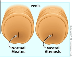
The urethral meatus is the opening through which urine leaves the body. Meatal stenosis means that this opening has become abnormally narrow and is interfering with urinary flow. It is most often discussed in boys and men, although narrowing of the female urethral opening can also occur.
An important point is that a meatus can look small without causing obstruction. Treatment should therefore be based on the complete picture, symptoms, the appearance of the opening, the urinary stream and, where appropriate, objective testing, not appearance alone.
What symptoms can it cause?
Typical symptoms include:
- a thin, forceful or upward-deflected stream;
- spraying or difficulty aiming the urine;
- taking longer to pass urine or needing to strain;
- burning or discomfort during urination;
- a small spot of blood at the meatus;
- urinary frequency, urgency or incomplete emptying; and
- recurrent urinary infection in selected patients.
In toilet-trained boys, an abnormal stream is often the most useful clue. Published patient-reported data show that improvement after meatotomy is most predictable when the preoperative problem is an abnormal or deflected stream. Frequency, urgency, wetting or dysuria may have another cause and should not automatically be attributed to a narrow-looking meatus.
Complete inability to pass urine is uncommon but requires urgent medical attention.
Congenital meatal stenosis
Congenital meatal stenosis is present from birth. True isolated congenital narrowing is uncommon and should be distinguished from normal variation in meatal size. It may also occur as part of another developmental urethral condition, including hypospadias, or following congenital urethral reconstruction.
In a baby or young child, symptoms can be difficult to recognise. The diagnosis becomes more apparent after toilet training, when a persistently narrow, spraying or markedly deflected stream can be observed.
Not every anatomically small meatus needs surgery. An asymptomatic child with a satisfactory stream, no urinary infections and normal bladder emptying can often be observed. Symptomatic obstruction, however, should be assessed by a paediatric urologist.
Acquired meatal stenosis
Acquired stenosis develops after birth. Its causes vary with age.
In boys
Meatal stenosis is recognised after circumcision, although published estimates vary substantially because studies use different definitions and methods of examination. Proposed mechanisms include irritation and inflammation of the exposed meatus, contact with wet nappies, meatal ulceration and subsequent scar formation. It often becomes clinically obvious between early childhood and school age rather than immediately after circumcision.
Other causes include inflammation, trauma, catheterisation and previous surgery, particularly repair of hypospadias. Following hypospadias surgery, narrowing may involve more than the external opening and must be assessed in the context of the reconstructed urethra.
In adolescents and adults
Important causes include:
- lichen sclerosus (also called balanitis xerotica obliterans or BXO), which can scar the foreskin, glans, meatus and more proximal urethra;
- repeated urethral instrumentation, catheterisation or endoscopic surgery;
- prior hypospadias repair or other penile surgery;
- trauma, infection or chronic inflammation; and
- previous radiotherapy or treatment affecting the urethra.
In adults, it is essential to determine whether narrowing is confined to the meatus or extends into the fossa navicularis or penile urethra. Treating only the visible opening will fail if more extensive scar disease has been overlooked.
Female meatal or urethral stenosis is uncommon. Symptoms may resemble recurrent urinary infection or other causes of bladder-outlet obstruction. Diagnosis should be made carefully, as urinary symptoms alone do not prove that the urethra is narrowed.
How is it diagnosed?
Assessment may include:
- A detailed history: including the direction and calibre of the stream, spraying, pain, infections, prior circumcision, catheterisation, surgery, trauma and skin disease.
- Examination: assessing the meatus and surrounding skin for scarring, pallor, inflammation, lichen sclerosus, hypospadias or surgical change.
- Observation of the urinary stream, particularly in a toilet-trained child.
- Uroflowmetry and post-void residual ultrasound when symptoms are unclear, the patient is older, or more extensive obstruction is suspected.
- Urinalysis or urine culture if pain, blood or infection is suspected.
- Urethral calibration, cystoscopy or urethrography selectively, especially in adults, recurrent disease, previous urethral surgery or suspected extension beyond the meatus.
Kidney and bladder ultrasound is not required for every straightforward case, but may be appropriate when there are recurrent infections, incomplete emptying, significant obstruction or concern about the upper urinary tract.
What is the correct management?
Management must match the patient’s symptoms, cause, age and extent of narrowing.
1. Observation
Observation is reasonable when the meatus is merely small in appearance but the patient has no relevant symptoms, passes a satisfactory stream and empties the bladder normally. Treating an incidental finding is unlikely to improve unrelated urgency, frequency or wetting.
2. Treat active skin or inflammatory disease
When lichen sclerosus or another inflammatory disorder is present, the underlying disease must be treated as well as the narrowing. Potent topical corticosteroid treatment is commonly used for genital lichen sclerosus under medical supervision. Circumcision may be indicated when the foreskin is affected, but established meatal or urethral scar may also require surgery.
Persistent or suspicious penile lesions may need biopsy. Long-term review can be appropriate because lichen sclerosus can recur, extend into the urethra and is associated with a small but important risk of penile malignancy.
3. Meatotomy or meatoplasty
For a short, symptomatic stenosis confined to the meatus, meatotomy or meatoplasty is usually the definitive treatment.
- A meatotomy enlarges the opening with a controlled incision.
- A meatoplasty reconstructs and sutures the edges to create a durable, appropriately shaped opening.
Both can provide excellent relief in appropriately selected children. A large paediatric series reported that meatotomy required more early manual spreading and had a higher reoperation rate than meatoplasty; technique and postoperative care therefore matter. In another study, 95% of families reported their child was at least somewhat improved after meatotomy, with the strongest benefit in boys treated for an abnormal stream.
The operation may be performed under local anaesthesia in selected cooperative patients or under general anaesthesia, particularly in younger children. Expected short-term effects include stinging, minor spotting of blood and temporary spraying while swelling settles. The surgeon may advise ointment and gentle separation of the meatal edges during early healing; instructions vary according to the procedure used.
4. Dilatation
Repeated blind dilatation is generally not a durable solution for dense scar-related meatal stenosis. It may cause further tearing and scarring and can commit a patient to repeated procedures. Carefully selected dilation or self-dilatation may have a role as temporary or palliative management, or as part of a specialist regimen for inflammatory disease, but it should not replace an appropriate reconstructive assessment in recurrent or complex disease.
5. Recurrent, adult or extended distal disease
If narrowing recurs, is associated with lichen sclerosus, or extends into the fossa navicularis/distal urethra, a simple repeat incision may not be sufficient. Options include formal meatoplasty or distal urethroplasty using an oral mucosal graft. The European Association of Urology recommends offering open meatoplasty or distal urethroplasty for meatal and distal urethral strictures; the precise operation should be individualised to stricture length, tissue quality and the patient’s priorities.
In lichen-sclerosus-related urethral disease, genital skin should not be used as a graft because the disease may recur in that tissue. Oral mucosa is generally preferred when graft reconstruction is required.
Follow-up and recurrence
Most patients with an isolated, properly treated meatal stenosis do well. Follow-up should assess:
- improvement in stream direction and calibre;
- pain, bleeding, infection or difficulty voiding;
- bladder emptying when clinically indicated; and
- restenosis or progression of an underlying condition such as lichen sclerosus.
Prompt reassessment is advisable if the stream narrows again, spraying persists after healing, voiding becomes painful or difficult, infections recur, or new skin changes appear.
The practical message
Meatal stenosis is not simply “a small hole.” Correct care begins by confirming that the narrowing is clinically important and identifying its cause and extent. A symptomatic, short stenosis confined to the meatus is usually treated successfully with meatotomy or meatoplasty. Recurrent stenosis, adult disease, previous hypospadias repair or lichen sclerosus requires a more detailed urethral assessment and sometimes formal reconstruction.
This article provides general educational information and does not replace an individual assessment. Seek urgent care if you or your child cannot pass urine, develops fever with urinary symptoms, or has significant bleeding or pain.
Selected references
- European Association of Urology. EAU Guidelines on Urethral Strictures: Disease Management in Males. 2026. https://uroweb.org/guidelines/urethral-strictures/chapter/disease-management-in-males
- European Association of Urology. EAU Guidelines on Urethral Strictures: Definition, Epidemiology, Aetiology and Prevention. 2026. https://uroweb.org/guidelines/urethral-strictures/chapter/definition-epidemiology-aetiology-and-prevention
- Wessells H, et al. Urethral Stricture Disease Guideline Amendment (2023). Journal of Urology. 2023. doi:10.1097/JU.0000000000003482.
- Dothan D, et al. Surgical Treatment of Meatal Stenosis: Lessons Learned from the Pediatric Urology Practice. Urology. 2023;171:220–224. PMID: 35981660.
- Varda BK, et al. Minor procedure, major impact: patient-reported outcomes following urethral meatotomy. Journal of Pediatric Urology. 2018;14(2):165.e1–165.e5. doi:10.1016/j.jpurol.2017.11.018.
- Godley SP, et al. Meatal stenosis: a retrospective analysis of over 4000 patients. Journal of Pediatric Urology. 2015;11(1):38.e1–38.e6. doi:10.1016/j.jpurol.2014.09.016.
- Morris BJ, Krieger JN. Does circumcision increase meatal stenosis risk? A systematic review and meta-analysis. Urology. 2017;110:16–26. doi:10.1016/j.urology.2017.07.027.
- Wang MH. Surgical management of meatal stenosis with meatoplasty. Journal of Visualized Experiments. 2010;(45):2213. doi:10.3791/2213.
Prepared for patient education.
Richard Marsden
Many of my longstanding patients in Bundaberg will remember Richard Marsden, a highly respected urologist who served the Bundaberg community for many years.
For much of that time, Richard was Bundaberg’s only urologist. He was deeply committed to regional Queensland and to ensuring that people living outside the major cities could receive high-quality specialist care close to home. This remarkable man passed away on 5 November.
I owe my own life-changing move from South Africa to Australia, in no small part, to Richard.
We first met in 2005 at the biennial UROSA conference, the Urological Association of South Africa meeting held at Sun City. We struck up a conversation, and by the end of it Richard had handed me his business card. He told me that he was preparing to retire and invited me to contact him if I might be interested in taking over his urology practice in Bundaberg.
At the time, I did not fully appreciate the significance of his offer. It took several weeks for me to realise what an extraordinary opportunity he had placed before me.
A few months later, my wife, Natasha, and I travelled to Bundaberg for what South Africans often call an “LSD trip” a “look, see and decide” visit undertaken before making the enormous decision to uproot a family and step into the unknown.
Australia represented the promise of safety, freedom and a hopeful future for our daughters. From the moment we arrived in Bundaberg, we felt an immediate connection with the town. Its ocean, sugarcane fields and humid subtropical climate reminded us of the east coast of South Africa and the former province of Natal.
Richard and his wife, Carol, welcomed us warmly. Richard generously shared his daily routine with me and introduced me to the operating theatre and ward staff. He gave us an honest and personal insight into both the practice and the community we were considering making our home.
We also discovered that Richard understood the immigrant journey firsthand. Born in the United Kingdom, he spent part of his childhood during the Second World War in Johannesburg, South Africa. After the war, he joined his parents in Melbourne before the family later returned to the United Kingdom. Richard went on to study medicine and establish his medical career in London.
Years later, he made his own courageous decision to relocate to Australia with his two daughters. In many ways, his journey mirrored the one Natasha and I were contemplating. His story, his generosity and the welcome extended by Richard and Carol helped us make one of the most difficult and ultimately most important decisions of our lives: Bundaberg was where we wanted to raise our girls.
Richard did far more than offer me a professional opportunity. He opened a door that changed the course of my career and transformed the future of my family.
I will always be grateful for his kindness, his trust and his commitment to the people of regional Queensland.
Thank you, Richard. Your legacy lives on in the community you served, the colleagues you inspired and the lives you changed—including mine.