Tag Archive for: wesley hospital urologist brisbane

Superficial Urothelial Carcinoma of the Bladder

Understanding non-muscle-invasive bladder cancer, TURBT, intravesical therapy and long-term surveillance

Being told that you have a bladder tumour is understandably alarming. The reassuring part is that many bladder cancers are discovered while they are still confined to the inner layers of the bladder and have not invaded the bladder muscle.

This is called non-muscle-invasive bladder cancer (NMIBC), historically referred to as superficial bladder cancer. NMIBC includes Ta tumours, T1 tumours and carcinoma in situ (CIS). These tumours behave very differently depending on their stage, grade and other pathological features, so treatment is tailored according to the risk of the cancer coming back (recurrence) or becoming more aggressive (progression).

The good news is that most NMIBC can initially be treated through the urethra without making an incision in the abdomen. The less convenient news is that bladder cancer has a habit of returning, which is why careful surveillance becomes an important part of treatment.


What is urothelial carcinoma?

The inside of the bladder is lined by specialised cells called urothelial cells. Cancer arising from these cells is known as urothelial carcinoma.

Urothelial carcinoma can develop anywhere along the urinary tract, including the:

  • renal collecting system
  • ureters
  • bladder
  • urethra

The bladder is by far the most common site.

When a bladder tumour has not invaded the muscular wall of the bladder, it is classified as non-muscle-invasive bladder cancer.

https://images.openai.com/static-rsc-4/y3Nkc3nnwnLWbduLwtOBo9wlGDPYvlRZ3hqmZmerWsmC4HUjhpIEvsXgWPs9IwZOQ9PLgJllQH1edLQmAni1U_538dO2IEbu48myP0AM4El4gyVQqzEuJjxyZdTq2Ak6qThyv190dh_MhP59fXKNJByHY8EL-AhSnzHK3LUJq2tbc5vhmHhLL8WBeJitn-Wb?purpose=fullsize


What causes bladder cancer?

There is rarely one identifiable cause. Instead, bladder cancer develops following genetic changes within urothelial cells, often influenced by environmental exposures over many years.

Smoking

Cigarette smoking is the most important preventable risk factor for bladder cancer.

Chemicals absorbed through cigarette smoke enter the bloodstream, are filtered by the kidneys and eventually become concentrated in the urine. The bladder lining is therefore repeatedly exposed to these carcinogens.

Stopping smoking after a bladder cancer diagnosis is strongly encouraged.

Other risk factors

These include:

  • increasing age
  • occupational exposure to certain industrial chemicals
  • previous pelvic radiotherapy
  • previous treatment with cyclophosphamide
  • chronic urinary tract irritation in selected circumstances
  • a personal history of urothelial carcinoma

Sometimes there is no obvious risk factor at all.


How does bladder cancer present?

Blood in the urine

The classic presentation is haematuria, or blood in the urine.

This may be:

Visible haematuria

The urine may suddenly become:

  • pink
  • red
  • burgundy
  • tea-coloured
  • or contain blood clots

Importantly, bleeding from a bladder tumour is frequently painless and intermittent.

The bleeding may disappear completely for days, weeks or even months. Its disappearance does not necessarily mean that the underlying problem has resolved.

Unexplained visible haematuria should always be investigated.

Microscopic haematuria

Sometimes blood is detected only on urine testing and cannot be seen with the naked eye.


Other possible symptoms

Some patients, particularly those with carcinoma in situ (CIS), may experience bladder irritation rather than obvious bleeding.

Symptoms can include:

  • urinary frequency
  • urgency
  • burning when passing urine
  • nocturia
  • pelvic or bladder discomfort
  • recurrent symptoms resembling a urinary tract infection

Persistent urinary symptoms with repeatedly negative urine cultures may therefore warrant further investigation.


Investigating suspected bladder cancer

The investigation usually involves a combination of urine testing, imaging and direct examination of the bladder.

Urine testing

A urine sample may be checked for:

  • blood
  • infection
  • inflammatory cells
  • abnormal malignant cells

Urine cytology

Urine cytology examines cells shed from the urinary tract under a microscope.

It is particularly useful for detecting high-grade urothelial carcinoma and CIS, but is considerably less sensitive for low-grade tumours.

A negative cytology therefore does not exclude bladder cancer.


Imaging the urinary tract

Patients with haematuria may require imaging of the kidneys, ureters and bladder.

Depending on the clinical circumstances, this may include:

  • renal tract ultrasound
  • CT urinary tract imaging
  • CT urography

CT urography is particularly useful when investigating haematuria because urothelial carcinoma can occasionally arise within the renal collecting system or ureters as well as the bladder.


Cystoscopy

A cystoscopy allows the urologist to look directly inside the bladder.

A thin flexible telescope is passed through the urethra, usually under local anaesthetic.

Most bladder tumours have a characteristic appearance. They may resemble delicate fronds, seaweed or a tiny underwater cauliflower attached to the bladder wall.

Flat lesions such as CIS can be much more difficult to see.

If a suspicious lesion is identified, the next step is usually a transurethral resection of bladder tumour (TURBT).


TURBT: biopsy and removal of the bladder tumour

TURBT stands for:

Transurethral Resection of Bladder Tumour

This procedure serves two important purposes:

  1. Treatment: removing all visible tumour where possible.
  2. Diagnosis and staging: providing tissue for the pathologist to determine exactly what type of tumour is present and how deeply it has invaded.

The procedure is usually performed under general or spinal anaesthesia.

A telescope called a resectoscope is passed through the urethra into the bladder. The tumour is carefully removed, usually using an electrical or bipolar resection system.

There is therefore generally no external incision or abdominal scar.


Why obtaining bladder muscle matters

An adequate TURBT should establish how deeply the tumour extends.

For many tumours, particularly T1 and high-grade lesions, the specimen should contain muscularis propria (detrusor muscle) so that the pathologist can determine whether the cancer has reached the muscle layer. The pathological report should document the presence and involvement of muscularis propria where applicable.

This distinction dramatically changes treatment.


Understanding bladder cancer staging

A simplified view of the bladder wall is:

Urine

↓

Urothelium

↓

Lamina propria

↓

Detrusor muscle

↓

Fat surrounding the bladder

The important early stages are:

Ta

The tumour is confined to the urothelial surface and has not invaded the supporting tissue underneath.

Tis: carcinoma in situ

CIS is a flat, high-grade malignant lesion confined to the urothelium.

Unlike the typical papillary bladder tumour, CIS may be difficult to identify visually.

Despite being superficial anatomically, CIS is biologically aggressive and requires appropriate treatment.

T1

The tumour has invaded into the lamina propria, but has not invaded the muscularis propria.

T1 disease, particularly high-grade T1 disease, carries a greater risk of recurrence and progression.

T2

The cancer has invaded the bladder muscle.

Once muscle invasion is identified, the disease is no longer classified as NMIBC and requires a different treatment pathway.


Stage and grade are not the same thing

This distinction frequently causes confusion.

Stage describes how deeply the cancer has travelled into the bladder wall.

Grade describes how abnormal and biologically aggressive the cancer cells appear under the microscope.

A tumour may therefore be superficial but still be high grade.

Broadly, urothelial tumours are classified as:

Low grade

These generally grow more slowly and have a relatively low risk of progressing to muscle-invasive cancer, although they may recur.

High grade

These cells look significantly abnormal and have a greater potential for:

  • recurrence
  • invasion
  • progression
  • spread beyond the bladder

Risk stratification

Following TURBT, the tumour is classified into a risk category.

Risk assessment considers factors such as:

  • stage
  • grade
  • tumour size
  • number of tumours
  • previous recurrence rate
  • presence of CIS
  • T1 disease
  • pathological features including lymphovascular invasion
  • certain variant histological subtypes

This classification helps determine whether the patient requires:

TURBT alone → intravesical chemotherapy → BCG → or, in selected very-high-risk situations, consideration of radical cystectomy.

Modern guidelines emphasise risk-adapted rather than one-size-fits-all management.


Do I need another TURBT?

Sometimes.

A second-look or re-staging TURBT may be recommended when:

  • the initial tumour was incompletely removed
  • adequate muscle was not present in an important specimen
  • high-grade disease is present in selected circumstances
  • T1 disease is identified
  • there is concern that the original tumour may have been understaged
  • certain variant histologies are identified

The aim is to ensure that residual tumour has not been left behind and, crucially, that muscle-invasive disease has not been missed.


Intravesical treatment

Intravesical simply means that medication is placed directly into the bladder through a catheter.

This allows the treatment to come into direct contact with the bladder lining while reducing systemic exposure compared with intravenous chemotherapy.

Two important treatments are:

Mitomycin C

and

BCG

They are not interchangeable and are used for different risk groups.


Intravesical Mitomycin C

Mitomycin C is a chemotherapy drug that can be placed directly into the bladder.

Immediate Mitomycin C following TURBT

For appropriate patients with suspected low- or intermediate-risk NMIBC, a single postoperative instillation of intravesical chemotherapy may be given soon after TURBT.

Current guidelines recommend that, when used, the immediate instillation should generally occur within 24 hours of TURBT.

Its purpose is to destroy microscopic tumour cells remaining in the bladder and tumour cells released during the resection.

This reduces the risk of recurrence.

When should immediate Mitomycin C be avoided?

It should generally not be administered if there is:

  • suspected bladder perforation
  • significant bleeding requiring bladder irrigation
  • an extensive or very deep resection where perforation is a concern

This is important because chemotherapy leaking outside the bladder can cause significant local tissue injury.


Further courses of intravesical chemotherapy

Selected patients with recurrent or intermediate-risk low-grade NMIBC may receive a course of intravesical chemotherapy rather than simply a single postoperative dose.

The exact drug, schedule and duration depend upon the tumour risk profile and local treatment protocols.


Side effects of intravesical Mitomycin C

Most patients tolerate treatment reasonably well.

Possible side effects include:

  • urinary frequency
  • urgency
  • burning
  • bladder discomfort
  • haematuria
  • chemical cystitis
  • skin irritation if the medication contacts the genital skin

Rarely, severe bladder inflammation or tissue injury can occur.


Intravesical BCG

BCG stands for Bacillus Calmette-Guérin.

Yes, it originated as a tuberculosis vaccine. In the bladder it performs a completely different job.

BCG stimulates a powerful local immune response against urothelial cancer cells and remains one of the most effective bladder-preserving treatments for high-risk NMIBC.


Who should receive BCG?

BCG is principally considered for patients with:

  • high-grade Ta tumours
  • high-grade T1 tumours
  • carcinoma in situ
  • other appropriately selected high-risk or intermediate-risk NMIBC

For high-risk NMIBC, full-dose BCG with maintenance treatment for one to three years remains a guideline-supported treatment, while immediate radical cystectomy should also be discussed in appropriate high-risk and particularly very-high-risk disease.


How is BCG given?

BCG is inserted into the bladder through a small catheter.

A typical induction course consists of:

One treatment per week for six weeks.

The solution is retained within the bladder for a prescribed period and then passed out in the urine.

Patients who respond may subsequently receive maintenance BCG.

A commonly used maintenance approach involves three weekly treatments at defined intervals after induction. In high-risk disease, guideline schedules may continue maintenance for up to three years depending upon tumour risk, treatment tolerance and BCG availability.


When should BCG not be given?

BCG is a live attenuated organism and must be administered carefully.

Treatment should generally be postponed in patients with:

  • visible haematuria
  • symptomatic urinary tract infection
  • traumatic catheterisation
  • very recent TURBT

The EAU lists the first two weeks following TURBT, visible haematuria, traumatic catheterisation and symptomatic urinary infection as absolute contraindications to an intravesical BCG instillation.


Side effects of BCG

A degree of bladder irritation is common.

Patients may experience:

  • frequency
  • urgency
  • burning
  • bladder discomfort
  • mild haematuria
  • fatigue
  • low-grade fever
  • flu-like symptoms

These symptoms usually settle.

Rarely, BCG can cause a more significant systemic infection or inflammatory reaction.

Persistent high fever, chills, marked deterioration or severe illness following BCG requires urgent medical assessment.


What if BCG does not work?

Persistent or recurrent high-grade cancer despite adequate BCG treatment requires careful reassessment.

This situation should not simply be managed by repeatedly giving more BCG indefinitely.

Patients meeting criteria for BCG-unresponsive NMIBC should be counselled regarding further treatment, and radical cystectomy remains the oncological standard for suitable patients with BCG-unresponsive high-risk disease. Bladder-preserving alternatives may be considered for patients who are medically unsuitable for cystectomy or decline surgery, ideally within appropriate specialist or clinical-trial pathways.


When should radical cystectomy be considered?

Most patients with superficial bladder cancer will never require removal of their bladder.

However, early radical cystectomy may be discussed for very-high-risk disease, including selected patients with:

  • aggressive high-grade T1 disease
  • associated CIS
  • variant histology
  • lymphovascular invasion
  • persistent high-grade disease
  • BCG-unresponsive disease
  • other features associated with a high risk of progression

The decision involves balancing the risks of major surgery against the danger of allowing biologically aggressive disease to progress.


Surveillance after treatment

Removing the tumour is only the first chapter.

NMIBC has a significant tendency to recur, and some high-risk tumours can progress. Consequently, regular cystoscopic surveillance is essential.

The first surveillance cystoscopy is generally performed approximately three months after TURBT.

After this, surveillance is tailored to the patient’s risk category.

A practical risk-adapted surveillance framework

Risk group Typical cystoscopy schedule Cytology Upper tract imaging
Low risk 3 months, 12 months, then annually Usually not routinely required Not routinely required
Intermediate risk 3 months, then approximately every 6 months for 2 years, then annually Risk-dependent Selected patients
High / very high risk Approximately every 3 months initially, with intervals gradually extended Usually included Periodic upper urinary tract imaging

The 2026 EAU guidance continues to recommend that follow-up intensity and duration be determined by the patient’s risk category, with the first cystoscopy at three months.

Individual surveillance schedules may differ depending on pathology, previous recurrences, treatment response, age, comorbidities and the treating urologist’s protocol.


Why is surveillance so important?

A recurrence does not automatically mean that treatment has failed or that the cancer has become life-threatening.

Low-grade papillary tumours may recur while remaining superficial and biologically low risk.

The purpose of surveillance is to identify recurrence early, while it can still be treated appropriately.

High-grade disease requires closer attention because the consequences of missing progression are considerably greater.


Can bladder cancer come back after years?

Yes.

This is why follow-up for intermediate- and particularly high-risk NMIBC can continue for many years.

The frequency of cystoscopy usually decreases when repeated examinations remain clear, but high-risk patients generally require prolonged surveillance.


What can I do after a diagnosis?

One of the most important modifiable factors is:

Stop smoking

Smoking cessation reduces exposure to the carcinogens responsible for many urothelial cancers and provides substantial additional cardiovascular, respiratory and general health benefits.

Patients should also:

  • attend every scheduled cystoscopy
  • report recurrent visible haematuria
  • complete prescribed intravesical treatment
  • report significant side effects from BCG or chemotherapy
  • maintain appropriate hydration unless medically restricted
  • discuss occupational chemical exposure where relevant

The bottom line

Non-muscle-invasive urothelial carcinoma is bladder cancer that has not invaded the muscular wall of the bladder.

The pathway typically involves:

Haematuria or urinary symptoms
↓
Urine tests + imaging
↓
Cystoscopy
↓
TURBT and pathological examination
↓
Stage + grade + risk classification
↓
Risk-adapted treatment

For some patients, TURBT followed by surveillance may be sufficient.

Others benefit from intravesical Mitomycin C or another intravesical chemotherapy to reduce recurrence.

Patients with high-grade disease, T1 cancer or CIS frequently require intravesical BCG, usually incorporating induction and maintenance treatment.

Very-high-risk or BCG-unresponsive disease may require consideration of radical cystectomy.

Most importantly, bladder cancer treatment does not finish when the initial tumour has been removed. Long-term cystoscopic surveillance is an integral part of treatment.

A final word

Bladder cancer can be a persistent visitor, but surveillance means we do not leave the front door unattended.

Early detection, complete TURBT, accurate pathological staging, appropriate intravesical therapy and structured follow-up provide the best opportunity to keep non-muscle-invasive bladder cancer under control.

This information is intended as general patient education and does not replace individual assessment or treatment advice from your urologist. Management should be tailored to the pathology, tumour risk category, general health and individual circumstances of each patient.

Urodynamic Studies: Understanding How Your Bladder Works

Bladder problems are not always as straightforward as they appear.

Two people may both complain of urinary urgency, leakage or difficulty emptying their bladder, yet the underlying causes can be completely different. One bladder may be contracting when it should be relaxing, another may not contract strongly enough, and sometimes the bladder is behaving perfectly well while the outlet is causing all the trouble.

This is where urodynamic studies, often simply called urodynamics, can help.

Think of urodynamics as a stress test for the bladder. Rather than simply looking at its anatomy, we observe how the bladder fills, stores urine and empties while simultaneously measuring pressures, urine flow and sometimes pelvic floor activity.

The aim is not merely to produce a collection of colourful graphs. The important question is:

What is the bladder actually doing, and will knowing this change the treatment?


What Are Urodynamic Studies?

Urodynamics refers to a group of tests that assess the function of the bladder, urethra and urinary sphincter.

Depending on the clinical situation, testing may evaluate:

  • How much urine the bladder can comfortably hold
  • Bladder sensation during filling
  • Whether the bladder remains relaxed while filling
  • Whether involuntary bladder contractions occur
  • Whether urinary leakage occurs and why
  • The pressure required to store urine
  • The strength of the bladder muscle during urination
  • Whether there is obstruction to urinary flow
  • Whether the bladder empties completely
  • How the urinary sphincter and pelvic floor behave during filling and voiding

Urodynamics therefore investigates function rather than simply structure.

An ultrasound or cystoscopy may show us what the urinary tract looks like. Urodynamics tells us what it is doing.


Who May Need Urodynamic Testing?

Not every patient with urinary symptoms requires urodynamics.

For many straightforward urinary problems, the diagnosis can be made from the history, examination, bladder diary, urine testing, flow studies and ultrasound.

Urodynamics becomes particularly useful when the diagnosis is uncertain, symptoms are complex, previous treatments have failed, neurological disease is present, or an invasive treatment is being considered where understanding bladder function may alter the choice of treatment.

Common indications include:

Urinary Incontinence

Urodynamics may be useful when assessing:

  • Stress urinary incontinence
  • Urgency urinary incontinence
  • Mixed urinary incontinence
  • Persistent leakage following previous continence surgery
  • Incontinence following prostate surgery
  • Complex or unexplained urinary leakage

In straightforward female stress urinary incontinence, urodynamics is not necessarily required before treatment. It becomes more useful when symptoms and clinical findings do not agree or when there are complicating factors.


Overactive Bladder

Patients with overactive bladder may experience:

  • Urinary urgency
  • Frequent urination
  • Nocturia
  • Urgency urinary incontinence

Urodynamics can sometimes demonstrate detrusor overactivity, where the bladder muscle contracts involuntarily during filling.

Importantly, a normal urodynamic study does not necessarily exclude overactive bladder. Symptoms and urodynamic findings do not always travel together.


Difficulty Emptying the Bladder

Patients may report:

  • Poor urinary flow
  • Hesitancy
  • Straining
  • Intermittent flow
  • Incomplete emptying
  • Urinary retention

The challenge is determining why.

Is there an obstruction?

Or is the bladder muscle simply not contracting strongly enough?

The symptoms can look remarkably similar, but the treatments can be very different.


Men With Lower Urinary Tract Symptoms

In men with prostate enlargement, urodynamics may help distinguish between:

Bladder outlet obstruction

and

Detrusor underactivity, where the bladder muscle has insufficient strength to empty effectively.

This distinction may be particularly valuable before prostate surgery when there is uncertainty about whether removing the obstruction will improve urinary flow.


Neurological Bladder Dysfunction

Urodynamics has an especially important role in patients with neurological conditions such as:

  • Spinal cord injury
  • Multiple sclerosis
  • Parkinson’s disease
  • Spina bifida
  • Stroke
  • Certain spinal disorders

In these patients, we are interested not only in urinary symptoms but also in whether bladder pressures could potentially damage the kidneys.


Before or After Certain Urinary Procedures

Urodynamics may also be considered:

  • Before complex continence surgery
  • Following unsuccessful continence surgery
  • Before some bladder outlet procedures
  • Following prostate surgery when symptoms persist
  • When considering treatments such as bladder Botox or sacral neuromodulation in selected complex patients

What Happens During a Urodynamic Study?

The test is usually performed as an outpatient procedure.

It generally takes approximately 30–60 minutes, although this varies depending on the type of study being performed.

You can usually eat, drink and take your normal medications unless specifically instructed otherwise.


Step 1: Arriving With a Comfortably Full Bladder

You may be asked to arrive with a reasonably full bladder.

You will urinate into a special toilet called a uroflowmeter.

This measures:

  • Urine volume
  • Maximum urinary flow rate
  • Average flow rate
  • The shape of the urinary flow curve
  • How long urination takes

An ultrasound may then measure how much urine remains in the bladder.

This is called the post-void residual.


Step 2: Placement of Small Catheters

A very fine catheter is gently passed through the urethra into the bladder.

A second small pressure catheter is usually placed into the rectum, or occasionally the vagina.

Why two pressure measurements?

Because pressure inside the abdomen affects pressure inside the bladder.

By measuring both, the computer can calculate the pressure generated specifically by the bladder muscle:

Detrusor pressure = bladder pressure − abdominal pressure

This simple equation sits at the heart of conventional urodynamics.


Step 3: Filling the Bladder

The bladder is slowly filled with sterile fluid.

During filling, you will be asked to describe what you feel.

We may record:

  • First sensation of bladder filling
  • First desire to urinate
  • Strong desire to urinate
  • Maximum bladder capacity

The pressure inside the bladder is continuously monitored.

Normal filling cystometrogram

https://images.openai.com/static-rsc-4/Hgriy5E9FGtRilFNQh3tQE53yrlbWRzHlZE-lg73YC5P10G0EFIrVgMREqgOL5uEui9m4vTZQa3aZdbm2IQos6eTVRAdTIXlg1d841nyByIJ3GZk8QFyf9avtCBCjESBk_UcSEGbJGzOSIvZvPI6wZtNpz7LHnbB3kQIYDHsibqiwXmWpERNixUhOeW6Tm2B?purpose=fullsize
https://images.openai.com/static-rsc-4/BXwVAcldUDjh2tAaV2En87FX0ONS1oomHAkm4Z6BLZyoexIiDmaACUh8t4rB7TDfao-guZf76ULaTYX-VsV554nagvCAPRpNzs7HNQvKKZYuX8N6oPO5c_n_74de2tABnoEt69kwxCY_fJyF86M5rduxVEOG1zrNzm0SXMSrnXPiadRMzOfo1hPUnKvP1ahH?purpose=fullsize
https://images.openai.com/static-rsc-4/ySZcZmtN_5IZCgz4scVLbu2KqbZD3OVebnVIjG7F41-xqaWl2-D1RqW5T2NMWjugPRIkt7H-S3JB2UGWLPXIYYyURXURsMwzaASQ1zMpc7f1yB8UJcFDYMc1HW3qPkSw7rWCM7V8cMPh-oI2CVWQYD1HhlPxcJ6eJY6HJvYEz3QSK5VgEbfQgqejsiCVUfkK?purpose=fullsize
4

Normal filling cystometrogram. During filling, the bladder accommodates increasing volume while detrusor pressure (Pdet) remains relatively stable. Coughs cause corresponding rises in Pves and Pabd without a true detrusor contraction. Sensations such as first sensation, first desire and strong desire can be marked on the tracing.


Step 4: Provocative Testing

You may be asked to:

  • Cough
  • Strain
  • Stand
  • Change position
  • Listen to running water

These manoeuvres are not designed to make the test unnecessarily theatrical. They help reproduce the circumstances under which your symptoms normally occur.

If urinary leakage occurs during coughing without an involuntary bladder contraction, this may demonstrate urodynamic stress incontinence.


Step 5: Voiding

Once the bladder is comfortably full, you will be asked to urinate.

During urination we simultaneously measure:

  • Urinary flow
  • Bladder pressure
  • Abdominal pressure
  • Detrusor pressure

This is called a pressure-flow study.

It can be extremely useful when determining whether poor urinary flow is caused by obstruction or weak bladder contraction.


What Does Urodynamics Measure?

Bladder Sensation

We assess when you first become aware that the bladder is filling and when the desire to urinate becomes stronger.

Sensation may be:

  • Normal
  • Increased
  • Reduced
  • Absent

Abnormal bladder sensation may occur in several neurological and functional bladder disorders.


Bladder Capacity

The amount of urine the bladder can comfortably hold is measured.

A small functional capacity may occur with conditions such as severe urgency or bladder hypersensitivity, although capacity must always be interpreted in clinical context.


Bladder Compliance

A healthy bladder should behave rather like a flexible reservoir.

As it fills, its volume should increase without a major rise in pressure.

Poor bladder compliance means pressure rises excessively as the bladder fills.

This is particularly important in some neurological bladder disorders because persistently elevated storage pressures can potentially threaten the upper urinary tract.


Detrusor Overactivity

If the bladder muscle contracts involuntarily during filling, this is called:

Detrusor overactivity.

It may be associated with:

  • Urgency
  • Urgency incontinence
  • Frequency
  • Nocturia

When associated with an underlying neurological condition, it may be described as neurogenic detrusor overactivity.

Examples of terminal, phasic, wet and dry detrusor overactivity.

https://images.openai.com/static-rsc-4/Snzbj3YLBffWTq3-7c6jgZQWHTjwV03Siqh8rQZor7nWrcUTSujo5I4h9H6QmPWIw5TQO8oSYgzzugdooybbroYfbxONiyj0mx1Qe1uBjCwkL4K0AR5hGhULd9PKwFrw7LhUIrL8ZxpKQO1sQjk0qtqph3By5IOK30vdjd9Iahom6SUGG32ajdo2KW48nbhP?purpose=fullsize
https://images.openai.com/static-rsc-4/kx0pZHTOwdE7kDcSE1Awe3tnf1oB9kJizr_DFgQV0kdb9o6N2KxN9PW2lGCO4Rxcyx__zDzECnRwDhOrCXcXYbHZKkw4yASruDH4PtFNhlq6LqHzLF1d6NmcZkyOnoMHG_lNoKxw1Y5hCVuog5TIJqiN5O_zT8m7FtEhZkE-uHlwJMGAWpqcUhXKZdvq-h6c?purpose=fullsize
https://images.openai.com/static-rsc-4/9Fkv2MrdLw18QItSBrvBMWrFM_bRYOzwvod8KcVnSNQcBgQhcEl7m90lhNqlnqb3sX1pkEmdrx3XvCJxwi0gyzVvPse-T2lyODyK7BrW5xC3GJ2ZMggrrj7i-iArmLIQ05FXaE_4Cl8FYZ8yrU3dqXP5YN2gEVqQjCLOqH_xcbXAxuyLiYNLNijADJM9eUNR?purpose=fullsize

Detrusor overactivity. During bladder filling there is an involuntary increase in Pdet that cannot be explained by a corresponding increase in abdominal pressure. The contraction may be associated with urgency and, when leakage occurs, urgency urinary incontinence.

View the LABORIE-based detrusor-overactivity tracings


Stress Urinary Incontinence

If urine leaks when coughing or straining without a detrusor contraction, this supports a diagnosis of:

Urodynamic stress incontinence.

Additional measurements such as leak-point pressures may occasionally contribute to assessment in selected patients.


Bladder Outlet Obstruction

During urination, a pattern of:

High bladder pressure + low urinary flow

may suggest obstruction.

In men, benign prostate enlargement is a common cause.

Other causes include:

  • Urethral stricture
  • Bladder neck obstruction
  • Previous continence surgery
  • Dysfunctional voiding

Bladder outlet obstruction: pressure-flow study

https://images.openai.com/static-rsc-4/Efx8v2pSLdy0yriNx_qrcdTAHacUkb8mu_Sb7IjS9JvC0wxMQnPCPhcGQqAt0MQUNQ5jvnXe4yfU0qsIfsNRYrR7VLV4HhGFH6Ko01-kkE8GfSflqXD25Q6gkF3bmxyKYN9iYNHRqCJ0bV-igYyNfPP-21Nuvrls3kleV1a2fqsUDDQTALKE8mVkjEwglUbr?purpose=fullsize
https://images.openai.com/static-rsc-4/tQjdaSX6-MApQNNeYZuCDcT--H0Z_StPvpdtjnAdqL29pMWjRBGXsV_1KAe9iBU0kahuMqsxvNOoXDeZHFfSLBLxXg9w9uWzWnQxuEBsHfk3yjNkiDLedV-AQMJTHLPoif8aUAghyfhiv605EryWDjmBxVWKeBTNK3ky_okFHyRdz9rN1G-HNnhJBUnvnQhG?purpose=fullsize

Bladder outlet obstruction. During voiding the bladder generates a relatively high detrusor pressure, but urinary flow remains reduced. This high-pressure, low-flow relationship is characteristic of increased bladder outlet resistance.

In men, pressure-flow studies allow calculation of the Bladder Outlet Obstruction Index (BOOI):

BOOI = PdetQmax − 2(Qmax)

For men, BOOI >40 is generally classified as obstructed, 20–40 as equivocal and <20 as unobstructed.


Detrusor Underactivity

Sometimes the urinary flow is poor because the bladder muscle does not generate sufficient pressure.

This is known as:

Detrusor underactivity.

This distinction matters.

Operating on the prostate of a patient whose main problem is a very weak bladder may not produce the expected improvement in urinary flow.

Urodynamics can therefore occasionally prevent us from treating the wrong end of the plumbing.


Dysfunctional Voiding

Some patients involuntarily contract their pelvic floor or urinary sphincter while attempting to urinate.

Instead of the outlet opening smoothly, it intermittently closes.

Pelvic floor electromyography may help identify this pattern in selected patients.


Understanding the Urodynamic Tracing

A urodynamic report typically contains several pressure and flow curves.

At first glance, the tracing can resemble an enthusiastic seismograph.

The important measurements usually include:

Pves: pressure measured inside the bladder.

Pabd: pressure measured within the abdomen.

Pdet: pressure generated by the bladder muscle.

Flow: the rate at which urine leaves the bladder.

The relationship between these measurements allows us to understand what is happening during bladder filling and emptying.


Common Findings and What They May Mean for Treatment

Urodynamic Finding Possible Interpretation Potential Treatment Direction
Normal storage and emptying Normal urodynamic study Treatment guided by symptoms and other investigations
Detrusor overactivity Overactive bladder physiology Bladder training, medication, Botox, neuromodulation
Stress leakage Stress urinary incontinence Pelvic floor therapy, bulking agents, sling or other continence surgery
High pressure + low flow Bladder outlet obstruction Treatment of obstruction where appropriate
Low pressure + low flow Detrusor underactivity Emptying strategies, intermittent catheterisation or selected treatments
Poor compliance High-pressure storage Pressure-reducing treatment and upper urinary tract surveillance
Pelvic floor activation during voiding Dysfunctional voiding Pelvic floor physiotherapy/biofeedback
Significant residual urine Incomplete bladder emptying Identify obstruction versus impaired bladder contraction

The urodynamic result is not a treatment plan by itself.

It must be interpreted alongside symptoms, examination findings, bladder diaries, ultrasound, cystoscopy and other investigations where appropriate.


How Urodynamics Can Guide Treatment

If Detrusor Overactivity Is Found

Initial treatment may include:

  • Bladder training
  • Fluid modification
  • Pelvic floor physiotherapy
  • Antimuscarinic medication
  • Beta-3 agonist medication

For persistent symptoms, options may include:

  • Intravesical Botox injections
  • Sacral neuromodulation
  • Other specialist treatments in selected patients

If Stress Incontinence Is Demonstrated

Treatment depends on severity, sex, previous surgery and individual circumstances.

Options may include:

  • Pelvic floor rehabilitation
  • Continence devices
  • Peri-urethral bulking agents
  • Sling procedures
  • Colposuspension in selected women
  • Male sling following prostate surgery
  • Artificial urinary sphincter in appropriate men

If Bladder Outlet Obstruction Is Demonstrated

Treatment depends on the underlying cause.

For benign prostate enlargement, options may include:

  • Medication
  • Minimally invasive prostate treatments
  • Laser prostate surgery
  • Transurethral prostate surgery
  • Other surgical procedures appropriate to prostate size and anatomy

A urethral stricture or bladder neck obstruction requires a different treatment strategy.


If the Bladder Muscle Is Weak

Treatment may involve:

  • Double voiding
  • Timed voiding
  • Reviewing medications
  • Treating reversible causes
  • Intermittent self-catheterisation when necessary
  • Long-term catheterisation in selected circumstances
  • Sacral neuromodulation in carefully selected patients with non-obstructive urinary retention

The key issue is avoiding unnecessary outlet surgery when obstruction is not actually the problem.


If Bladder Storage Pressures Are High

This is particularly important in neurological bladder disease.

Treatment may include:

  • Medication
  • Intermittent catheterisation
  • Intravesical Botox
  • Regular renal imaging
  • Monitoring kidney function
  • Repeat urodynamic assessment
  • Reconstructive bladder surgery in uncommon severe cases

Here the objective extends beyond improving symptoms. We may also be trying to protect kidney function over the long term.


Are Urodynamic Studies Uncomfortable?

Most patients tolerate the investigation well.

Passing the small catheter can cause brief discomfort, and there may be some mild burning when urinating afterwards.

Having an audience while your bladder performs under laboratory conditions is admittedly not anyone’s preferred afternoon entertainment, but the clinical staff performing these studies do this routinely and will make the process as private and comfortable as possible.


Possible Side Effects and Complications

Urodynamic testing is generally safe.

Possible side effects include:

  • Temporary burning during urination
  • Urinary urgency
  • Minor urethral discomfort
  • A small amount of blood in the urine
  • Urinary tract infection
  • Temporary difficulty urinating

Significant complications are uncommon.

Patients should seek medical advice if they develop fever, chills, worsening urinary symptoms, significant bleeding or an inability to urinate following the test.


Does Everyone With Bladder Symptoms Need Urodynamics?

No.

This is an important point.

Urodynamics should generally be performed when the result is likely to answer a clinically relevant question or potentially alter management.

The investigation is particularly valuable when:

  • Symptoms are complex
  • The diagnosis remains uncertain
  • Symptoms and examination findings disagree
  • Previous treatment has failed
  • Neurological bladder dysfunction is suspected
  • There is significant urinary retention
  • Complex surgery is being considered
  • Understanding bladder pressure or contractility could change treatment

Testing simply because “we haven’t done one yet” is not a particularly compelling indication.


Urodynamics: Turning Symptoms Into Physiology

Patients often arrive describing urgency, leakage, poor flow or difficulty emptying their bladder.

These symptoms tell us what is happening.

Urodynamics can sometimes tell us why.

By measuring how the bladder stores and releases urine, we can distinguish between problems involving the bladder muscle, urinary sphincter, pelvic floor and bladder outlet.

Most importantly, urodynamics can help ensure that treatment is directed at the underlying problem rather than simply the symptom.

The Bottom Line

Urodynamics is not necessary for every bladder problem. But when the diagnosis is uncertain or treatment decisions depend on understanding bladder function, it can provide exceptionally useful information.

A good urodynamic study does more than generate graphs.

It helps answer the question that matters most:

What treatment is most likely to work for this particular bladder?


This information is intended for general patient education and does not replace individual medical assessment. The need for urodynamic testing and interpretation of results should be discussed with your treating urologist or continence specialist.

The Long Foreskin: When a Little Extra Skin Is Just… Extra

Foreskins come in all shapes and sizes. Some are short, some sit neatly over the tip of the penis, and others extend well beyond the glans, occasionally giving the penis the appearance that it has ordered the extra-long sleeve option.

A long foreskin, sometimes described medically as a redundant or elongated prepuce, is usually simply a normal anatomical variation. Length alone is not a disease and does not automatically require treatment.

The important question is not “How long is it?” but rather:

Does it cause any problems?

What is a long or redundant foreskin?

The foreskin, or prepuce, is the fold of skin covering the glans (head) of the penis. Its length varies considerably between men.

In some men, the foreskin extends only slightly beyond the glans when the penis is flaccid. In others, there may be a considerable amount of skin extending beyond the tip.

The appearance can also change substantially between the flaccid and erect states. A foreskin that looks impressively generous when flaccid may retract quite normally during an erection.

There is therefore no particular measurement at which a foreskin suddenly becomes “too long.”

If it retracts comfortably, causes no symptoms and can be kept clean, there is generally no medical reason to shorten it.

Can a very long foreskin cause problems?

Most men with a long foreskin have no problems whatsoever.

Occasionally, however, additional foreskin can contribute to several issues.

Hygiene

A longer foreskin may make cleaning beneath the foreskin slightly more important.

Smegma, dead skin cells and moisture can accumulate underneath the foreskin if it is not regularly retracted and washed. This may produce irritation or an unpleasant smell.

Fortunately, the solution is usually wonderfully low-tech: water, gentle washing and regular hygiene.

Aggressive scrubbing, strong soaps, antiseptics and fragranced products can actually irritate this rather sensitive real estate.

Recurrent balanitis

Balanitis is inflammation of the glans. When both the glans and foreskin are inflamed, the condition is called balanoposthitis.

Symptoms may include:

  • Redness
  • Swelling
  • Itching or burning
  • Discomfort
  • Discharge
  • Unpleasant odour
  • Difficulty retracting the foreskin

Recurrent inflammation deserves assessment because there may be an underlying cause such as infection, dermatitis, diabetes or a chronic inflammatory skin condition.

Difficulty retracting the foreskin

A long foreskin and a tight foreskin are not the same thing.

A very long foreskin that retracts easily is generally harmless. If the opening is too narrow to retract comfortably over the glans, however, this is phimosis.

Phimosis can cause discomfort, recurrent infections, painful erections or difficulty with sexual activity.

Paraphimosis

Occasionally a tight foreskin is pulled behind the glans and becomes trapped there. The foreskin swells and cannot be returned to its normal position.

This is called paraphimosis.

Unlike having a long foreskin, paraphimosis is not merely an interesting anatomical conversation starter. It can compromise blood flow to the glans and requires urgent medical attention.

Problems during sex

Some men with considerable redundant foreskin notice excessive movement or bunching of the skin during intercourse.

Others experience:

  • Irritation
  • Small tears
  • Discomfort
  • Difficulty using condoms
  • Foreskin becoming trapped behind the glans
  • Problems associated with an accompanying short frenulum

Importantly, many men with long foreskins have completely normal and comfortable sexual function.

Urination

A long foreskin may occasionally balloon during urination or cause the urinary stream to spray.

A little post-urination dribbling from urine retained beneath a long foreskin can also occur.

If the foreskin opening is normal and there are no other symptoms, this is often more inconvenient than medically significant. Persistent ballooning associated with a very narrow opening, pain or difficulty passing urine should be assessed.

Does a long foreskin increase the risk of cancer?

Having a long foreskin by itself does not mean that a man will develop penile cancer.

Penile cancer is uncommon, particularly in Australia. Important risk factors include persistent HPV infection, smoking, chronic inflammation and certain foreskin disorders such as lichen sclerosus.

Any persistent penile ulcer, lump, thickened area, bleeding lesion, unusual discharge or change in the skin should therefore be examined rather than waiting for it to disappear.

The penis, regrettably, has never been particularly good at diagnosing itself.

Does a long foreskin need treatment?

Usually, no.

If the foreskin:

  • Retracts comfortably
  • Can be cleaned easily
  • Does not cause recurrent infections
  • Does not interfere with urination
  • Does not cause pain during erections or intercourse
  • Does not bother the patient cosmetically

then no treatment is required.

The medical principle here is refreshingly simple:

If it isn’t causing a problem, there may be nothing to fix.

Conservative management

When symptoms are minor, treatment may involve improving foreskin hygiene and treating any underlying inflammation or infection.

Where there is associated mild phimosis, a doctor may recommend a course of topical corticosteroid cream combined with gentle stretching.

Forceful stretching should be avoided. Small tears can produce scarring, and scarring can make the foreskin progressively tighter, turning a relatively minor problem into a considerably more stubborn one.

Surgical options

Surgery may be considered when there are significant or recurrent problems.

Circumcision

Circumcision removes the foreskin and permanently exposes the glans.

It is a reliable treatment for recurrent balanitis, troublesome phimosis and certain chronic foreskin diseases.

Potential complications include bleeding, infection, altered sensation, cosmetic dissatisfaction, wound problems and, rarely, more significant complications.

Foreskin-preserving surgery

Not every foreskin problem requires complete circumcision.

Depending upon the anatomy and underlying problem, procedures such as preputioplasty can widen a tight foreskin while preserving much of the foreskin.

In selected men with excessive redundant skin but an otherwise healthy foreskin, surgical shortening may also be discussed.

The appropriate operation depends upon the reason for treatment rather than simply the number of centimetres of foreskin involved.

What about appearance?

Some men simply dislike having a long foreskin.

That is a legitimate concern to discuss with a urologist, but cosmetic surgery deserves careful consideration because surgery permanently changes normal anatomy.

It is particularly important to distinguish between a genuinely troublesome anatomical issue and anxiety created by comparisons with pornography, photographs or other men.

There is an enormous range of normal penile anatomy.

Normal does not come in one factory setting.

When should you see a urologist?

Consider seeking medical advice if you have recurrent infections, difficulty retracting or replacing the foreskin, painful erections, tearing or bleeding, problems during intercourse, difficulty passing urine, persistent skin changes or simply uncertainty about whether your foreskin is normal.

A foreskin that suddenly becomes swollen and trapped behind the glans requires urgent assessment, particularly if the glans becomes increasingly swollen, painful or discoloured.

The bottom line

A very long foreskin is usually a variation of normal anatomy rather than a medical condition.

There is no prize for having the shortest foreskin, and fortunately no excess-baggage fee for having the longest.

What matters is function.

If your foreskin retracts normally, stays healthy, allows comfortable erections and intercourse, and can be kept clean, its length alone is unlikely to require treatment.

If it becomes tight, painful, repeatedly inflamed or simply troublesome, however, a urological assessment can determine whether conservative treatment or surgery is appropriate.

When it comes to foreskins, longer isn’t necessarily better or worse. Sometimes it is simply… longer.

Vasectomy: A Guide to Permanent Male Contraception

Vasectomy is a safe, effective and permanent method of male contraception. It is a relatively minor surgical procedure that prevents sperm from entering the semen by interrupting the vas deferens, the tubes that carry sperm from the testicles.

For men and couples who are confident that their family is complete, vasectomy provides highly reliable contraception without the need for ongoing medication or contraceptive devices.

What Happens During a Vasectomy?

Sperm are produced in the testicles and mature within the epididymis. During ejaculation, sperm normally travel through a tube called the vas deferens, where they ultimately mix with fluid produced by the prostate and seminal vesicles to form semen.

During a vasectomy, each vas deferens is divided and sealed so that sperm can no longer reach the ejaculate.

Importantly, vasectomy does not stop the testicles from producing sperm. The sperm that continue to be produced are naturally broken down and absorbed by the body.

Who Should Consider a Vasectomy?

Vasectomy may be appropriate for men who:

  • Have completed their family and do not want more children.
  • Do not wish to have children in the future.
  • Want a highly effective permanent form of contraception.
  • Have a partner for whom pregnancy would represent a significant health risk.
  • Prefer male sterilisation rather than their partner undergoing a more invasive sterilisation procedure.
  • Wish to avoid the need for long-term hormonal or barrier contraception.

The most important consideration is that vasectomy should be regarded as permanent.

Although vasectomy reversal is possible, successful reversal cannot be guaranteed. Men who are uncertain about future fertility should carefully consider alternative contraception or, in selected circumstances, sperm banking before proceeding.

How Is Vasectomy Performed?

Vasectomy is usually performed as a day procedure, commonly under local anaesthetic, although sedation or general anaesthesia may occasionally be appropriate.

A conventional or no-scalpel technique may be used.

The vas deferens is identified through a small opening in the scrotal skin. A short segment of the vas may be removed, and the divided ends are then sealed using techniques such as cautery, ligation and/or fascial interposition.

The procedure generally takes approximately 15–30 minutes.

Patients are usually able to return home shortly afterwards.

What Should I Expect After the Procedure?

Mild discomfort, bruising and swelling of the scrotum are common during the first few days.

Simple measures can help recovery, including:

  • Wearing supportive underwear.
  • Using simple analgesia as recommended.
  • Avoiding strenuous physical activity and heavy lifting for several days.
  • Keeping the wound clean and dry according to your surgeon’s instructions.
  • Avoiding sexual activity until discomfort and swelling have settled.

Most men can return to sedentary work relatively quickly, while men involved in heavy physical work may require a longer recovery period.

Vasectomy Does Not Work Immediately

This is one of the most important facts to understand about vasectomy.

A man is not sterile immediately after the procedure.

Sperm that were already present beyond the site of the vasectomy may remain within the reproductive tract for a period after surgery.

For this reason, another reliable method of contraception must be used until a post-vasectomy semen analysis confirms that the procedure has been successful.

Your urologist will advise when the semen test should be performed according to the protocol being followed.

Do not stop contraception simply because a certain number of weeks or ejaculations have passed. Wait until appropriate semen testing has confirmed success.

How Effective Is Vasectomy?

Vasectomy is one of the most effective forms of contraception available.

However, no contraceptive procedure can be regarded as absolutely 100% effective. Rarely, the divided ends of the vas deferens can reconnect, a process known as recanalisation.

Recanalisation may occur early after surgery and be detected on semen testing, or very rarely occur later after an initially successful vasectomy.

This is one reason why post-vasectomy semen analysis is an essential part of the procedure rather than an optional extra.

Does Vasectomy Affect Testosterone?

No.

Vasectomy does not remove or damage the testicles, and testosterone production continues normally.

A vasectomy should therefore not cause:

  • Loss of masculinity.
  • Reduced testosterone levels.
  • Loss of facial or body hair.
  • Reduced muscle mass.
  • Premature ageing.

Does Vasectomy Affect Erections or Sexual Function?

Vasectomy does not normally affect the ability to obtain or maintain an erection.

It also does not interfere with orgasm.

The nerves and blood vessels responsible for erections are not interrupted during a routine vasectomy.

Will Ejaculation Be Different?

Most of the volume of semen comes from the prostate and seminal vesicles, rather than from sperm.

Consequently, ejaculation continues after vasectomy and the appearance and volume of the semen are generally essentially unchanged.

The difference is microscopic: once the vasectomy has been successful, sperm are no longer present in the ejaculate.

What Are the Possible Complications?

Vasectomy is generally a low-risk procedure, but complications can occur.

Bleeding and Haematoma

Bleeding into the scrotum can result in swelling, bruising or a collection of blood known as a haematoma.

Small haematomas usually settle with conservative management. Large or expanding haematomas occasionally require further treatment.

Infection

Infection of the skin or deeper scrotal tissues is uncommon but may occur. Symptoms can include increasing pain, redness, swelling, discharge or fever.

Pain and Swelling

Some degree of discomfort and swelling is expected during the initial recovery period and usually settles.

Sperm Granuloma

Sperm may occasionally leak from the divided end of the vas and produce a small inflammatory lump known as a sperm granuloma.

These are often harmless but can occasionally be tender.

Epididymal Congestion

Some men develop aching or discomfort around the epididymis following vasectomy, possibly related to pressure within the sperm-carrying system.

This is usually temporary but can occasionally persist.

Post-Vasectomy Pain Syndrome

A small proportion of men experience persistent or recurrent testicular or scrotal pain lasting for months or longer after vasectomy.

This is known as chronic post-vasectomy pain or post-vasectomy pain syndrome.

Treatment depends upon severity and may include:

  • Anti-inflammatory or pain medication.
  • Pelvic floor or other targeted therapy where appropriate.
  • Nerve-directed treatment.
  • Spermatic cord procedures.
  • Epididymectomy in selected cases.
  • Vasectomy reversal in carefully selected patients.

Further surgery is rarely required, but persistent pain is an important complication to discuss before deciding on vasectomy.

Vasectomy Failure and Recanalisation

Occasionally sperm remain present because the vas deferens has reconnected or the procedure has otherwise failed to achieve sterility.

Persistent sperm on semen analysis may require further testing and, occasionally, a repeat vasectomy.

Very rarely, late recanalisation can occur after a previously satisfactory semen analysis, meaning that pregnancy remains possible even after initial clearance.

Does Vasectomy Increase the Risk of Prostate Cancer?

Vasectomy has been extensively studied in relation to prostate cancer and other long-term health concerns.

Current evidence has not established vasectomy as a cause of prostate cancer, and vasectomy is not generally considered a reason to alter routine prostate cancer screening or assessment.

Men should continue appropriate prostate health checks based on their age, family history, symptoms and individual risk factors.

Can a Vasectomy Be Reversed?

Yes, but reversal is considerably more complex than the original vasectomy.

Vasectomy reversal involves microsurgically reconnecting the reproductive tract, usually with a vasovasostomy or, when necessary, a vasoepididymostomy.

Success depends on several factors, including:

  • The length of time since vasectomy.
  • The condition of the vas deferens and epididymis.
  • The surgical technique required.
  • The experience of the microsurgeon.
  • The fertility and age of both partners.

Successful return of sperm to the semen does not necessarily guarantee pregnancy.

For this reason, a vasectomy should never be undertaken on the assumption that it can simply be reversed later.

Alternatives to Vasectomy

Men who are uncertain about permanent contraception should consider reversible alternatives.

Depending upon individual circumstances, these may include condoms or contraceptive methods used by the female partner, including hormonal contraception, intrauterine devices and contraceptive implants.

The choice should take into account effectiveness, side effects, medical history, personal preference and future fertility plans.

When Should I Seek Medical Attention After Vasectomy?

Contact your doctor or urologist if you develop:

  • Increasing rather than improving scrotal pain.
  • Significant or rapidly increasing swelling.
  • Heavy bleeding.
  • Increasing redness or discharge from the wound.
  • Fever or feeling systemically unwell.
  • Persistent testicular pain.
  • A concerning new scrotal lump.

The Bottom Line

Vasectomy is a highly effective, relatively simple and permanent form of male contraception.

For appropriately selected men who have completed their families, it can remove the need for ongoing contraception while leaving testosterone production, erections, orgasm and ejaculation essentially unchanged.

The key points are to understand that vasectomy should be considered permanent, complications such as chronic scrotal pain and failure can occur, and the procedure does not provide immediate contraception.

Most importantly, continue contraception after vasectomy until your post-vasectomy semen analysis has confirmed that it is safe to stop.

This information is intended for general education and does not replace individual medical advice. Your urologist can discuss whether vasectomy is appropriate for you, the technique used, expected recovery and the post-vasectomy semen-testing protocol.

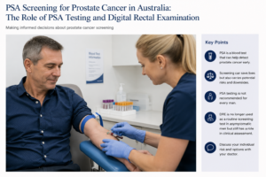
PSA Screening for Prostate Cancer in Australia: Should You Have a PSA Test?

Prostate cancer is one of the most commonly diagnosed cancers in Australian men. One of the difficulties with prostate cancer is that early prostate cancer usually causes no symptoms. By the time symptoms develop, some cancers may already have progressed beyond the prostate.

A simple blood test called the prostate-specific antigen (PSA) test can help identify men who may be at increased risk of prostate cancer. However, PSA testing is not a perfect cancer test and deciding whether to have regular PSA testing involves balancing the potential benefits of early diagnosis against the risks of unnecessary investigation and treatment.

In Australia, there is currently no national population-based prostate cancer screening program. Instead, PSA testing is based on an informed discussion between a man and his doctor, taking into account age, family history, overall health, life expectancy and personal preferences.


What is PSA?

PSA, or prostate-specific antigen, is a protein produced by prostate cells. A small amount normally enters the bloodstream and can be measured with a simple blood test.

An elevated PSA does not automatically mean that you have prostate cancer.

PSA can rise because of:

  • prostate cancer
  • benign enlargement of the prostate (BPH)
  • inflammation or infection of the prostate
  • urinary infection
  • recent ejaculation or sexual activity
  • recent instrumentation of the urinary tract
  • manipulation of the prostate.

Conversely, some men with prostate cancer can have a PSA within the expected range. PSA is therefore best considered a marker of prostate activity and prostate cancer risk rather than a test that diagnoses cancer.


Who Should Consider PSA Testing in Australia?

Current Australian guidance supports informed, individualized PSA testing rather than universal population screening.

For men at average risk who have been informed about the potential benefits and harms and decide to undergo regular testing, current RACGP guidance recommends offering PSA testing every two years between the ages of 50 and 69 years.

Men with a significant family history may be advised to start earlier. Depending on the strength of the family history, testing may begin from approximately 40–45 years of age and continue every two years to age 69.

A stronger family history includes having a father, brother or multiple close relatives diagnosed with prostate cancer, particularly when prostate cancer occurred at a younger age.

Other factors, including ancestry, genetic predisposition and an individual’s general health and life expectancy, may also influence the decision about when to begin testing.

Importantly, these recommendations apply to men without symptoms. Men with symptoms or clinical findings that raise concern about prostate cancer require individual assessment rather than simply entering a screening program.


What Does an Elevated PSA Mean?

An elevated PSA is not a diagnosis of prostate cancer.

For men aged 50–69 undergoing early detection testing, a PSA above 3.0 ng/mL generally prompts further assessment under current Australian guidance. An abnormal result will often be repeated because PSA levels can fluctuate and temporary elevations are common.

Depending on the PSA level, age, prostate size, family history and other risk factors, further assessment may include:

Repeat PSA → assessment of PSA trend and other risk factors → specialist review → prostate MRI → possible prostate biopsy

Modern prostate cancer assessment has changed substantially. An abnormal PSA no longer necessarily means proceeding directly to a prostate biopsy. Multiparametric MRI (mpMRI) can help identify suspicious areas within the prostate and assist the urologist in deciding whether biopsy is necessary.


The Potential Benefits of PSA Screening

The principal advantage of PSA testing is straightforward:

It may detect a significant prostate cancer while it is still curable.

Aggressive prostate cancer can spread to lymph nodes, bones and other organs. Once metastatic prostate cancer develops, treatment is generally aimed at controlling rather than curing the disease.

Regular PSA testing provides an opportunity to identify some clinically important cancers years before they would otherwise become apparent.

Large screening trials have produced differing results, which is one reason PSA screening remains controversial. European screening data have demonstrated a reduction in prostate cancer mortality associated with PSA-based screening, while other trials have shown less convincing mortality benefits.

For an individual man, therefore, the potential benefit is significant: early detection may identify an aggressive cancer at a stage when curative treatment remains possible.


What Are the Downsides of PSA Screening?

The difficulty is that prostate cancer is not one disease.

Some prostate cancers are aggressive and potentially life-threatening. Others grow extremely slowly and may never cause symptoms during a man’s lifetime.

PSA testing can detect both.

False-positive PSA results

An elevated PSA may lead to anxiety, repeat blood tests, MRI scans and sometimes biopsy even when no cancer is ultimately found.

Overdiagnosis

PSA testing can identify a prostate cancer that would never have caused illness or shortened the man’s life.

This is known as overdiagnosis.

Overtreatment

If an insignificant cancer is diagnosed and subsequently treated, the man can potentially experience treatment complications without gaining a meaningful survival benefit.

Potential consequences of prostate cancer treatment include:

  • erectile dysfunction
  • urinary incontinence
  • urinary symptoms
  • ejaculatory changes
  • bowel symptoms following some forms of radiotherapy.

This is one of the major reasons Australia does not currently have a universal PSA population-screening program.

Importantly, however, diagnosing prostate cancer does not automatically mean treating it.

Many men with low-risk prostate cancer are now managed with active surveillance, involving PSA monitoring, MRI and selective repeat biopsy. Treatment is reserved for evidence that the cancer is becoming more significant.


What Are the Implications of Not Having PSA Screening?

Choosing not to undergo PSA testing is also a legitimate decision, particularly after considering the benefits and disadvantages.

The advantage is avoiding the potential cascade of:

PSA testing → abnormal result → anxiety → MRI → biopsy → diagnosis of an insignificant cancer → possible unnecessary treatment.

However, there is another side to this decision.

Because early prostate cancer frequently produces no symptoms, a man who does not undergo PSA testing may remain unaware that he has prostate cancer.

For most men who never develop clinically significant disease, this causes no harm.

For a smaller group who develop aggressive prostate cancer, however, the first indication of the disease may occur after the cancer has grown substantially or spread beyond the prostate.

At that stage, the opportunity for curative treatment may have been reduced or lost.

The decision is therefore not simply:

“Do I want to know whether I have prostate cancer?”

A more useful question is:

“Given my age, health, family history and personal priorities, do the potential benefits of finding a significant prostate cancer early outweigh the potential harms of investigating and possibly diagnosing a cancer that may never affect me?”

This is why shared decision-making with your GP or urologist is important.


What About a Digital Rectal Examination?

The digital rectal examination, or DRE, involves a doctor gently inserting a gloved, lubricated finger into the rectum to examine the back surface of the prostate.

The doctor assesses the prostate for abnormalities such as:

  • unusual hardness
  • nodules
  • asymmetry
  • irregularity
  • loss of the normal contour of the gland.

Historically, DRE was routinely combined with PSA testing for prostate cancer screening.

This has changed.

DRE is no longer recommended as a routine screening test in asymptomatic men

Current Australian guidance states that DRE should not routinely be added to PSA testing for early detection in asymptomatic men in primary care.

There are several reasons.

A finger can examine only part of the prostate. Small cancers may not be palpable, and cancers in areas of the prostate that cannot be reached during examination can be missed. A normal DRE therefore does not exclude prostate cancer.

Conversely, benign prostate enlargement and other abnormalities can sometimes produce an abnormal examination.


Does DRE Still Have a Role?

Yes.

Not recommending DRE as a routine screening test does not mean that the examination has become obsolete.

A urologist may perform a DRE when assessing a man with:

  • an elevated or rising PSA
  • urinary symptoms
  • suspected prostate disease
  • an abnormal MRI
  • concern about locally advanced prostate cancer
  • other pelvic or rectal symptoms.

Occasionally, a prostate cancer may produce a distinctly hard or irregular prostate even when the PSA is not dramatically elevated. An abnormal DRE can therefore remain clinically important and may prompt further investigation. Cancer Council Australia notes that urologists will commonly perform DRE as part of prostate assessment even though GPs no longer routinely use it for screening asymptomatic men.

The key distinction is:

DRE has a role in clinical examination and investigation, but it is not recommended as a routine population-screening test in an otherwise well man undergoing PSA testing.


PSA Screening Is About Risk, Not Just a Number

One of the most important changes in modern prostate cancer detection is moving away from treating PSA as simply “normal” or “abnormal.”

A PSA result should be interpreted in context.

Your doctor or urologist may consider:

your age + PSA level + change in PSA over time + family history + prostate size + symptoms + examination findings + MRI findings + overall health

Together, these provide a much more meaningful assessment of prostate cancer risk than a single PSA result.


Should I Have a PSA Test?

There is no single answer that is appropriate for every man.

For a healthy man in the appropriate age group, particularly someone with a family history of prostate cancer, the potential benefit of identifying an aggressive cancer while it remains curable may be important.

For an older man with substantial medical problems or limited life expectancy, the likelihood that PSA screening will provide meaningful benefit becomes considerably smaller. Current Australian guidance advises against PSA testing in men whose life expectancy is likely to be less than approximately seven years because a mortality benefit from early detection is unlikely to occur within that period.

The decision should therefore be individual rather than automatic.


The Bottom Line

PSA testing can save lives by detecting clinically significant prostate cancer at an earlier and potentially curable stage, but it can also detect cancers that would never have caused harm.

Australia therefore does not currently operate a universal PSA screening program. Instead, men are encouraged to make an informed decision about PSA testing after discussing their individual risk and the potential benefits and harms with their doctor.

For men who choose screening, current Australian guidance generally supports PSA testing every two years from age 50–69 for men at average risk, with earlier testing considered for men with a significant family history.

A digital rectal examination is no longer recommended as a routine addition to PSA screening in asymptomatic men, but it remains an important component of specialist prostate assessment in selected circumstances.

Concerned about your prostate cancer risk?

If you are considering PSA testing, have a family history of prostate cancer, have noticed a change in your PSA, or have urinary symptoms, discuss your individual risk with your GP or urologist.

Early detection should not simply be about finding more prostate cancers. The goal is to identify the cancers that matter, at a time when something useful can still be done about them.

This information is intended for general education and does not replace individual medical advice. Recommendations may differ according to your age, family history, medical conditions and previous PSA results.

Further information

Australian patient information and guidance are available from Cancer Council Australia: Prostate cancer screening and early detection and the RACGP Guidelines for preventive activities in general practice.

Fascia Lata Allograft as a Mid-Urethral Sling for Female Stress Urinary Incontinence

A “new/revamped” kid on the block – Fascia Lata Allograft: A biological alternative to synthetic mesh

Stress urinary incontinence (SUI) is one of the most common forms of urinary incontinence in women. It occurs when urine leaks with activities that increase abdominal pressure, such as coughing, sneezing, laughing, exercising, lifting or running.

For women whose symptoms remain troublesome despite pelvic floor muscle training and appropriate conservative treatment, surgical treatment can provide substantial and durable improvement.

For many years, the mid-urethral sling has been one of the most effective minimally invasive surgical treatments for SUI. Synthetic mesh mid-urethral slings remain available in Australia and have extensive evidence supporting their effectiveness. However, concerns surrounding synthetic mesh have led many women to ask whether there is a biological alternative.

One such option is a fascia lata allograft, including the fascia lata allograft supplied by Samson Medical Technologies in Australia.

The important distinction is that the Samson product is not a synthetic mesh. It is human donor fascia lata, processed for use as a biological graft. Samson describes its current fascia lata allograft as 100% human-derived, without storage media, non-irradiated and processed using Super Critical CO₂ technology.

However, there is an important caveat: the published clinical evidence for fascia lata allografts in SUI largely relates to earlier cadaveric fascia products and pubovaginal sling techniques, rather than Samson’s current specific product. Therefore, the historical allograft results should not be presented as if they were Samson-specific clinical trial results.


What is an allograft sling?

An allograft is tissue obtained from a human donor rather than from the patient herself.

Traditional biological pubovaginal slings have commonly used the patient’s own rectus fascia or fascia lata. This provides a strong, durable biological support but requires an additional incision to harvest the patient’s tissue.

An allograft avoids this donor-site surgery.

The fascia lata is positioned beneath the urethra and secured to provide a supportive platform. During coughing, sneezing or physical exertion, the urethra is compressed against this support, helping prevent urine leakage.

The concept is therefore quite different from a synthetic polypropylene mid-urethral sling. Instead of leaving a permanent synthetic mesh implant, the surgeon is using processed human connective tissue.

Samson currently supplies fascia lata allografts in several dimensions, including 20 × 100 mm and 20 × 120–140 mm configurations.


How effective are fascia lata allografts?

This is where the evidence requires some careful interpretation.

There is a substantial historical literature examining cadaveric fascia lata as a sling material, but the results have been inconsistent.

Early studies

Some early studies reported excellent short-term results.

One study of 26 women treated with solvent-dehydrated cadaveric fascia lata reported that 77% were completely dry and 92% used one pad or fewer per day at a mean follow-up of 15 months.

Another series of 31 women reported complete resolution of SUI in 93% at approximately one year.

A larger series of 104 women using freeze-dried, non-irradiated cadaveric fascia lata demonstrated substantial reductions in pad use, although the study’s follow-up was relatively short.

These studies created considerable enthusiasm for allograft tissue because it offered the advantages of a biological sling without the morbidity of harvesting the patient’s own fascia.

But there was also a warning signal

Other studies produced considerably less encouraging results.

A study of 18 women using solvent-dehydrated, gamma-irradiated fascia lata found that 27.8% experienced significant failure, with recurrent incontinence occurring within 3–6 months. The authors concluded that this particular allograft material was unreliable.

A subsequent comparative study of 71 women found a concerning difference in objective urodynamic recurrence: urodynamic stress incontinence was present in 41.7% of women who received Tutoplast allograft compared with none of those receiving autologous fascia lata at follow-up. Patient-reported outcomes, however, were more favourable.

Other series have reported much better results. In a 134-patient comparison, 63 women received allograft fascia and 71 received autologous fascia. There was no statistically significant difference in overall cure of stress and urge incontinence at approximately two years, while the allograft group experienced less postoperative pain and disability.

What does this mean?

The historical evidence suggests that allograft fascia can work well, but the durability and consistency of older allograft products were variable.

This variability is important when discussing the Samson allograft. There is currently insufficient published comparative evidence to state that the Samson product has a specific cure rate such as 80%, 90% or 95%.

The more scientifically defensible statement is:

Samson fascia lata provides a contemporary biological allograft option for sling surgery, but Samson-specific long-term comparative clinical outcome data are still developing.

That distinction is important when counselling patients.


How does Samson allograft differ from older allografts?

The earlier allograft literature predominantly involved freeze-dried, solvent-dehydrated and/or gamma-irradiated cadaveric fascia lata.

The Samson product currently described by the manufacturer is different in its processing. Samson reports that its fascia lata is:

  • 100% human-derived
  • non-irradiated
  • supplied without storage media
  • processed using Super Critical CO₂ technology
  • available in several sizes.

The rationale behind modern tissue processing is to preserve the structural characteristics of the collagenous tissue while making the graft suitable for implantation.

Nevertheless, a different processing technology does not automatically establish superior clinical continence outcomes. Laboratory characteristics, tensile strength and histological appearance are not substitutes for long-term clinical trials.

This is one of the areas where future Australian outcome data will be particularly valuable.


How does an allograft compare with synthetic mid-urethral mesh?

Synthetic mid-urethral slings have the largest evidence base of any minimally invasive SUI operation.

A Cochrane review including 81 trials and more than 12,000 women found that more than 80% of women were cured or significantly improved following mid-urethral sling surgery, with good results maintained for up to five years.

Retropubic and transobturator approaches have broadly comparable effectiveness, although their complication profiles differ.

For example, retropubic slings have a higher risk of bladder perforation, whereas transobturator procedures have more groin pain. Vaginal mesh exposure is uncommon but recognised with both approaches.

The Australian situation

It is important to distinguish mid-urethral slings from transvaginal mesh used for pelvic organ prolapse.

The Australian TGA has removed certain transvaginal mesh products from the market, particularly mesh used for prolapse and single-incision mini-slings. However, mid-urethral sling devices for SUI remain approved in Australia.

Indeed, a 2026 Australian Government review concluded that mid-urethral sling devices should remain on the Prescribed List, with their existing listing unchanged.

Therefore, synthetic MUS should not simply be described as “banned mesh” in Australia. It remains an accepted treatment for appropriately selected women.

Potential advantages of an allograft

A biological fascia lata sling potentially offers:

Advantages

  • No synthetic polypropylene mesh
  • No fascial harvest from the patient’s abdomen or thigh
  • Avoidance of donor-site pain
  • Biological rather than permanent synthetic material
  • Potentially useful where avoidance of synthetic mesh is an important patient preference

Potential disadvantages

  • Less robust long-term evidence than synthetic MUS
  • Historical allograft studies have shown variable durability
  • It is still a surgical sling and can cause obstruction, urinary retention, infection, bleeding or pain
  • It requires more extensive dissection than a conventional MUS
  • It should not currently be marketed as proven superior to synthetic MUS

What about the Burch colposuspension?

The Burch colposuspension is the classic non-mesh surgical treatment for SUI.

Rather than placing a sling beneath the urethra, sutures are placed beside the bladder neck and proximal urethra and attached to supporting pelvic tissues.

It can be performed by open or laparoscopic surgery.

Long-term studies demonstrate that Burch colposuspension can be highly effective. One classic study found an 81.6% cure rate at 5–10 years.

More contemporary comparative data are reassuring. In a large study with more than 1,300 women and long-term follow-up, the proportion reporting no ongoing SUI was 83.0% after Burch colposuspension compared with 85.0% following retropubic MUS, with no significant difference.

However, Burch surgery is more invasive than a mid-urethral sling and is generally no longer the first surgical option for uncomplicated primary SUI.

It can nevertheless be an attractive option in selected women, particularly when avoiding synthetic mesh is important.


What about peri-urethral bulking agents?

Urethral bulking agents occupy a completely different part of the treatment spectrum.

A material is injected into the urethral wall around the bladder neck or proximal urethra to increase coaptation and improve continence.

In Australia, polyacrylamide hydrogel such as Bulkamid is an established example.

The major attraction is that it is substantially less invasive than sling surgery.

It can often be performed under local anaesthesia or sedation and has a relatively favourable safety profile.

The trade-off is effectiveness and durability.

A 2024 study of Bulkamid reported that at 12 months 69% of women described themselves as cured or improved, with 40% reporting cure, while 23% underwent subsequent treatment, including repeat Bulkamid injection or MUS.

Longer-term data are more modest. In a cohort of 357 women followed for a median of approximately 2.5 years, subjective cure was 30.9%, while 52.1% reported improvement. Minor adverse events occurred in 21%, with no serious adverse events attributed to the procedure.

Current guideline evidence therefore generally considers bulking agents less effective than MUS, Burch colposuspension or autologous fascial sling surgery, and repeat injections may be necessary.


Putting the options together

Treatment Approximate effectiveness Main advantages Main disadvantages
Samson fascia lata allograft sling Samson-specific long-term cure rate not yet established; historical allograft studies range widely Biological tissue; no synthetic mesh; no tissue harvest Limited contemporary comparative evidence; historical allograft durability concerns; surgical sling risks
Synthetic mid-urethral sling Generally >80% cure/improvement in medium-term evidence Minimally invasive, rapid recovery, extensive evidence Mesh-specific complications including exposure, pain and voiding dysfunction
Burch colposuspension Approximately 70–85% long-term continence in many series No vaginal mesh; durable native-tissue operation More invasive; longer recovery; risk of prolapse and voiding problems
Autologous fascial sling Generally high effectiveness, comparable with other established sling procedures No synthetic mesh; patient’s own tissue Requires tissue harvest; more postoperative morbidity
Bulking agent Lower cure rate, but meaningful improvement for many women Minimally invasive; useful for frail/high-risk patients or those avoiding sling surgery Less durable; repeat injections commonly required

The Australian Government’s own review of MUS specifically considered colposuspension, native-tissue pubovaginal slings and urethral bulking agents as important comparators.


Who might consider an allograft sling?

A fascia lata allograft may be particularly attractive for a woman who:

  • has objectively demonstrated SUI;
  • has failed appropriate conservative treatment;
  • wishes to avoid a permanent synthetic mesh implant;
  • does not want an abdominal or thigh incision to harvest her own fascia;
  • understands that the long-term evidence base for contemporary allograft products is smaller than that for synthetic MUS;
  • accepts that biological tissue does not eliminate the risks associated with sling surgery.

It may also have a role in selected women with recurrent SUI, particularly where previous synthetic mesh surgery makes another synthetic procedure unattractive. However, recurrent SUI is a more complex clinical situation and requires individual assessment.


The important question: is Samson allograft better than synthetic mesh?

At present, there is not enough evidence to say that it is.

The strongest evidence remains with synthetic mid-urethral slings. Their effectiveness has been demonstrated in thousands of women and across multiple randomised trials and systematic reviews.

The potential attraction of Samson fascia lata is different.

It offers a biological alternative for women who would prefer to avoid synthetic mesh while also avoiding the morbidity of harvesting their own fascia.

That makes it an interesting addition to the surgeon’s toolbox rather than a replacement for the established mid-urethral sling.

The key unanswered question is long-term durability.

Historical cadaveric allograft studies demonstrate that some allografts performed extremely well, while others experienced substantial early failure.

Consequently, contemporary prospective data specifically evaluating Samson’s processing technology, clinical continence outcomes, complications and durability would be valuable.


What should a woman expect from the procedure?

As with any continence operation, the goal is not simply to make a stress test negative. The aim is to improve the woman’s quality of life while preserving normal bladder emptying and sexual function.

Potential complications of sling surgery include:

  • temporary or persistent difficulty emptying the bladder;
  • urinary retention requiring catheterisation;
  • urinary tract infection;
  • bleeding or haematoma;
  • pain;
  • de novo urgency or worsening overactive bladder;
  • recurrent or persistent SUI;
  • vaginal wound problems;
  • infection;
  • rarely, injury to the bladder, urethra or surrounding structures.

Synthetic mesh has additional mesh-specific risks, including vaginal exposure/erosion and chronic pain. The TGA specifically requires information regarding complications such as severe chronic pain, groin pain and bladder perforation in the instructions for use of Australian MUS devices.

An allograft avoids synthetic mesh exposure, but it does not make the operation risk-free.


The bottom line

For a woman with bothersome stress urinary incontinence, there is no single operation that is right for everyone.

Synthetic mid-urethral slings remain the most extensively studied minimally invasive surgical treatment and have excellent success rates.

Burch colposuspension remains a durable non-mesh alternative, although it is more invasive.

Bulking agents offer the least invasive surgical option but generally have lower and less durable success rates.

Fascia lata allograft represents an intriguing middle ground: a biological sling that avoids both synthetic mesh and the need to harvest the patient’s own fascia.

The Samson allograft is particularly interesting because its contemporary processing differs from several of the older cadaveric fascia products that generated the historical literature. Samson describes its current graft as non-irradiated human fascia lata processed using Super Critical CO₂ technology.

However, the enthusiasm should be matched by scientific caution. The historical literature demonstrates that allografts can achieve good continence outcomes, but it also demonstrates that not all allograft materials have behaved identically over time.

For patients considering this option, the most accurate discussion is therefore not “this is a better sling than mesh.” It is:

“This is a biological alternative to synthetic mesh, with encouraging historical experience but less mature long-term clinical evidence for the contemporary product.”

As further Australian experience and prospective follow-up accumulate, Samson fascia lata allograft may become an increasingly useful option in the treatment of female stress urinary incontinence.

Important note

This article is intended for general information and does not replace an individual assessment by a urologist or uro-gynaecologist. The choice between conservative management, bulking agent, synthetic MUS, biological or autologous sling and Burch colposuspension depends on the type and severity of incontinence, previous surgery, urethral function, pelvic anatomy, patient preference and overall health.

Post-Prostatectomy Urinary Incontinence

Men’s Guide to Lower Urinary Tract Symptoms: What’s Causing Them and What Can Be Done?

Medical note: This is patient education rather than individual medical advice. The investigation and treatment approach should be tailored to the individual, particularly where there is haematuria, urinary retention, recurrent infection, neurological disease or concern about prostate cancer. The treatment information above reflects contemporary EAU guidance, including the 2026 guideline update.

Blood in the Urine (Haematuria): When Should You See a Urologist Brisbane?

Discovering blood in your urine can be alarming. While there are many possible causes, blood in the urine should never be ignored. Known medically as haematuria, this symptom can sometimes indicate an underlying condition requiring specialist assessment. Consulting an experienced Urologist Brisbane patients rely on can help identify the cause and provide appropriate treatment.

What Is Haematuria?

Haematuria refers to the presence of blood in the urine. It can occur in two forms:

Visible Haematuria

The urine appears pink, red or brown due to the presence of blood.

Microscopic Haematuria

Blood cells are detected only during laboratory testing and may not be visible to the naked eye.

Both forms should be assessed by a healthcare professional.

Common Causes of Blood in the Urine

There are numerous possible causes of haematuria, including:

Urinary Tract Infections

Infections affecting the bladder, kidneys or urinary tract can cause irritation and bleeding.

Kidney Stones

Kidney stones can damage the lining of the urinary tract, resulting in blood appearing in the urine.

Enlarged Prostate

An enlarged prostate can place pressure on the urinary system and may contribute to urinary symptoms and bleeding.

Vigorous Exercise

In some cases, intense physical activity can lead to temporary blood in the urine.

Kidney Disease

Various kidney conditions may result in haematuria and require specialist management.

Urinary Tract Cancers

Although less common, blood in the urine can sometimes be associated with cancers affecting the bladder, kidney or urinary tract. This is one reason why prompt evaluation by a Urologist Brisbane specialist is important.

Symptoms That May Accompany Haematuria

Depending on the underlying cause, patients may also experience:

  • Pain during urination
  • Frequent urination
  • Lower abdominal discomfort
  • Back or flank pain
  • Difficulty passing urine
  • Fever or chills

These symptoms can provide valuable clues during diagnosis.

How Does a Urologist Brisbane Diagnose Haematuria?

A comprehensive evaluation may include:

Medical History and Physical Examination

Your specialist will discuss symptoms, medical history and risk factors.

Urine Testing

Urinalysis helps identify infection, blood cells and other abnormalities.

Blood Tests

Blood tests may assess kidney function and identify underlying conditions.

Imaging Studies

Ultrasound or CT imaging can help detect stones, tumours or structural abnormalities.

Cystoscopy

A cystoscopy allows a Urologist Brisbane specialist to examine the inside of the bladder using a small camera.

Why Early Investigation Matters

While haematuria can sometimes result from minor conditions, it may also be an early warning sign of a more serious problem. Early diagnosis often leads to more effective treatment and improved outcomes.

Delaying assessment can allow certain conditions to progress and become more difficult to treat.

When Should You Seek Medical Advice?

You should arrange an appointment if:

  • You notice visible blood in your urine
  • Blood is detected during routine testing
  • Haematuria occurs repeatedly
  • You experience pain or urinary symptoms
  • You have a history of smoking or urinary tract conditions

Speak with a Urologist Brisbane Today

If you have noticed blood in your urine or have concerns about your urinary health, our experienced team is here to help. We provide thorough assessments, advanced diagnostic services and personalised treatment plans to identify the cause and protect your long-term health. Contact our clinic today to book a consultation with a trusted Urologist Brisbane specialist.

Kidney Stones: Symptoms, Causes and Treatment Options Explained

Kidney stones: symptoms, treatment and prevention

Kidney stones can cause sudden, intense pain, but some are discovered by chance and cause no symptoms. The right treatment depends on where a stone sits, its size, whether urine can drain, whether infection is present and how well the kidneys are working. If you have suspected stone pain, an assessment can establish which approach is safest for you.

What is a kidney stone?

A kidney stone forms when substances in urine crystallise and grow. Calcium-based stones are common; other types include uric acid, infection-related (struvite) and cystine stones. A stone may remain in the kidney or move into the ureter, the tube carrying urine to the bladder. A ureteric stone can obstruct urine flow and cause renal colic.

Symptoms to recognise

  • Severe pain in the side or back that may travel towards the lower abdomen or groin, often in waves
  • Blood in the urine, which may be visible or detected on a test
  • Nausea or vomiting
  • Urgency, frequent urination or discomfort passing urine, particularly when a stone is near the bladder

Cloudy urine, fever or chills may indicate infection. These symptoms also have other possible causes, so imaging and tests are important.

Seek urgent care at an emergency department if you have fever or chills with suspected stone pain, cannot pass urine, cannot keep fluids down, have pain that cannot be controlled, or have a single functioning kidney or known kidney disease with possible obstruction. An infected, obstructed kidney is a urological emergency: antibiotics and urgent drainage with a ureteric stent or nephrostomy may be needed before definitive stone treatment.

Why do stones form?

Low fluid intake, heavy sweating, a high-salt diet, previous stones and family history can increase risk. Other contributors include certain medicines and conditions such as gout, bowel disease or recurrent infection. The causes differ by stone type. Finding the type matters when planning prevention.

How are kidney stones diagnosed?

Assessment usually includes your symptoms and medical history, urine testing for blood and infection, and blood tests to check kidney function when appropriate. A low-dose CT scan without contrast is often used for adults with suspected renal colic because it shows the size and position of a stone and signs of obstruction. Ultrasound may be preferred first in pregnancy and some other circumstances. If you pass or have a stone removed, laboratory analysis can guide future prevention.

What are the treatment options?

Observation and pain relief. Many small ureteric stones pass without a procedure. This is reasonable when pain is controlled, there is no infection or concerning obstruction, and kidney function is safe. Your clinician will arrange follow-up to confirm that the stone has passed or the obstruction has resolved. Pain relief is tailored to your health; anti-inflammatory medicines may be useful but are unsuitable for some people, including those with impaired kidney function. Drink normally and avoid dehydration; forcing large amounts of water during acute colic does not push a blocked stone through.

Medication to help passage. An alpha blocker may improve passage for selected stones in the lower ureter, especially those around 5–10 mm. This use is off-label and should be discussed alongside possible adverse effects, such as dizziness. It is not suitable as a substitute for urgent treatment if infection, uncontrolled pain or worsening kidney function develops.

Shock wave lithotripsy (SWL or ESWL). Shock waves delivered from outside the body break a suitable stone into fragments. Its success depends on factors including stone size, position and density; further treatment can sometimes be needed.

Ureteroscopy and laser treatment. A fine telescope passes through the urethra and bladder into the ureter or kidney. The stone can be removed or fragmented with a laser. A temporary ureteric stent is sometimes placed and can cause urinary discomfort until removal.

Percutaneous nephrolithotomy (PCNL). For some large or complex kidney stones, instruments are passed through a small opening in the back to remove the stone. This is more invasive than ureteroscopy or shock wave treatment and has its own bleeding and infection risks.

Your urologist will discuss the expected chance of clearing the stone, anaesthetic needs, recovery, possible stenting and risks of each appropriate option.

Reducing the chance of another stone

For most people, drinking enough fluid to keep urine dilute is a useful starting point, particularly in Queensland heat. If you have heart failure or kidney disease, follow your treating team’s fluid advice instead of increasing intake on your own. Reduce excess salt, maintain a balanced diet and avoid unnecessarily restricting normal dietary calcium. Further dietary advice or preventive medication should be based on the stone’s composition and, for recurrent or higher-risk stones, blood and 24-hour urine testing. Follow-up imaging may be recommended to check for residual or new stones.

When should you see a urologist in Brisbane?

Arrange an assessment if you have a diagnosed stone that has not passed, recurrent stones, persistent pain, blood in the urine, or concerns about kidney function. If you are acutely unwell or have fever with stone symptoms, go to an emergency department rather than waiting for a routine appointment.

This article is general information and does not replace individual medical advice.

References

  1. European Association of Urology. EAU Guidelines on Urolithiasis (2026).
  2. National Institute for Health and Care Excellence. Renal and ureteric stones: assessment and management (NG118).
  3. Healthdirect Australia. Kidney stones: symptoms, diagnosis and treatment.
  4. Kidney Health Australia. Kidney stones.