
Tag Archive for: dr jo schoeman

‘Bag-of-Worms” Varicocele: Causes, Symptoms, Fertility and Treatment Options

A varicocele is an enlargement of the veins surrounding the testicle. It is similar to a varicose vein in the leg, although it occurs within the scrotum.

Varicoceles are common and often harmless. However, in some men they may cause discomfort, reduce the size or function of the affected testicle, or contribute to fertility problems. Most varicoceles do not require treatment, and the decision to intervene depends on symptoms, testicular development, semen quality and plans for future fertility.
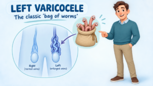
What causes a varicocele?

Blood leaves each testicle through a network of small veins called the pampiniform plexus. These veins normally carry blood upwards towards larger veins in the abdomen.

A varicocele develops when blood pools within these veins. This is usually related to poorly functioning venous valves, together with the anatomy and pressure within the testicular veins.

Around 80–90% of clinically detected varicoceles occur on the left side. This is because the left testicular vein is longer and enters the left renal vein at a right angle, making venous drainage less efficient. A varicocele may occur on both sides, while an isolated right-sided varicocele is much less common.

Most varicoceles develop gradually during puberty and have no dangerous underlying cause.

A new varicocele in an older man, particularly one that is right-sided, develops suddenly or does not disappear when lying down, may require abdominal imaging to exclude obstruction of the vein by an abdominal or retroperitoneal condition. This is uncommon but important to recognise.

How common are varicoceles?

A varicocele is found in approximately 15% of adolescent and adult men. Many men never know that they have one.

Varicoceles are more frequently identified among men being investigated for infertility. They may be present in approximately:

  • 35–40% of men with primary infertility
  • Up to 70–80% of men with secondary infertility, where a previous pregnancy has occurred, but the couple is now having difficulty conceiving

Importantly, most men with a varicocele remain fertile. Finding a varicocele does not automatically mean that it is causing infertility.

What are the symptoms?

Many varicoceles cause no symptoms and are discovered during a routine examination, fertility assessment or scrotal ultrasound.

When symptoms occur, they may include:

  • A dull ache, dragging sensation or heaviness in the scrotum
  • Discomfort that becomes worse after standing, exercising or later in the day
  • Improvement in discomfort when lying down
  • Visible or enlarged veins above the testicle
  • A soft swelling sometimes described as feeling like a “bag of worms”
  • Reduced size or slower growth of the affected testicle
  • Abnormal semen-analysis results
  • Difficulty conceiving with a partner

A varicocele generally does not cause erectile dysfunction or difficulty urinating. Its relationship with testosterone production remains under investigation, although some men with a clinically significant varicocele may have impaired testicular function.

Sudden severe testicular pain is not typical of a varicocele and requires urgent medical assessment to exclude testicular torsion or another acute scrotal condition. An abdominal ultrasound is also required to exclude a possible lesion in the abdomen obstructing the veins.

How is a varicocele diagnosed?

Diagnosis usually begins with a physical examination. The scrotum is examined while the patient is standing and lying down. You may be asked to take a breath and gently bear down, the Valsalva manoeuvre, to make enlarged veins easier to feel.

Clinical varicoceles are commonly graded as:

  • Grade 1: Felt only during the Valsalva manoeuvre
  • Grade 2: Felt while standing without straining
  • Grade 3: Easily visible through the scrotal skin

A scrotal ultrasound may be recommended if the examination is uncertain, the testicles differ in size, pain is present, or another scrotal problem needs to be excluded. Ultrasound can measure the veins, assess backward blood flow and examine the testicles.

For fertility assessment, investigations may include:

  • One or more semen analyses
  • Testicular volume measurement
  • Hormone tests when indicated
  • Assessment of both partners, because fertility is a shared consideration

An ultrasound-only or “subclinical” varicocele that cannot be felt on examination is not usually treated for infertility.

How can a varicocele affect fertility?

The testicles work best at a temperature slightly below core body temperature. The pampiniform veins help cool the arterial blood entering the testicle.

Pooling of warm blood in a varicocele may interfere with this cooling system. Increased temperature, venous pressure, oxidative stress and altered blood flow may affect sperm production and testicular function.

Possible semen changes include:

  • Reduced sperm concentration
  • Reduced sperm movement or motility
  • Increased abnormal sperm forms
  • Increased sperm DNA damage

The degree of impact varies considerably. A large varicocele can be associated with normal fertility, while a smaller palpable varicocele may be significant in another man. The diagnosis must therefore be interpreted together with semen results, the couple’s fertility history, the female partner’s age and other fertility factors.

Does every varicocele require treatment?

No. Observation is appropriate when a varicocele:

  • Causes no troublesome symptoms
  • Is not affecting testicular growth
  • Is associated with normal semen parameters
  • Is found incidentally on ultrasound but cannot be felt
  • Is unlikely to be the main cause of the couple’s fertility difficulty

Simple measures such as supportive underwear, avoiding activities that consistently aggravate discomfort and occasional appropriate pain relief may help mild symptoms. Regular review may be recommended for adolescents with a difference in testicular size.

When should treatment be considered?

Treatment may be considered when there is:

Infertility with abnormal semen parameters

Repair is most likely to benefit a couple when the man has:

  • A varicocele that can be felt on examination
  • Difficulty conceiving
  • One or more abnormal semen parameters
  • No better explanation for the infertility
  • A reasonable opportunity for natural conception following treatment

Both the AUA/ASRM and European guidance support considering repair in men attempting conception who have a palpable varicocele and abnormal semen parameters. Treatment is not routinely recommended for a varicocele detected only by ultrasound. AUA/ASRM male infertility guideline and EAU male infertility guideline

Persistent scrotal pain

Repair may be reasonable when typical varicocele-related discomfort persists despite conservative measures and other causes of scrotal pain have been excluded.

Reduced testicular growth in an adolescent

Treatment may be discussed if the affected testicle is persistently smaller, growth is impaired, the varicocele is large or semen parameters are abnormal in an appropriately selected older adolescent.

Progressive testicular dysfunction

Selected men with deteriorating semen quality or evidence of impaired testicular function may benefit from repair after individual assessment.

What treatment procedures are available?

Treatment works by blocking or dividing the abnormal veins. Blood then returns through healthy alternative veins while the testicular artery and lymphatic drainage are preserved.

Management option How it is performed Advantages Disadvantages and risks
Observation Clinical review, with semen analysis or testicular measurements when appropriate No procedure or anaesthetic; suitable for most asymptomatic men; avoids unnecessary treatment Does not correct the varicocele; symptoms or testicular changes may require future review
Conservative symptom management Scrotal support, activity modification and appropriate pain relief Simple and non-invasive; may adequately control mild discomfort Does not remove the varicocele or improve abnormal semen parameters; long-term anti-inflammatory medication may not be suitable for everyone
Microsurgical subinguinal varicocelectomy A small incision is made near the groin. An operating microscope helps identify and divide abnormal veins while protecting the testicular artery and lymphatics Usually the lowest recurrence and hydrocele rates; no abdominal incision; effective for unilateral or bilateral disease; commonly preferred for fertility treatment Requires an anaesthetic and microsurgical expertise; temporary bruising, swelling or discomfort; uncommon risks include infection, recurrence, hydrocele or testicular artery injury
Microsurgical inguinal varicocelectomy Similar microsurgical repair through a slightly higher groin incision Good visualisation and low recurrence when performed microsurgically Requires opening the external oblique fascia; recovery may involve slightly more groin discomfort
Laparoscopic varicocelectomy Veins are clipped or divided through small abdominal keyhole incisions Both sides can be treated during the same procedure; useful in selected cases General anaesthetic; entry into the abdomen; generally higher hydrocele risk than lymphatic-sparing microsurgery; rare abdominal or vascular complications
Open high ligation The testicular vein is divided through an abdominal or groin incision without microsurgical magnification Established technique; may be effective where microsurgery is unavailable Higher recurrence and hydrocele rates than modern microsurgical repair; less precise preservation of small arteries and lymphatics
Radiological embolisation or sclerotherapy An interventional radiologist passes a catheter into the testicular vein and blocks it using coils, plugs or a sclerosant No surgical incision in the scrotum or groin; often performed with local anaesthesia or sedation; relatively quick recovery; useful for recurrence after surgery The vein cannot always be accessed; radiation and contrast exposure; recurrence or persistence may occur; coils can rarely migrate; availability depends on local expertise

How effective is treatment?

The results depend on why treatment is being performed, the technique used and whether other fertility or pain-related factors are present.

Improvement in semen quality

Following repair of a clinically significant varicocele, approximately 60–80% of appropriately selected men demonstrate improvement in at least one semen parameter. Improvement is not guaranteed, and some men show little or no measurable change.

Because sperm production takes approximately three months, semen analysis is usually repeated about three months after treatment and may be reassessed again at six months.

Pregnancy and fertility outcomes

Varicocele repair may increase the chance of natural pregnancy in selected infertile couples, but it does not guarantee conception. Studies commonly report spontaneous pregnancy in roughly 25–40% of couples after repair, although rates vary substantially according to female partner factors, duration of infertility, baseline semen quality and study design.

Treatment may also improve semen quality sufficiently to allow a couple to use a less invasive assisted-reproduction option. In other cases, IVF or intracytoplasmic sperm injection may still be required.

The couple’s overall fertility timeline is important. If the female partner has reduced ovarian reserve or advancing reproductive age, waiting several months for semen improvement may not be appropriate. Varicocele repair and assisted reproductive treatment should therefore be considered as part of a shared plan.

Relief of pain

When pain is typical of a varicocele, a dull ache or heaviness made worse by standing or activity, approximately 70–90% of appropriately selected men experience meaningful improvement after repair.

Surgery is less predictable when pain is sharp, constant or atypical, and pain may occasionally persist despite technically successful treatment.

Testicular growth

In adolescents with a persistently smaller affected testicle, repair may allow “catch-up” growth. Decisions should be based on repeated measurements rather than a single ultrasound result.

What are the possible complications?

Varicocele procedures are generally safe, but potential complications include:

  • Bruising, swelling or wound discomfort
  • Infection or bleeding
  • Persistence or recurrence of the varicocele
  • Hydrocele, caused by fluid collecting around the testicle
  • Ongoing or occasionally altered scrotal pain
  • Injury to the testicular artery, which is uncommon but potentially serious
  • Testicular shrinkage, which is rare
  • Anaesthetic complications
  • Technical failure, contrast reaction, radiation exposure or coil-related problems following embolisation

Using microsurgical magnification and preserving the lymphatic vessels usually reduces the risks of recurrence, hydrocele and arterial injury.

Recovery after varicocele repair

Most men return home on the day of treatment. Bruising, mild swelling and groin or scrotal discomfort are expected during the early recovery period.

Patients are commonly advised to:

  • Wear supportive underwear
  • Keep the wound clean and dry
  • Avoid heavy lifting and strenuous exercise for the period advised by the surgeon
  • Resume sexual activity when comfortable and medically cleared
  • Attend follow-up to assess healing and symptom improvement
  • Repeat semen analysis after approximately three months when fertility is the reason for treatment

Specific recovery instructions vary according to the procedure and the individual patient.

When should you seek medical advice?

Arrange a medical assessment if you notice:

  • A new lump or swelling in the scrotum
  • Persistent scrotal discomfort or heaviness
  • A difference in testicular size
  • Difficulty conceiving
  • A new right-sided varicocele
  • A swelling that remains when lying down

Seek urgent medical attention for sudden severe testicular pain, rapid swelling, nausea or a high-riding testicle. These features may indicate testicular torsion, which is an emergency.

The bottom line

A varicocele is common and, for most men, causes no significant harm. Treatment is not based on the ultrasound appearance alone. The most important considerations are symptoms, testicular development, semen quality and the couple’s overall fertility situation.

For men with persistent typical pain, impaired testicular growth or a palpable varicocele associated with infertility and abnormal semen parameters, treatment can be worthwhile. Microsurgical varicocelectomy generally offers the most favourable balance between effectiveness, recurrence and complication risk, while embolisation is an effective minimally invasive alternative in selected patients.

A consultation with a urologist allows the varicocele—and the patient rather than simply the scan—to be assessed before deciding whether observation, surgery or embolisation is the most appropriate approach.

This article provides general information and does not replace an individual medical assessment. Treatment recommendations depend on examination findings, semen results, symptoms, age and the fertility circumstances of both partners.

So, if this is something that you have found on your regular scrotal examinations, and you have concerns, come see your Brisbane urologist, Dr Jo Schoeman for advice.

Conservative Management of a Distal Ureteric Stone: When Can You Wait and When Is Surgery Needed?

A distal ureteric calculus is a stone located in the lower part of the ureter—the tube carrying urine from the kidney to the bladder. These stones are often close to the ureterovesical junction, where the ureter enters the bladder.

As the stone moves towards the bladder, it can cause severe intermittent pain known as renal colic. Pain commonly begins in the side or back and travels into the lower abdomen, groin, testicle or labia. Blood in the urine, nausea, vomiting, urinary urgency and frequency may also occur.

Many small distal ureteric stones pass naturally. However, conservative management is only safe when pain is controlled, infection is absent, kidney function is satisfactory and appropriate follow-up is arranged.

When is a ureteric stone an emergency?

A stone obstructing an infected kidney is a urological emergency. Antibiotics alone may be insufficient because infected urine cannot drain normally.

Seek urgent medical assessment if stone symptoms are accompanied by:

  • Fever or shaking chills
  • Feeling severely unwell, weak, confused or drowsy
  • Persistent vomiting or inability to drink
  • Pain that remains severe despite medication
  • Passing very little or no urine
  • Known poor kidney function
  • A solitary functioning kidney
  • Pregnancy with significant pain or fever

The obstructed kidney may need urgent drainage with a ureteric stent or nephrostomy tube. Definitive stone treatment is usually delayed until the infection has been controlled. The European Association of Urology recommends immediate antibiotics and urgent drainage for sepsis associated with an obstructing stone. EAU Guidelines on Urolithiasis

Australian Healthdirect similarly advises patients with renal colic and fever to attend a doctor or hospital emergency department promptly. Healthdirect Australia—Kidney stones

What determines whether a stone will pass naturally?

The likelihood of spontaneous passage depends mainly on:

  • Stone size
  • Position within the ureter
  • Degree of obstruction
  • Ureteric anatomy
  • Whether the stone is impacted
  • Previous stone history
  • Associated swelling and inflammation
  • The duration for which the stone has remained in the same position

Smaller stones located close to the bladder have the greatest likelihood of passing.

The EAU reports that distal ureteric stones collectively have a spontaneous passage rate of approximately 68–83%. A distal ureteric stone smaller than 5 mm has an estimated passage rate of approximately 89%. The likelihood decreases as stone size increases.

These percentages describe groups of patients and cannot predict exactly what will happen to an individual stone. A 4 mm stone may occasionally become impacted, while a larger stone may sometimes pass without surgery.

The average reported passage time is approximately 17 days, although passage can occur sooner or take several weeks. EAU Guidelines on Urolithiasis

Who may be suitable for conservative management?

Conservative management, also called observation, expectant management or a trial of passage, may be appropriate when:

  • The stone is small enough to have a reasonable chance of passing
  • The stone is in the distal ureter
  • Pain can be controlled with oral medication
  • There is no fever or evidence of urinary infection
  • Kidney function is stable
  • The patient can drink and keep medication down
  • There is no complete obstruction threatening kidney function
  • The patient is passing urine normally
  • There is no high-risk situation involving a solitary kidney or bilateral obstruction
  • The patient understands the warning symptoms
  • Follow-up and repeat imaging can be arranged
  • The patient is comfortable waiting for the stone to pass

Stones of 5 mm or less are particularly suitable for observation when no complications are present.

Selected distal stones between 5 and 10 mm may also be managed conservatively. These stones are less likely to pass than smaller stones, but some patients may avoid surgery with careful observation and medical expulsive therapy.

Stones larger than 10 mm are substantially less likely to pass naturally and are more commonly treated with an intervention.

What assessment is required?

Before recommending conservative management, the diagnosis and severity of obstruction should be established.

Assessment may include:

  • A detailed history and physical examination
  • Urine dipstick testing
  • Urine culture when infection is suspected
  • Kidney-function blood tests
  • Full blood count and inflammatory markers
  • Non-contrast CT scan of the kidneys, ureters and bladder
  • Ultrasound in selected patients
  • A plain abdominal X-ray when the stone is visible on X-ray
  • Pregnancy testing when clinically relevant

A low-dose non-contrast CT scan is often the most accurate test for determining the stone’s size and location. It can also assess hydronephrosis, the swelling of the kidney caused by obstruction and identify alternative causes of pain.

Ultrasound is particularly useful when radiation should be avoided, including during pregnancy, although it may not show every ureteric stone.

What does conservative management involve?

Pain relief

Non-steroidal anti-inflammatory medicines, NSAIDs, are often the most effective initial treatment for renal colic when they are medically safe.

Depending on the patient, treatment may include:

  • Ibuprofen
  • Diclofenac
  • Paracetamol
  • A prescribed opioid for breakthrough pain
  • Medication for nausea or vomiting

NSAIDs may be unsuitable for people with:

  • Reduced kidney function
  • Previous stomach ulcers or gastrointestinal bleeding
  • Certain cardiovascular conditions
  • Anticoagulant treatment
  • NSAID allergy
  • Some stages of pregnancy

Pain medication should be selected according to the patient’s medical history rather than taken indiscriminately.

Hydration

Patients should generally remain normally hydrated and avoid becoming dehydrated.

Drinking excessive volumes of water during an episode of acute obstruction does not necessarily force the stone through and may increase discomfort. The aim is steady, sensible fluid intake unless a doctor has advised otherwise.

Straining the urine

Passing urine through a stone strainer can help recover the calculus. A captured stone can be sent for laboratory analysis, which may guide future prevention.

Pain disappearing does not always prove that the stone has passed. Occasionally a stone stops causing pain while obstruction remains, so follow-up imaging may still be required.

Activity

Normal gentle activity is usually reasonable if the patient feels well. Some patients find walking helpful, although exercise cannot guarantee stone passage.

Driving, remote travel and hazardous work may be unsafe while unpredictable severe pain or medication-related drowsiness remains possible.

Medical expulsive therapy

Medical expulsive therapy—usually abbreviated to MET, uses medication to help a ureteric stone pass.

The most frequently used medicines are alpha-blockers, including:

  • Tamsulosin
  • Silodosin
  • Alfuzosin

Tamsulosin is commonly selected because it is widely available and familiar to urologists.

Alpha-blockers are primarily used to improve urinary symptoms caused by prostate enlargement. Their use for ureteric stone passage is generally off-label, meaning that stone passage is not necessarily the indication listed in the medicine’s formal registration.

Off-label use does not mean that treatment is experimental or prohibited. It means that the potential benefits, limitations and side effects should be discussed before prescribing it.

How do alpha-blockers affect the distal ureter?

The ureter contains smooth muscle and alpha-adrenergic receptors. These receptors are particularly relevant in the distal ureter.

When a stone enters the ureter, the surrounding muscle can contract and spasm. Swelling may develop around the stone, increasing resistance to its movement.

Alpha-blockers may:

  • Relax distal ureteric smooth muscle
  • Reduce ureteric spasm
  • Reduce pressure below and around the stone
  • Increase the ureter’s ability to accommodate the calculus
  • Improve the likelihood of stone passage
  • Shorten passage time in some patients
  • Reduce episodes of renal colic and analgesic requirements in selected cases

An alpha-blocker does not:

  • Dissolve most stones
  • Make the stone physically smaller
  • Treat a urinary infection
  • Remove a completely impacted stone
  • Protect a kidney from prolonged significant obstruction
  • Replace drainage of an infected obstructed kidney

How effective are alpha-blockers?

Research into alpha-blockers has produced mixed results.

When all ureteric stones are grouped together, some large studies have shown little or no overall benefit. This may be because very small stones frequently pass without medication and very large or impacted stones remain unlikely to pass despite treatment.

The clearest benefit appears to be in patients with distal ureteric stones between approximately 5 and 10 mm.

The EAU recommends offering an alpha-blocker as one treatment option for conservatively managed distal ureteric stones in this size range, while explaining that treatment is off-label. EAU Guidelines on Urolithiasis

For a stone smaller than 5 mm, the natural passage rate is already high, so an alpha-blocker may add relatively little benefit. Treatment decisions should therefore be individualised.

Side effects and precautions with alpha-blockers

Possible side effects include:

  • Dizziness
  • Light-headedness on standing
  • Low blood pressure
  • Weakness or fatigue
  • Headache
  • Nasal congestion
  • Palpitations
  • Reduced semen volume
  • Failure of ejaculation or retrograde ejaculation

Extra caution is required in patients who:

  • Already have low blood pressure
  • Have a history of fainting or falls
  • Take several blood-pressure medicines
  • Use medication for erectile dysfunction
  • Have significant cardiovascular disease
  • Are planning cataract or glaucoma surgery

Tamsulosin has been associated with intraoperative floppy iris syndrome during cataract surgery. Patients should tell their ophthalmologist about current or previous use.

Alpha-blocker treatment should be stopped and medical advice obtained if infection, uncontrollable pain or deterioration in kidney function develops.

How long can conservative management continue?

There is no single safe waiting period for every patient. The decision depends on symptoms, obstruction, kidney function, stone movement and the likelihood of passage.

Many stones that pass naturally do so within two to four weeks. A monitored trial of passage may sometimes continue for up to approximately four to six weeks, provided that:

  • Pain remains manageable
  • Infection does not develop
  • Kidney function remains stable
  • Obstruction is not causing progressive harm
  • Follow-up imaging is performed
  • The patient still prefers conservative treatment

Observation should not become open-ended. A painless obstructing stone can still impair kidney function.

Follow-up may involve an X-ray, ultrasound or low-dose CT scan. The most suitable test depends on whether the stone was visible on the original X-ray and whether ongoing obstruction needs to be assessed.

When has conservative management failed?

Conservative management should be reconsidered when:

  • The stone has not passed within an appropriate observation period
  • Repeat imaging shows that the stone has not moved
  • Pain remains severe despite adequate medication
  • Repeated emergency presentations are required
  • Nausea or vomiting prevents oral fluids or medication
  • Fever or urinary infection develops
  • Kidney function deteriorates
  • Hydronephrosis persists or worsens
  • Urine output falls significantly
  • The stone has a low likelihood of spontaneous passage
  • The patient has a solitary kidney or bilateral obstruction
  • The patient cannot safely continue waiting
  • Work, caring responsibilities or travel make unpredictable colic unacceptable
  • The patient prefers definitive removal

The EAU identifies persistent pain, persistent obstruction, impaired kidney function and a low likelihood of spontaneous passage as indications for active stone removal. EAU Guidelines on Urolithiasis

Failure of conservative management is not a personal failure. It usually means that the stone is too large, impacted, anatomically trapped or producing complications that make further waiting unsafe.

Surgical and procedural treatment

The two main definitive treatments for a distal ureteric calculus are:

  • Ureteroscopy with laser lithotripsy or stone extraction
  • Shock wave lithotripsy

The most appropriate option depends on stone size, density and position, as well as the patient’s anatomy, medical health, preferences and treatment availability.

Ureteroscopy and laser lithotripsy

Ureteroscopy is commonly used for distal ureteric stones and generally provides the best chance of becoming stone-free after one procedure.

Under anaesthesia, a small telescope is passed through:

  1. The urethra
  2. The bladder
  3. The opening of the ureter
  4. The ureter to the level of the stone

The stone may be removed intact with a small basket or fragmented using a holmium or thulium laser. The pieces are then extracted or allowed to pass naturally.

No external incision is usually required.

Is a ureteric stent required?

A temporary ureteric stent may be inserted when:

  • The ureter is swollen
  • Access was difficult
  • Infection is a concern
  • Stone fragments remain
  • There was ureteric trauma
  • Reliable drainage is required
  • Kidney function is vulnerable

A stent extends from the kidney to the bladder. It can cause:

  • Urinary frequency and urgency
  • Bladder discomfort
  • Pain in the kidney during urination
  • Blood in the urine
  • Discomfort during activity
  • A sensation of incomplete emptying

Stents must be removed or exchanged at the planned time. A forgotten stent can become encrusted and cause serious complications.

Risks of ureteroscopy

Possible complications include:

  • Urinary infection or sepsis
  • Bleeding
  • Failure to reach or remove the stone
  • Residual fragments
  • Ureteric perforation
  • Ureteric narrowing or stricture
  • Need for a further procedure
  • Anaesthetic complications

Major ureteric injury is uncommon but can require additional surgery.

Compared with shock wave treatment, ureteroscopy generally offers a higher chance of clearing the stone in one procedure, although it is more invasive and has a higher complication rate. EAU Guidelines on Urolithiasis

Shock wave lithotripsy

Shock wave lithotripsy, SWL or ESWL, uses externally generated shock waves focused onto the stone. These waves fragment the calculus into smaller pieces that can pass down the ureter.

Advantages may include:

  • No telescope passing up the ureter
  • No surgical incision
  • Lower procedural morbidity
  • Day-treatment suitability in many patients

Limitations include:

  • Lower single-treatment stone-free rates than ureteroscopy
  • Possible need for repeat treatment
  • Pain while fragments pass
  • Residual fragments
  • Difficulty targeting some distal stones
  • Reduced success with dense or impacted stones
  • Reduced effectiveness with greater skin-to-stone distance
  • Possible need for subsequent ureteroscopy

SWL may not be suitable during pregnancy, with untreated infection, uncorrected bleeding disorders, an aneurysm near the treatment field or an anatomical obstruction below the stone.

The American Urological Association recognises both ureteroscopy and shock wave lithotripsy as options when active treatment is required for a distal ureteric stone. AUA Surgical Management of Kidney and Ureteral Stones Guideline

Emergency drainage with a ureteric stent

A ureteric stent may be inserted urgently to bypass an obstructing stone and allow urine to drain from the kidney.

This is particularly important when there is:

  • Infection or sepsis
  • Deteriorating kidney function
  • A solitary obstructed kidney
  • Bilateral obstruction
  • Uncontrollable pain
  • Severe obstruction where immediate stone removal is unsuitable

Emergency stenting relieves the obstruction but does not always remove the stone. Definitive ureteroscopy or SWL may be scheduled later.

Percutaneous nephrostomy

A nephrostomy tube is inserted through the skin of the back directly into the kidney under imaging guidance.

It may be used when:

  • Rapid drainage of an infected kidney is required
  • A ureteric stent cannot be inserted
  • The patient is too unwell for a longer procedure
  • Ureteric anatomy prevents retrograde access

Both ureteric stenting and nephrostomy provide effective emergency drainage. The choice depends on clinical circumstances, local expertise and the patient’s condition.

Open, laparoscopic or robotic stone removal

Open or keyhole ureterolithotomy is now rarely required for an isolated distal ureteric stone.

It may occasionally be considered for:

  • A very large impacted stone
  • Unusual urinary anatomy
  • Failure of less invasive treatments
  • A stone requiring treatment during another planned reconstruction

Most distal stones can be treated successfully with ureteroscopy or SWL.

Which treatment is best?

There is no single procedure that is best for every patient.

Ureteroscopy may be preferred when:

  • Rapid and reliable stone clearance is important
  • The stone is impacted
  • The stone is dense or unlikely to fragment with SWL
  • Previous SWL has failed
  • The patient has significant obesity
  • The stone is difficult to target externally
  • The patient wishes to minimise the likelihood of repeat treatment

SWL may be preferred when:

  • The stone can be targeted clearly
  • The stone has favourable size and density
  • The patient wishes to avoid ureteroscopy
  • Anaesthetic considerations favour a less invasive approach
  • The patient accepts the possibility of repeat treatment

The decision should include a discussion of success rates, anaesthesia, stent requirements, recovery, complications and the possible need for another procedure.

Preventing another stone

Once the acute episode has resolved, prevention becomes important.

General measures may include:

  • Drinking enough fluid to produce at least 2–2.5 litres of urine daily, unless medically restricted
  • Reducing excessive salt intake
  • Maintaining normal dietary calcium rather than eliminating calcium
  • Moderating excessive animal-protein intake
  • Maintaining a healthy body weight
  • Avoiding recurrent dehydration
  • Capturing the stone for analysis
  • Completing blood and urine testing when indicated

Patients with recurrent stones, a solitary kidney, childhood stone disease, unusual stone types or a strong family history may require a formal metabolic evaluation, including 24-hour urine testing.

Prevention should be tailored to the stone composition and the patient’s metabolic risk factors.

The bottom line

Many small distal ureteric stones can be managed safely without surgery. Stones smaller than 5 mm near the bladder have the greatest likelihood of passing naturally.

Conservative treatment usually involves appropriate pain relief, sensible hydration, urine straining, follow-up imaging and—in selected patients—an alpha-blocker such as tamsulosin.

Alpha-blockers relax the smooth muscle of the distal ureter. Their greatest likely benefit is for distal stones between approximately 5 and 10 mm. They do not dissolve the stone and should not delay treatment when infection, uncontrolled pain, persistent obstruction or declining kidney function develops.

Ureteroscopy with laser treatment provides the most reliable single-procedure clearance for many distal stones. Shock wave lithotripsy is less invasive but may require repeat treatment. An infected obstructed kidney requires urgent drainage rather than continued observation.

This article provides general information and does not replace individual medical assessment. Anyone with renal colic and fever, inability to pass urine, persistent vomiting or uncontrollable pain should seek urgent medical care.

Need help deciding whether your ureteric stone can safely pass?

If you have been diagnosed with a distal ureteric calculus, appropriate management depends on more than its size alone. Stone position, pain, infection, kidney function and the degree of obstruction must all be considered.

Your local Brisbane urologist, Dr Jo Schoeman, can assess whether observation, medical expulsive therapy, ureteroscopy or shock wave treatment is the most appropriate option for you.

References and further reading

Balanitis Xerotica Obliterans and Lichen Sclerosus of the Foreskin

Balanitis xerotica obliterans—usually abbreviated to BXO, is the traditional urological name for male genital lichen sclerosus.

Lichen sclerosus is a chronic inflammatory skin condition that most commonly affects the foreskin and head of the penis. It can cause whitening, inflammation, splitting and progressive scarring of the foreskin. In some men it also narrows the urinary opening or extends into the urethra.

BXO is not simply an infection or ordinary balanitis. Early recognition and treatment can relieve symptoms and reduce permanent scarring. Long-term observation is also important because genital lichen sclerosus is associated with a small but genuine risk of penile squamous cell carcinoma.

Is BXO different from lichen sclerosus?

The names are often used interchangeably:

  • Lichen sclerosus is the preferred modern medical term.
  • Balanitis xerotica obliterans describes lichen sclerosus affecting the glans penis and foreskin, particularly when scarring has developed.

The condition may involve:

  • The inner and outer foreskin
  • The glans penis
  • The frenulum
  • The urinary opening—the meatus
  • The fossa navicularis and penile urethra

The disease is usually confined to the genital region in men, although lichen sclerosus can occasionally affect skin elsewhere.

What causes lichen sclerosus?

The exact cause remains uncertain. It is probably produced by a combination of inflammation, immune-system dysfunction, genetic susceptibility, skin injury and local environmental factors.

Possible associations include:

  • Autoimmune or inflammatory activity
  • Chronic exposure of susceptible skin to trapped urine
  • Repeated friction or minor trauma
  • A tight foreskin
  • Previous inflammation beneath the foreskin
  • Obesity and a “buried” penis
  • Diabetes or metabolic disease
  • Cigarette smoking
  • Genetic susceptibility

The moist, occluded environment beneath the foreskin may contribute to continuing inflammation in susceptible men.

Lichen sclerosus is:

  • Not caused by poor personal character or behaviour
  • Not usually a sexually transmitted infection
  • Not contagious
  • Not something that can be passed to a partner through ordinary sexual contact

Yeast or bacterial infection may occur at the same time, particularly in men with diabetes, but infection is not the underlying cause of lichen sclerosus.

Who can develop it?

Male genital lichen sclerosus can occur at any age.

It is recognised in:

  • Boys with acquired or severe scarring phimosis
  • Adolescents
  • Younger and middle-aged men
  • Older men
  • Men with a previously retractable foreskin that has gradually tightened

It is much less common in men who were circumcised early in life, although circumcision does not make the risk absolutely zero.

How does lichen sclerosus present?

The appearance varies considerably. Early disease can be subtle and may initially be mistaken for thrush, dermatitis, recurrent balanitis or ordinary phimosis.

Possible signs include:

  • White, pale or porcelain-coloured foreskin
  • A white, firm ring around the foreskin opening
  • Thickened or hardened skin
  • Shiny, crinkled or “cigarette-paper” skin
  • Red or inflamed areas
  • Small cracks or splits
  • Easy bleeding or bruising
  • Painful erosions
  • Adhesions between the foreskin and glans
  • Progressive tightening of the foreskin
  • Shortening or scarring of the frenulum
  • Whitening around the urinary opening
  • Narrowing of the meatus
  • Loss of the normal shape or definition of the glans and foreskin

Some men have significant visible disease with surprisingly little discomfort.

Symptoms patients may notice

Symptoms can include:

  • Difficulty retracting the foreskin
  • Inability to replace or retract the foreskin fully
  • Pain during erections
  • Splitting or bleeding during intercourse
  • Itching, burning or soreness
  • Recurrent “balanitis”
  • Pain or reduced enjoyment during sexual activity
  • Spraying or deflection of the urinary stream
  • A weak or narrowed stream
  • Straining to urinate
  • Dribbling after urination
  • Ballooning of the foreskin
  • Recurrent urinary infections
  • Difficulty emptying the bladder

A narrow urinary opening may be the first sign that the disease has extended beyond the foreskin.

Lichen sclerosus and phimosis

Phimosis means that the foreskin cannot be retracted comfortably behind the glans.

A non-retractile foreskin is normal in young boys and does not automatically indicate disease. However, a previously retractable foreskin that becomes tight, particularly when accompanied by a white scarred ring is suspicious for lichen sclerosus.

Forceful stretching of a scarred foreskin is not recommended. Repeated tearing may cause additional inflammation and scarring.

Paraphimosis, where a retracted tight foreskin becomes trapped behind the glans, requires urgent medical attention.

How is lichen sclerosus diagnosed?

The diagnosis is often made from the history and characteristic appearance.

Assessment should include examination of:

  • The complete foreskin
  • Glans penis
  • Frenulum
  • Urinary opening
  • Palpable tissue beneath abnormal skin
  • Urinary stream and bladder emptying when relevant
  • Groin lymph nodes if cancer is suspected

Other conditions that can resemble lichen sclerosus include:

  • Fungal or bacterial balanitis
  • Irritant or allergic dermatitis
  • Psoriasis
  • Lichen planus
  • Zoon balanitis
  • Vitiligo
  • Genital warts
  • Penile intraepithelial neoplasia: PeIN
  • Squamous cell carcinoma

Diabetes testing may be appropriate in men with recurrent inflammation or other risk factors.

Is a biopsy always necessary?

Not every typical case requires a biopsy before treatment. However, biopsy is important when:

  • The diagnosis is uncertain
  • Treatment has not produced the expected response
  • An erosion or ulcer does not heal
  • There is a persistent red or thickened area
  • A lump or hardened area can be felt
  • The skin bleeds easily without an obvious cause
  • Pigmentation changes unexpectedly
  • Penile intraepithelial neoplasia or cancer is suspected

When circumcision is performed for suspected lichen sclerosus, the foreskin should usually be sent for pathological examination.

A biopsy should be taken from any suspicious persistent lesion rather than relying only on the appearance.

Medical management

The goals of medical treatment are to:

  • Suppress inflammation
  • Relieve discomfort and itching
  • Prevent further scarring
  • Preserve foreskin and urinary function where possible
  • Identify patients who require surgery or biopsy

General skin care

Helpful measures include:

  • Washing gently with water or a non-soap cleanser
  • Avoiding perfumed soaps, antiseptics and harsh products
  • Drying the glans and foreskin gently after urination
  • Applying a bland barrier ointment or emollient
  • Avoiding forceful foreskin retraction
  • Using adequate lubrication during sexual activity
  • Stopping smoking
  • Managing diabetes carefully
  • Addressing obesity or a buried penis where relevant

Good hygiene means gentle care. Repeated scrubbing or strong antiseptics can worsen inflamed genital skin.

Potent topical corticosteroids

A prescribed potent or ultrapotent topical corticosteroid is the usual first-line medical treatment.

Clobetasol propionate 0.05% ointment is commonly used in specialist practice. A typical initial course may involve a small amount applied to the affected skin once daily for one to three months, but the exact regimen should be determined by the treating doctor.

Patients should be shown:

  • Exactly where to apply the medication
  • How much to use
  • How long to continue
  • Whether treatment should be reduced gradually
  • When follow-up is required

When used correctly for a defined course, topical corticosteroids can reduce inflammation, soreness and early tightening. They cannot always reverse established dense scar tissue.

Possible steroid-related effects include irritation, secondary infection, skin thinning or visible small blood vessels, although these are uncommon with appropriate genital use and medical supervision.

Other topical treatments

Topical calcineurin inhibitors such as tacrolimus or pimecrolimus may occasionally be considered by a dermatologist or experienced specialist when corticosteroids are unsuitable.

They are not generally preferred over potent topical corticosteroids as initial treatment. They can cause burning or irritation, and persistent suspicious lesions must be biopsied rather than repeatedly treated empirically.

Antifungal or antibiotic medication is only useful when a secondary infection is actually present. It does not treat the underlying lichen sclerosus.

When should circumcision be considered?

Circumcision is an important and often definitive treatment for foreskin lichen sclerosus.

It should be considered when there is:

  • Established scarring phimosis
  • Painful or recurrent splitting
  • Difficulty with hygiene
  • Recurrent balanitis
  • Paraphimosis
  • Failure to respond adequately to a prescribed steroid course
  • Rapid recurrence after apparently successful medical treatment
  • Significant functional or sexual difficulty
  • Concern about premalignant or malignant change
  • A need to obtain tissue for diagnosis

Complete circumcision removes the diseased foreskin and eliminates the moist, urine-exposed environment beneath it. Many men experience long-term resolution when the disease is limited to the foreskin.

Partial circumcision or simply making a slit in the scarred foreskin is generally less reliable because affected skin remains and may scar again.

Will circumcision cure lichen sclerosus?

Circumcision is often highly effective when the condition is limited to the foreskin. However, it is not an absolute guarantee of cure.

Disease may persist or recur on the:

  • Glans
  • Coronal sulcus
  • Frenulum
  • Urinary opening
  • Urethra

Ongoing symptoms or abnormal skin after circumcision should therefore be reviewed rather than assumed to be normal postoperative scarring.

Circumcision appears to reduce the risk associated with chronic inflammation and phimosis, but it does not completely eliminate the future risk of penile cancer.

Surgery for meatal narrowing

Lichen sclerosus can scar the urinary opening, producing meatal stenosis.

Symptoms may include:

  • A thin urinary stream
  • Spraying or deflection
  • Prolonged urination
  • Straining
  • Discomfort
  • Incomplete emptying

Treatment may require a meatotomy or meatoplasty, in which the opening is enlarged and reconstructed.

Simple dilatation may provide temporary improvement but often does not control active scar disease. Medical treatment of the surrounding lichen sclerosus and continued surveillance may still be required.

What if the disease involves the urethra?

In some men, scarring extends from the meatus into the penile urethra and occasionally further towards the bulbar urethra.

Assessment may include:

  • Urinary flow-rate testing
  • Post-void residual ultrasound
  • Flexible cystoscopy
  • Retrograde urethrogram
  • Voiding cystourethrogram
  • Urethral ultrasound in selected cases

Short strictures may sometimes be managed endoscopically, but repeated dilatation or urethrotomy has a significant recurrence risk when active lichen sclerosus remains.

Longer or recurrent strictures may require urethroplasty. Oral lining tissue, usually buccal mucosa from the inside of the cheek, is commonly used for reconstruction.

The European Association of Urology recommends that genital skin should not be used as a graft for lichen-sclerosus-related urethral strictures, because the disease can affect the graft and recurrence rates are poor.

Complex cases are best managed by a urologist experienced in reconstructive urethral surgery.

Other surgical options

Depending on the extent of disease, specialist procedures may include:

  • Frenuloplasty in carefully selected cases without extensive LS
  • Complete circumcision
  • Meatotomy or meatoplasty
  • Excision or biopsy of suspicious lesions
  • Glans resurfacing when premalignant disease is present
  • Oral-mucosa graft urethroplasty
  • Staged urethral reconstruction
  • Perineal urethrostomy for severe or recurrent extensive stricture disease

The operation should be tailored to the location and activity of the condition. Preserving diseased genital skin simply to avoid circumcision may produce repeated scarring and further procedures.

Lichen sclerosus and penile cancer

Male genital lichen sclerosus is associated with squamous cell carcinoma of the penis and with penile intraepithelial neoplasia, which is a precancerous change.

Published studies have reported widely differing cancer rates among men with genital lichen sclerosus, from close to zero in some groups to approximately 12.5% in highly selected specialist series. Frequently quoted estimates place the risk at roughly 2–6%, but the precise lifetime risk for an individual man remains uncertain.

This does not mean that most men with lichen sclerosus will develop penile cancer. Penile cancer remains rare, and appropriate treatment of inflammation and phimosis is expected to reduce risk.

It is also reported that lichen sclerosus is found in a substantial proportion of penile cancer specimens. This association cannot be reversed to mean that the same proportion of men with lichen sclerosus will develop cancer.

Risk is likely to be greater when there is:

  • Long-standing uncontrolled inflammation
  • Severe phimosis
  • Persistent ulceration or thickening
  • Poor visibility of the glans
  • Cigarette smoking
  • Immune suppression
  • Coexisting penile intraepithelial neoplasia
  • Failure to attend follow-up

Warning signs that require prompt review

A man with current or previous lichen sclerosus should seek medical assessment for:

  • A persistent ulcer or sore
  • A new lump or firm area
  • Skin thickening
  • An irregular red or velvety patch
  • A warty or cauliflower-like growth
  • Unexplained bleeding
  • Offensive discharge
  • Increasing pain
  • A lesion that does not respond to prescribed steroid treatment
  • A new lump in the groin

These findings do not necessarily mean cancer, but they should not be treated repeatedly as thrush or simple inflammation without further investigation.

Follow-up and self-examination

Lichen sclerosus can recur, and cancer-related changes may develop many years after initial treatment.

Patients should become familiar with the normal appearance of their penis and perform regular self-examination. When possible, the entire glans, foreskin and urinary opening should be inspected.

Medical follow-up should be tailored to disease severity. More frequent review is appropriate during active treatment, after surgery or when urethral involvement is present. Once stable, periodic or annual review may be appropriate, together with lifelong self-monitoring.

Circumcised men should still report new or persistent abnormalities.

The outlook

When recognised early, male genital lichen sclerosus can often be controlled successfully with appropriate topical corticosteroid treatment, good skin care and follow-up.

Established scarring is less likely to resolve with medication alone. Circumcision is usually highly effective for disease confined to the foreskin, while meatal and urethral disease may require more specialised reconstruction.

The important message is not to ignore a foreskin that is becoming progressively white, scarred or tight. Early assessment may prevent painful erections, urinary obstruction and more complex surgery.

The bottom line

Balanitis xerotica obliterans is the older name for male genital lichen sclerosus involving the foreskin and glans.

It commonly presents with white or scarred skin, acquired phimosis, splitting, painful erections or urinary-stream changes. Potent prescribed topical corticosteroids are the first medical treatment for suitable early disease, while circumcision is often required for established phimosis, recurrent disease or failure of medical treatment.

Meatal or urethral involvement requires urological assessment and may need reconstructive surgery.

The risk of penile squamous cell carcinoma is small but real. Persistent thickening, ulceration, bleeding, a lump or a lesion that does not respond to treatment requires prompt examination and often biopsy.

This article provides general information and does not replace personalised medical assessment. Do not use potent topical corticosteroids on genital skin without appropriate medical advice.

So guys, if you cannot retract the foreskin and has the typical appearance discussed in this article, take the time to review with your GP to have this checked out. There is effective management for this which will also then reduce your risk for penile cancer. Do not vary your head in the send and ignore this, have it seen to and let your GP refer you to your local Brisbane urologist, Dr Jo Schoeman.

References and further reading

Enlarged Prostate (BPH): Understanding Prescription Combination Therapy

Benign prostatic hyperplasia (BPH), also called benign prostate enlargement, becomes more common as men get older. An enlarged prostate may narrow the urethra or affect the way the bladder works.

Possible symptoms include:

  • a slow or intermittent urinary stream
  • difficulty starting urination
  • straining to urinate
  • a feeling that the bladder has not emptied fully
  • urinary frequency or urgency
  • waking at night to urinate
  • dribbling after urination
  • urinary retention

These symptoms are not always caused by an enlarged prostate. Infection, overactive bladder, urethral narrowing, bladder stones, neurological conditions and prostate or bladder cancer can sometimes produce similar symptoms. An appropriate clinical assessment is therefore important before treatment is selected.

What treatments are available?

Management depends on symptom severity, prostate size, bladder emptying, general health and personal preferences. Options may include:

  • monitoring and lifestyle measures
  • prescription medicines
  • minimally invasive procedures
  • prostate surgery

No single option is best for every patient. A treatment that improves symptoms in one man may be unsuitable for another.

How prescription combination therapy works

For selected men, a doctor may prescribe two types of medicine together:

  1. An alpha blocker, such as tamsulosin, relaxes smooth muscle in the prostate and bladder neck. This may improve urine flow and symptoms relatively quickly, although response varies.
  2. A 5-alpha-reductase inhibitor, such as dutasteride or finasteride, alters the hormonal pathway involved in prostate growth. Over time, it may reduce prostate volume and lower the risk of urinary retention or BPH-related surgery in appropriately selected men.

These medicines are available only on prescription. They may be prescribed separately or, in some circumstances, in a fixed-dose combination. This article does not recommend a particular medicine or brand.

The prostate-reducing component acts gradually. Any benefit is generally assessed over months rather than days. Combination therapy is commonly considered as a longer-term treatment, but regular review remains important.

Who might be considered for combination therapy?

A doctor may consider combination treatment for a man who has moderate-to-severe urinary symptoms associated with a demonstrably enlarged prostate and an increased risk of BPH progression.

The decision may take account of:

  • how troublesome the symptoms are
  • prostate size and shape
  • PSA results, interpreted in clinical context
  • urinary flow rate
  • the amount of urine left in the bladder after voiding
  • previous urinary retention
  • the expected benefits, limitations and adverse effects of treatment
  • other illnesses and medicines
  • the patient’s preferences, including the importance of preserving sexual and ejaculatory function

Prostate size alone should not determine treatment. Some men with large prostates have few symptoms, while a smaller prostate can still cause obstruction because of its shape, a tight bladder neck or another condition.

Assessment before treatment

Depending on the individual, assessment may include:

  • a urinary, medical and medication history
  • a validated symptom questionnaire
  • physical examination, which may include a digital rectal examination
  • urine testing
  • PSA testing after discussion of its benefits and limitations
  • kidney-function blood tests
  • urinary flow measurement
  • ultrasound assessment of prostate size and post-void residual urine
  • a bladder diary
  • cystoscopy or urodynamic testing in selected cases

The purpose is to confirm the likely cause of the symptoms and identify complications or conditions that require a different approach.

Possible benefits and limitations

An alpha blocker may improve urine flow and some symptoms within days or weeks. It does not substantially shrink the prostate.

A 5-alpha-reductase inhibitor works more slowly. In men with prostate enlargement, it may reduce prostate volume and lower the long-term risk of acute urinary retention or BPH-related surgery. Not every patient will experience a worthwhile improvement.

Combination treatment may improve symptoms more than either medicine alone in appropriately selected patients, but it also exposes the patient to the adverse effects of both medicine classes. It controls BPH rather than permanently curing it. Symptoms may recur or progress, and a procedure may still be required.

Clinical-trial averages cannot predict an individual patient’s result. Benefits and harms should be reviewed with the prescribing doctor.

Possible adverse effects

Adverse effects differ between people. The Consumer Medicine Information supplied with the prescribed medicine should be read, and new or troublesome symptoms should be discussed with the prescriber or pharmacist.

Sexual and ejaculatory effects

Possible effects include:

  • reduced semen volume or absent ejaculation
  • semen passing backwards into the bladder
  • reduced sexual desire
  • difficulty achieving or maintaining an erection
  • changes in fertility or semen characteristics

Some sexual adverse effects have been reported to persist after a 5-alpha-reductase inhibitor is stopped, although their frequency, cause and predictors remain uncertain. Persistent symptoms deserve appropriate assessment.

Dizziness and low blood pressure

Alpha blockers may cause dizziness, light-headedness, weakness or, less commonly, fainting—particularly when standing up. The risk may be greater when treatment begins or when combined with other medicines that lower blood pressure. Patients who become dizzy should take care to avoid falls and seek medical advice.

Breast and other changes

5-alpha-reductase inhibitors may cause breast tenderness or enlargement. A breast lump, nipple discharge or persistent one-sided change requires medical assessment rather than being assumed to be a medicine effect.

Other reported adverse effects can include headache, tiredness, palpitations, nasal congestion, gastrointestinal discomfort, skin reactions, testicular symptoms and mood changes. The exact safety information depends on the medicine prescribed.

Seek urgent help

Urgent medical attention is appropriate for symptoms such as:

  • swelling of the face, tongue or throat, or difficulty breathing
  • collapse or fainting
  • a painful, prolonged erection
  • a severe blistering or peeling skin reaction
  • suicidal thoughts or severe psychological distress
  • inability to pass urine, especially with increasing lower abdominal pain

In Australia, call Triple Zero (000) in an emergency. Lifeline is available on 13 11 14 for crisis support. These services do not replace assessment by the treating clinician.

PSA monitoring

5-alpha-reductase inhibitors commonly lower PSA. This does not eliminate the possibility of prostate cancer.

A new PSA baseline may need to be established after treatment begins. Later results must be interpreted with knowledge that the patient is taking this type of medicine. A confirmed increase from the lowest PSA reached may require further assessment, even if the result falls within a laboratory’s usual reference range.

Patients should tell any clinician ordering or interpreting a PSA test about all current and recent prostate medicines.

Cataract or glaucoma surgery

Tamsulosin and some other alpha blockers are associated with intraoperative floppy iris syndrome, which may complicate cataract or glaucoma surgery.

Patients should tell their ophthalmologist about current or previous alpha-blocker use. They should not stop a prescribed medicine solely for an eye operation unless advised by the relevant treating clinicians, because stopping it may not remove the surgical risk.

Mood and persistent symptoms

Sexual, cognitive and mood symptoms have been reported during or after treatment with 5-alpha-reductase inhibitors. The evidence about persistent syndromes, their frequency and causation continues to evolve.

This uncertainty should be discussed without either assuming that persistent symptoms will occur or dismissing symptoms that a patient experiences. New depression, marked anxiety or sexual dysfunction should be reviewed. Suicidal thoughts require urgent assistance.

Monitoring treatment

Follow-up may include review of:

  • symptom response and quality of life
  • dizziness and postural blood pressure
  • sexual and ejaculatory function
  • mood changes
  • urinary flow and post-void residual urine
  • PSA results
  • kidney function when clinically indicated

Lack of early improvement does not necessarily mean the slower-acting component has failed. However, severe or worsening symptoms should not be managed by continuing medicine indefinitely without reassessment.

When might a procedure or surgery be considered?

A procedure may be discussed when there is:

  • recurrent or persistent urinary retention
  • catheter dependence
  • recurrent infection associated with poor bladder emptying
  • bladder stones
  • recurrent visible bleeding attributed to BPH
  • kidney or upper urinary tract effects from obstruction
  • a high or increasing post-void residual volume
  • persistent troublesome symptoms despite appropriate conservative treatment
  • unacceptable medicine adverse effects
  • a preference for a procedural option after informed discussion

Procedural choices include established operations and selected minimally invasive treatments. Suitability depends on prostate anatomy, bladder function, general health, bleeding risk, expected durability and the importance of preserving ejaculation. Each option has potential benefits, limitations and complications.

Questions to discuss with your doctor

  • What is the most likely cause of my urinary symptoms?
  • Is my prostate enlarged, and am I at risk of progression?
  • What are the reasonable alternatives, including observation, medicines and procedures?
  • How likely is each option to help in my circumstances?
  • What sexual, ejaculatory, blood-pressure and mood effects should I consider?
  • How will treatment affect the interpretation of my PSA?
  • When should treatment be reviewed or changed?
  • Which symptoms require urgent assessment?

The bottom line

Prescription combination therapy can be one option for selected men with bothersome urinary symptoms, prostate enlargement and a risk of BPH progression. It is not suitable for every urinary problem and does not guarantee that surgery will be avoided.

The choice should follow an individual assessment and a balanced discussion of expected benefit, possible harm, alternatives and the option of no immediate treatment. Do not start, stop or change a prescription medicine without advice from the prescribing clinician.

This article provides general educational information. It is not medical advice and does not promote or recommend a particular prescription medicine or brand. Treatment decisions should be made with an appropriately qualified health professional after individual assessment.

References and further reading


Superficial Bladder Cancer: Diagnosis, Treatment and the Risk of Progression

“Superficial bladder cancer” is an older term for cancer confined to the bladder lining or the tissue immediately beneath it. The preferred modern term is non–muscle-invasive bladder cancer, usually abbreviated to NMIBC.

Although these cancers have not invaded the bladder muscle, they do not all behave in the same way. Some are small, low-grade tumours with a relatively low risk of causing serious harm. Others, particularly high-grade T1 cancer and carcinoma in situ, can recur frequently and may progress into the bladder muscle.

Accurate staging and risk classification are therefore essential when deciding between surveillance, intravesical treatment and removal of the bladder.

What is non-muscle-invasive bladder cancer?

The bladder wall consists of several layers. Most bladder cancers begin in the urothelium, the specialised lining on the inside of the bladder.

NMIBC includes three main stages:

  • Ta: a papillary tumour growing from the bladder lining without invading the supporting tissue
  • T1: cancer that has invaded the connective tissue beneath the lining but has not reached the bladder muscle
  • Carcinoma in situ/CIS or Tis: a flat, usually high-grade cancer confined to the bladder lining

CIS can be difficult to see because it may look like a red or inflamed area rather than a typical bladder tumour. Despite being confined to the surface, CIS is biologically aggressive and requires active treatment.

Once cancer enters the bladder muscle, it becomes muscle-invasive bladder cancer—stage T2 or higher. This usually requires a different and more intensive treatment approach.

How common is superficial bladder cancer?

Approximately 70–75% of bladder cancers are non–muscle-invasive when first diagnosed. The remaining patients generally have muscle-invasive or metastatic disease at presentation.

Bladder cancer is considerably more common in men than women and occurs most frequently in people over 60. Women sometimes experience delays in diagnosis when blood in the urine is initially attributed to urinary infection.

Most bladder cancers are urothelial carcinomas. Less common types include squamous cell carcinoma, adenocarcinoma and small-cell or neuroendocrine carcinoma.

What symptoms can bladder cancer cause?

The most common presentation is visible blood in the urine, haematuria.

The urine may appear:

  • Pink
  • Red
  • Rust-coloured
  • Tea-coloured
  • Normal between episodes

The bleeding is often painless and may disappear for days or weeks. Its disappearance does not mean the underlying problem has resolved.

Other possible symptoms include:

  • Microscopic blood detected on a urine test
  • Urinary frequency
  • A sudden need to urinate
  • Burning or discomfort when passing urine
  • Recurrent symptoms resembling a urinary tract infection
  • Difficulty emptying the bladder
  • Pelvic discomfort

CIS may cause urinary urgency, frequency and burning without producing a large visible tumour.

Blood in the urine should always be investigated, particularly in an older adult or someone with a history of smoking. Infection, stones and benign prostate enlargement are common alternative explanations, but bladder and upper urinary tract cancers must be excluded.

What causes bladder cancer?

Bladder cancer develops when genetic damage causes cells in the bladder lining to grow abnormally. In many patients there is no single identifiable cause.

Cigarette smoking

Smoking is the most important preventable risk factor. Carcinogens from tobacco enter the bloodstream, are filtered by the kidneys and remain in contact with the bladder lining in the urine.

The risk increases with the amount and duration of smoking. Stopping smoking remains valuable even after diagnosis because continued smoking may increase the risk of recurrence and progression.

Occupational chemical exposure

Long-term exposure to certain aromatic amines and industrial chemicals can increase risk. Historically, higher-risk industries have included:

  • Dye and pigment manufacturing
  • Rubber and leather production
  • Painting
  • Printing
  • Metal processing
  • Petroleum and chemical industries

Modern workplace protections have reduced—but not eliminated—these exposures.

Other risk factors

Additional risk factors include:

  • Increasing age
  • Male sex
  • Previous pelvic radiotherapy
  • Previous cyclophosphamide chemotherapy
  • Chronic bladder irritation or inflammation
  • Long-term urinary catheterisation
  • Certain inherited cancer syndromes, particularly Lynch syndrome
  • A personal history of cancer elsewhere in the urinary tract

Bladder cancer is not generally considered hereditary, although familial and genetic risks exist in a minority of patients.

How is bladder cancer investigated?

Medical history and urine testing

Assessment begins with a history of the bleeding, urinary symptoms, smoking and occupational exposure. Urine testing may identify blood, infection or abnormal cells.

A negative urine test after an episode of visible haematuria does not remove the need for investigation.

Urine cytology

Urine cytology examines shed urinary cells under a microscope.

It is most useful for detecting:

  • High-grade urothelial cancer
  • Carcinoma in situ
  • Cancer elsewhere in the urinary tract

Cytology is less sensitive for low-grade tumours, so a negative result does not exclude bladder cancer.

Urinary molecular-marker tests may occasionally provide additional information, but they do not usually replace cystoscopy.

Imaging of the urinary tract

A CT urogram is commonly used to assess:

  • Kidneys
  • Renal pelvises
  • Ureters
  • Bladder
  • Enlarged lymph nodes or other abnormalities

An ultrasound may be appropriate for selected patients, particularly when CT contrast or radiation should be avoided. However, ultrasound cannot reliably exclude small bladder tumours or CIS.

Flexible cystoscopy

A flexible cystoscope is passed through the urethra under local anaesthetic to inspect the bladder directly.

If a suspicious lesion is found, the next step is generally a formal resection under anaesthesia.

Transurethral resection of bladder tumour: TURBT

TURBT is the central procedure for diagnosing and treating NMIBC.

A rigid telescope is passed through the urethra, and the visible tumour is removed using an electrical loop, bipolar instrument or other resection technique. Tissue is sent to a pathologist to determine:

  • Cancer type
  • Tumour grade
  • Depth of invasion
  • Whether bladder muscle is present in the specimen
  • Whether muscle invasion has occurred
  • Whether variant histology or lymphovascular invasion is present

A complete TURBT should remove all visible tumour where safely possible and include adequate sampling of the underlying bladder muscle.

Enhanced cystoscopy using blue-light fluorescence or narrow-band imaging may help identify small tumours or CIS in selected patients.

When is a second TURBT required?

A repeat resection, usually within approximately two to six weeks, may be recommended when:

  • The first resection was incomplete
  • No bladder muscle was present in the specimen, apart from selected clearly low-risk Ta tumours
  • The tumour is high-grade T1
  • There is uncertainty about staging
  • Residual tumour is suspected

Repeat TURBT may find residual cancer and occasionally identifies previously unrecognised muscle invasion. It can therefore materially change treatment.

Understanding low-, intermediate-, high- and very-high-risk disease

Treatment is based on more than the word “superficial.” Important risk factors include:

  • Ta, T1 or CIS stage
  • Low-grade or high-grade pathology
  • Number of tumours
  • Tumour size
  • First occurrence or recurrence
  • Frequency of previous recurrences
  • Presence of CIS
  • Depth and extent of T1 invasion
  • Variant histology
  • Lymphovascular invasion
  • Involvement of the prostatic urethra
  • Response to previous BCG treatment

Low-risk NMIBC

This usually involves a first, solitary, small, low-grade Ta tumour without CIS.

These cancers commonly recur but have a very low risk of progressing to muscle-invasive disease.

Intermediate-risk NMIBC

This is a broad group between low and high risk. It may include recurrent, multiple or larger low-grade tumours and selected other tumours without high-risk features.

The pattern and frequency of recurrence help determine treatment intensity.

High-risk NMIBC

High-risk disease includes most:

  • High-grade T1 tumours
  • Carcinoma in situ
  • High-grade Ta tumours with adverse features
  • Tumours with other aggressive pathological findings

These cancers have a meaningful risk of entering the bladder muscle and require more intensive treatment and surveillance.

Very-high-risk NMIBC

Very-high-risk disease may include combinations such as extensive high-grade T1 cancer with CIS, lymphovascular invasion, certain aggressive variant histologies or involvement of the prostatic urethra.

For these patients, early radical cystectomy may provide the best chance of cure.

Initial treatment after TURBT

Surveillance for selected low-risk disease

For a completely removed low-risk tumour, treatment may consist of:

  • TURBT
  • A single immediate dose of intravesical chemotherapy when safe
  • Follow-up cystoscopy

Small, recurrent low-grade tumours may sometimes be treated with office fulguration or carefully selected surveillance, depending on the patient and tumour history.

Intravesical treatment

“Intravesical” means that a medication is placed directly into the bladder through a catheter. The medicine is retained for a prescribed time and then drained or passed in the urine.

Because the treatment remains mainly inside the bladder, it generally causes fewer whole-body effects than intravenous chemotherapy.

The role of intravesical mitomycin C

Mitomycin C is a chemotherapy medicine that damages the DNA of rapidly dividing cancer cells.

A single immediate postoperative dose

A single dose may be placed into the bladder shortly after TURBT—preferably within 24 hours—when the procedure has been uncomplicated.

Its purpose is to destroy floating tumour cells and reduce the chance that they implant elsewhere in the bladder. It also treats microscopic tumour cells remaining at the resection site.

This treatment is particularly useful for low-risk tumours and selected intermediate-risk tumours.

Mitomycin should not be administered immediately when there is:

  • Suspected bladder perforation
  • A very deep or extensive resection
  • Significant ongoing bleeding
  • A need for continuous bladder irrigation
  • Concern that the drug could leak outside the bladder

A course of mitomycin

Patients with intermediate-risk disease may receive weekly mitomycin treatments followed by a variable maintenance schedule. The exact schedule depends on tumour characteristics, previous recurrence pattern and local protocol.

Side effects of mitomycin

Possible side effects include:

  • Burning when urinating
  • Urinary frequency and urgency
  • Bladder discomfort
  • Blood in the urine
  • Chemical cystitis
  • Skin irritation or a rash involving the hands or genital region
  • Reduced bladder capacity after repeated severe inflammation
  • Infection
  • Rare injury if the medication leaks outside the bladder

Patients should follow the treatment unit’s instructions regarding fluid intake, urine handling and washing after treatment.

The role of intravesical BCG

BCG, Bacillus Calmette–Guérin, is a live, weakened form of Mycobacterium bovis. It was originally developed as a tuberculosis vaccine but also stimulates a powerful immune response against bladder cancer cells.

BCG is generally the preferred bladder-preserving treatment for:

  • Carcinoma in situ
  • High-risk high-grade Ta cancer
  • High-grade T1 cancer after adequate resection
  • Selected recurrent or aggressive intermediate-risk tumours

How is BCG given?

The usual initial course consists of one bladder instillation each week for six weeks. This is called induction BCG.

Patients who respond may then receive maintenance BCG. For high-risk disease, treatment may continue intermittently for one to three years, depending on tolerance, availability and individual risk.

Maintenance therapy is important because induction BCG alone provides less durable protection against recurrence and progression.

Side effects of BCG

Common short-term effects include:

  • Burning when urinating
  • Frequency and urgency
  • Mild blood in the urine
  • Bladder discomfort
  • Fatigue
  • Low-grade fever
  • Flu-like symptoms

These effects usually settle within one or two days.

Less common but potentially serious complications include:

  • Severe bacterial urinary infection
  • Prostatitis
  • Epididymo-orchitis
  • Granulomatous inflammation
  • Joint inflammation
  • Hepatitis or pneumonitis
  • Systemic BCG infection or sepsis

A high or persistent fever, shaking chills, breathing difficulty, confusion or severe illness after BCG requires urgent medical assessment.

When should BCG be postponed or avoided?

BCG should not be given:

  • Within the early healing period after TURBT, generally the first two weeks
  • After traumatic catheterisation
  • When visible haematuria is present
  • During a symptomatic urinary tract infection
  • When bladder perforation is suspected
  • In some patients with significant immune suppression
  • When previous BCG caused a severe systemic reaction

BCG is handled differently from routine chemotherapy because it contains live bacteria. Patients must follow the treatment centre’s hygiene and urine-disposal instructions.

Mitomycin or BCG: which is better?

Neither treatment is best for every patient.

  • Low-risk disease: a single immediate chemotherapy instillation is usually sufficient after complete TURBT.
  • Intermediate-risk disease: a course of chemotherapy or one year of BCG may be considered according to recurrence and progression risk.
  • High-risk disease: induction and maintenance BCG is generally preferred when bladder preservation is appropriate.
  • Very-high-risk disease: early radical cystectomy should be discussed, although BCG may remain an option in carefully selected patients who understand the risk.

BCG is more effective than chemotherapy for preventing recurrence and progression in appropriately selected high-risk disease, particularly when maintenance BCG is completed. It also tends to cause more local and systemic side effects.

What is the chance of developing muscle-invasive cancer?

There is no single percentage that applies to every NMIBC patient.

Across all NMIBC categories, approximately 10–20% of patients may eventually develop muscle-invasive disease, but this average hides enormous differences between low- and high-risk tumours.

Using contemporary EAU risk categories, estimated five-year progression risks can range approximately from:

  • Around 1% or less for low-risk disease
  • Several per cent for intermediate-risk disease
  • Around 10% or higher for high-risk disease
  • Approximately 40% or more for very-high-risk disease

At ten years, the estimated risk in very-high-risk patients may exceed 50% without effective additional treatment. These figures are estimates from risk models and do not precisely predict an individual patient’s outcome. BCG, repeat resection, early cystectomy and other treatments can substantially change the risk.

Progression risk is particularly concerning with:

  • Persistent or recurrent high-grade T1 cancer
  • T1 cancer associated with CIS
  • Extensive or multifocal CIS
  • Deep invasion into the lamina propria
  • Lymphovascular invasion
  • Aggressive variant histology
  • Prostatic urethral involvement
  • Failure to respond to adequate BCG
  • Early high-grade recurrence following BCG

Recurrence and progression are different. A small low-grade Ta tumour may recur several times without becoming muscle invasive, while a high-grade T1 tumour may progress after relatively few visible recurrences.

What is BCG-unresponsive bladder cancer?

BCG-unresponsive disease is a specific high-risk situation in which high-grade cancer persists or returns despite an adequate course of BCG within a defined period.

Continuing the same BCG treatment in genuinely BCG-unresponsive disease is unlikely to provide meaningful benefit and could delay curative surgery.

For a patient fit enough for major surgery, radical cystectomy is generally the preferred oncological treatment for BCG-unresponsive high-risk NMIBC.

Alternative bladder-preserving treatments or clinical trials may be considered when a patient:

  • Is medically unfit for cystectomy
  • Declines cystectomy after informed discussion
  • Has a strong preference for bladder preservation and accepts the additional risk

However, the possibility of losing the optimal window for curative surgery must be discussed clearly.

When should removal of the bladder be considered?

Radical cystectomy means removing the bladder, nearby lymph nodes and certain adjacent organs, followed by creating a new way for urine to leave the body.

It may be considered for NMIBC when there is:

  • Very-high-risk NMIBC at initial diagnosis
  • Persistent high-grade T1 cancer after repeat TURBT
  • High-grade T1 cancer with CIS
  • Lymphovascular invasion
  • Aggressive variant histology, such as micropapillary, plasmacytoid or selected sarcomatoid differentiation
  • Extensive CIS that does not respond adequately to BCG
  • High-grade recurrence following adequate BCG
  • BCG-unresponsive disease
  • Tumour involvement of the prostatic urethra or ducts
  • Disease that cannot be completely controlled endoscopically
  • Frequent, extensive high-grade recurrences
  • Progression to muscle-invasive bladder cancer

Cystectomy may sound excessive for a cancer described as “superficial,” but high-grade T1 disease can already possess the biological ability to spread. Delaying surgery until muscle invasion or metastasis develops can reduce the chance of cure.

What does radical cystectomy involve?

In men, surgery commonly removes the:

  • Bladder
  • Prostate
  • Seminal vesicles
  • Pelvic lymph nodes

In women, surgery is tailored individually and may involve removal of the bladder, pelvic lymph nodes and selected reproductive organs. Organ-preserving approaches may be possible in carefully selected patients.

Urinary reconstruction options include:

  • Ileal conduit: urine drains through a short segment of bowel to a stoma and external bag
  • Orthotopic neobladder: bowel is used to create an internal reservoir connected to the urethra
  • Continent catheterisable reservoir: an internal pouch is emptied using a catheter through a small abdominal opening

The most appropriate option depends on cancer location, kidney function, bowel health, manual dexterity, general fitness and patient preference.

Radical cystectomy is major surgery. Potential effects on urinary, sexual and bowel function must be balanced against the danger of progression.

Why lifelong surveillance is important

NMIBC has a strong tendency to recur, even after apparently complete treatment. Follow-up commonly includes:

  • Regular cystoscopy
  • Urine cytology in higher-risk patients
  • Periodic upper urinary tract imaging
  • Biopsy or repeat TURBT when abnormalities are found
  • Monitoring for late treatment complications

Low-risk patients generally require less intensive surveillance. High-risk patients need frequent cystoscopy and cytology, particularly during the first two years, followed by long-term or lifelong monitoring.

The exact schedule should be tailored to the patient’s EAU risk group, pathology, treatment response and general health.

Can recurrence be prevented?

Not every recurrence can be prevented, but patients can improve their general and bladder health by:

  • Stopping smoking
  • Avoiding occupational carcinogen exposure
  • Completing recommended intravesical treatment
  • Attending every surveillance cystoscopy
  • Reporting recurrent blood in the urine promptly
  • Treating urinary infections appropriately
  • Maintaining good hydration unless medically restricted

Smoking cessation remains the most important modifiable step.

The bottom line

Most bladder cancers are diagnosed before they enter the bladder muscle, but the term “superficial” should not be mistaken for harmless.

Low-grade Ta tumours frequently recur but rarely progress. High-grade T1 cancer and CIS behave much more aggressively and require complete TURBT, appropriate intravesical therapy and close surveillance.

Mitomycin C is particularly useful for reducing recurrence after TURBT and treating selected low- or intermediate-risk disease. BCG is the main bladder-preserving treatment for high-risk NMIBC and CIS.

Radical cystectomy should be discussed early—not only after muscle invasion—in patients with very-high-risk features, persistent high-grade T1 cancer or BCG-unresponsive disease. For these patients, timely surgery may offer the best chance of cure.

This article provides general information and does not replace individual medical advice. Treatment should be based on formal pathology review, complete staging, medical fitness and multidisciplinary discussion.

References and further reading

So, if you are experiencing blood in your urine and have been identified by your GP as having a possible bladder cancer, come see your Urologist in Brisbane, Dr Jo Schoeman to discuss options with you.


Focal Therapy for Prostate Cancer: Targeting the Cancer While Preserving the Prostate

Established management of localised prostate cancer includes active surveillance for suitable low-risk disease and whole gland treatment with surgery or radiotherapy when treatment is indicated. ProFocal® is a newer focal approach that uses laser energy to destroy a selected cancerous area while leaving much of the surrounding prostate untreated.

This approach is known as focal laser therapy or focal laser ablation. Its intended aim is to control the treated cancer focus while reducing urinary and sexual side effects. Whether it provides cancer control equivalent to established whole-gland treatments over the long term has not been demonstrated.

Early Australian research is encouraging. However, ProFocal remains an investigational treatment, and important questions about its long-term cancer control have not yet been answered.

What is ProFocal therapy?

ProFocal is an Australian-developed focal therapy system designed to treat a carefully selected area of prostate cancer.

A fine laser applicator is inserted through the skin between the scrotum and anus, the perineum, and guided into the prostate using imaging. Laser energy heats the targeted tissue to a temperature that causes cancer-cell death.

The system incorporates cooling and real-time temperature monitoring. This is intended to make the area of ablation more predictable and to limit unintended heat damage to nearby structures such as the:

  • Urinary sphincter
  • Urethra
  • Bladder neck
  • Rectum
  • Neurovascular bundles involved in erections

Unlike radical prostatectomy, ProFocal does not remove the prostate. Unlike conventional radiotherapy, it does not expose the whole prostate to radiation.

Is ProFocal approved by the TGA?

This requires careful clarification.

As at September 2026, the manufacturer states that ProFocal is not included in the Australian Register of Therapeutic Goods, ARTG. It therefore does not have general TGA market authorisation for routine supply and use in Australia.

This is different from saying that the treatment has received unrestricted “TGA approval.”

ProFocal may be accessible in Australia through a clinical trial or a specific TGA pathway for an unapproved therapeutic good, such as the Authorised Prescriber Scheme or Special Access Scheme. These pathways permit access in defined circumstances; they are not equivalent to ARTG inclusion or routine TGA market authorisation. The applicable approval, governance and consent arrangements should be confirmed for the individual patient.

Authorised Prescriber access does not mean that the device has been entered on the ARTG or endorsed as routine standard treatment. Regulatory status, trial availability and funding arrangements should always be confirmed directly before treatment.

Medicare, private insurance and professional oversight in Australia

The Medicare Benefits Schedule (MBS) lists professional medical services subsidised by the Australian Government. As at September 2026, no specific MBS item for “ProFocal” or focal laser ablation of prostate cancer was identified in the current public MBS material reviewed for this article. This should not be interpreted as a definitive ruling on every component of an episode of care: consultations, anaesthesia, imaging, pathology or hospital services may each have different billing arrangements.

An MBS rebate for an associated service would not establish that ProFocal itself is TGA-approved, guideline-endorsed or proven effective. Conversely, lawful access through a TGA pathway does not guarantee Medicare or private-insurance funding. The Department of Health and Aged Care’s Medical Services Advisory Committee (MSAC) is the independent body that advises government on whether public funding of medical services and technologies is supported by evidence of safety, clinical effectiveness and cost-effectiveness.

The Australian Medical Association (AMA) is a professional representative body, not the regulator of therapeutic goods and not the agency that creates Medicare items. No AMA clinical guideline specifically recommending ProFocal was identified for this review. It would therefore be inappropriate to imply AMA endorsement. Relevant professional duties instead arise from the Medical Board of Australia’s code of conduct: patients should receive understandable information about the proposed treatment, reasonable alternatives, material risks, uncertainties, costs and any practitioner conflicts of interest so that consent is genuinely informed.

Who may be considered for ProFocal therapy?

ProFocal is principally being studied in men with localised, non-metastatic and MRI-visible prostate cancer.

In the first prospective phase II ProFocal study, eligible men had:

  • Prostate cancer confined to the prostate
  • An MRI-visible cancer target
  • ISUP Grade Group 2 or 3 disease
  • A PSA of 15 ng/mL or lower
  • A clinical stage of T2c or lower
  • Biopsy findings corresponding with the abnormality seen on MRI

Outside a clinical trial, suitability would need to be assessed individually by a multidisciplinary prostate cancer team.

A potential candidate generally requires a cancer that can be clearly identified, biopsied and safely surrounded by an adequate treatment margin. Detailed assessment usually includes:

  • Multiparametric prostate MRI
  • Targeted and systematic transperineal biopsies
  • PSA and PSA-density assessment
  • Clinical staging
  • Consideration of PSMA PET/CT in selected men
  • Review of the MRI and pathology at a multidisciplinary meeting

When may ProFocal be unsuitable?

ProFocal would generally not be considered appropriate when there is:

  • Metastatic prostate cancer
  • High-risk or locally advanced disease requiring comprehensive treatment
  • Cancer outside the prostate
  • Extensive cancer involving several areas of the gland
  • Cancer that cannot be reliably identified on MRI
  • A tumour position where an adequate and safe treatment margin cannot be achieved
  • Significant uncertainty about the true extent or grade of the cancer
  • An inability or unwillingness to undergo ongoing MRI scans and repeat biopsies
  • A medical condition that makes anaesthesia or the procedure unacceptably risky

Men with low-risk Grade Group 1 prostate cancer may be better managed with active surveillance, avoiding treatment and its potential complications altogether.

Focal therapy should not be regarded as an easier substitute for appropriate surgery or radiotherapy in men with aggressive, high-volume or advanced prostate cancer.

How is ProFocal treatment performed?

ProFocal is usually performed as a day procedure under general anaesthesia.

Treatment planning

The cancer identified on MRI and biopsy is mapped carefully. The treatment plan includes the visible tumour and an additional safety margin intended to treat microscopic cancer immediately around it.

Placement of the laser applicator

With the patient under anaesthesia, a transrectal ultrasound probe is used to visualise the prostate. A fine laser applicator is inserted through the perineum and positioned within the selected treatment area.

The route is similar to that used for a transperineal prostate biopsy.

Laser ablation

Controlled laser energy heats and destroys the targeted prostate tissue. Temperature monitoring helps the surgeon assess treatment delivery and protect surrounding structures. More than one applicator position may be needed to cover the planned treatment zone.

The published phase II study reported a median treatment time of approximately 60 minutes, although the complete anaesthetic and theatre procedure may take longer.

Recovery

A urinary catheter may be required temporarily because prostate swelling can make urination difficult. Many patients can return home on the day of treatment or after a short admission, depending on their recovery and ability to pass urine.

What are the potential advantages?

The proposed advantages of ProFocal include:

  • Treatment directed at the known cancer rather than the whole prostate
  • No abdominal incision
  • A transperineal, minimally invasive approach
  • Short hospital stay
  • Faster initial recovery than major surgery
  • No ionising radiation
  • Preservation of untreated prostate tissue
  • A potentially lower risk of persistent urinary incontinence
  • A potentially lower risk of erectile dysfunction than whole gland treatment
  • The possibility of further focal or whole-gland treatment if cancer remains or returns

These are potential benefits and should not be interpreted as guarantees.

How does it compare with established alternatives?

There is no mature randomised evidence showing that ProFocal gives the same long-term protection from metastasis or prostate-cancer death as radical prostatectomy or radiotherapy. A balanced consultation should therefore compare all reasonable options:

  • Active surveillance: avoids or delays treatment side effects and is preferred for many men with low-risk disease, but requires PSA testing, MRI and repeat biopsy, with treatment if the cancer progresses.
  • Radical prostatectomy: removes the prostate and provides complete surgical pathology. It has long-term cancer-control data, but carries risks including urinary incontinence, erectile dysfunction, loss of ejaculation and surgical complications.
  • Radiotherapy: has established long-term cancer-control data and avoids surgery, but may cause urinary, bowel and sexual adverse effects; androgen-deprivation therapy may be advised for some risk groups.
  • ProFocal/focal laser ablation: may offer quicker recovery and less short-term urinary or sexual morbidity for carefully selected men, but leaves untreated prostate tissue, requires MRI and repeat-biopsy surveillance, and has uncertain long-term comparative cancer-control outcomes.

The most appropriate option depends on cancer grade, volume and location; PSA and stage; age and general health; baseline urinary and sexual function; personal priorities; and willingness to accept surveillance or treatment uncertainty. A multidisciplinary opinion and, where useful, separate discussions with a urologist and radiation oncologist can reduce treatment-selection bias.

What did the early ProFocal study find?

The first published phase II trial included 100 men with localised, MRI-visible Grade Group 2 or 3 prostate cancer.

At the three-month biopsy:

  • 84% had no clinically significant Grade Group 2 or higher cancer within the treated area
  • Approximately 16% therefore had residual clinically significant cancer within the treatment zone
  • Erectile dysfunction was reported in 12%
  • The average sexual-function scores decreased by approximately 15%
  • Urinary-domain scores decreased by approximately 4.5%
  • No significant deterioration was reported in the other measured functional outcomes

These results are promising, but they represent very early follow-up. The study had no surgery, radiotherapy or active surveillance control group. It therefore cannot establish whether ProFocal provides equivalent long-term protection against recurrence, metastasis or death from prostate cancer.

Possible side effects and complications

Short-term effects may include:

  • Bruising or discomfort in the perineum
  • Blood in the urine
  • Blood in the semen
  • Burning or discomfort when urinating
  • Urinary frequency or urgency
  • Temporary difficulty passing urine
  • Temporary catheterisation
  • Urinary tract infection
  • Pelvic or rectal discomfort
  • Fatigue following anaesthesia

Potential longer-term or less common problems include:

  • New or worsening erectile dysfunction
  • Reduced ejaculatory volume
  • Retrograde ejaculation
  • Urinary incontinence
  • Urethral or bladder-neck scarring
  • Persistent urinary symptoms
  • Damage to tissue outside the intended treatment zone
  • Infection or abscess
  • A fistula involving the urinary tract and rectum, expected to be rare
  • Incomplete cancer treatment
  • Cancer developing or becoming apparent elsewhere in the untreated prostate
  • A requirement for repeat focal therapy, radiotherapy or radical prostatectomy

The risk of sexual or urinary dysfunction depends partly on the size and location of the tumour. A lesion near the neurovascular bundles, urethra, urinary sphincter or bladder neck may be more difficult to treat without affecting function.

The untreated prostate remains important

Prostate cancer is frequently multifocal, meaning that separate areas of cancer may exist within the same prostate. MRI is very useful but cannot detect every small or biologically significant tumour.

ProFocal treats the selected target, not every prostate cell.

This creates two possible patterns of treatment failure:

  1. In-field disease: cancer remains or returns inside the treated area.
  2. Out-of-field disease: cancer is subsequently found elsewhere in the untreated prostate.

A successful early scan does not prove that all clinically significant cancer has been eliminated.

Follow-up after ProFocal therapy

Follow-up is more intensive than simply checking the PSA.

Because the prostate remains in place, PSA will not normally fall to an undetectable level. There is also no universally accepted PSA threshold that defines successful focal treatment or recurrence.

Follow-up may include:

  • Regular PSA testing
  • Clinical review and symptom assessment
  • Multiparametric MRI
  • Quality-of-life and erectile function assessment
  • Repeat targeted biopsy of the treated area
  • Systematic biopsy of the untreated prostate
  • Additional imaging when recurrence is suspected

The Prostate Cancer Foundation of Australia cautions that PSA testing alone is not sufficient to exclude recurrent cancer after focal therapy. Patients must be willing to undergo long-term imaging and, when recommended, further prostate biopsies.

Can treatment be repeated?

Repeat focal treatment may be possible when residual or recurrent cancer remains localised and clearly targetable.

Depending on the findings, subsequent options may include:

  • Repeat focal ablation
  • Radical prostatectomy
  • External-beam radiotherapy
  • Another focal therapy technique
  • Active surveillance for selected low-volume disease
  • Systemic treatment if the cancer has spread

Salvage surgery or radiotherapy may still be possible after focal therapy, but treatment can sometimes be technically more complex because of scarring and tissue changes. The likely salvage options should therefore be discussed before proceeding with ProFocal.

Important limitations

Patients considering ProFocal should understand that:

  • ProFocal is not currently included on the Australian ARTG
  • It is not established as routine standard-of-care treatment
  • Published ProFocal evidence currently comes from a small number of men
  • The pivotal study was single-arm and had very short follow-up
  • Long-term rates of metastasis-free, cancer-specific and overall survival are unknown
  • There are no mature randomised comparisons with prostatectomy, radiotherapy or modern active surveillance
  • Approximately 16% of men in the initial study had clinically significant cancer remaining in the treated area at three months
  • Cancer may be missed elsewhere in the prostate
  • Repeat MRI scans and biopsies are required
  • Further cancer treatment may be needed
  • Access may be limited to trials or special regulatory pathways
  • Medicare or private health insurance may not cover treatment or follow-up costs

What do Australian and international guidance sources say?

The Prostate Cancer Foundation of Australia (PCFA) describes focal therapies as emerging and experimental. It notes that focal treatment may reduce side effects but that the prostate remains in place, PSA monitoring is less straightforward, and ongoing MRI and biopsy are required.

Australian evidence-based resources regard active surveillance, radical prostatectomy and radiotherapy as established pathways for appropriately selected localised disease. ProFocal has not yet acquired the same evidence base or standard-of-care status. The absence of a treatment-specific recommendation should not be reframed as support.

The European Association of Urology (EAU) states that focal therapy has favourable functional outcomes but that definitive evidence of long-term oncological benefit remains unavailable. Its guideline recommends focal HIFU or cryotherapy only within a prospective registry and other ablative methods—including focal laser therapy—only within a well-designed prospective clinical trial.

The American Urological Association and American Society for Radiation Oncology advise clinicians that comparative evidence for focal ablation is lacking and that patients must be informed that further treatment may be required. Focal or whole-gland ablation should not be offered for high-risk prostate cancer outside a clinical trial.

The UK National Institute for Health and Care Excellence (NICE) guidance for focal HIFU and focal cryoablation—not ProFocal specifically—also emphasises special governance, consent, audit/research arrangements and uncertainty about long-term cancer control. This is relevant context for focal therapy but must not be represented as device-specific approval of ProFocal.

The Prostate Cancer Foundation of Australia similarly describes focal therapies as experimental and emphasises the need for continuing MRI, biopsies and careful long-term monitoring.

The bottom line

ProFocal is an Australian-developed investigational technology that may eventually provide selected men with an additional option between surveillance and whole-gland treatment.

Its early results suggest that precisely delivered cooled laser therapy can destroy an MRI-visible prostate cancer target with relatively limited short-term urinary morbidity. However, early cancer clearance is not the same as proven long-term cancer control.

At present, ProFocal should be considered an investigational focal therapy. Consistent with current guidance for focal laser ablation, its most defensible use is within a well-designed prospective clinical trial with ethics and regulatory oversight, multidisciplinary assessment, explicit informed consent, independent outcome reporting and mandatory long-term follow-up.

The decision should be made only after comparing ProFocal with all appropriate alternatives, including active surveillance, radical prostatectomy and radiotherapy.

This article provides general information and does not replace individual medical advice. Regulatory status and treatment availability can change and should be confirmed at the time of consultation.

References and further reading

Hydrocoele in Younger Men: Causes, Fertility and Treatment Options

A hydrocoele is a collection of clear fluid around the testicle. It usually causes a painless swelling on one side of the scrotum, although both sides can occasionally be affected.

Hydrocoeles are common in babies and older men, but they can also develop during adolescence or young adulthood. In a younger man, the important question is not only “Is this a hydrocoele?” but also “Why has it developed?”

Most hydrocoeles are benign and do not threaten the testicle. Nevertheless, any new scrotal swelling should be examined because conditions such as an inguinal hernia, infection, testicular torsion or testicular tumour can sometimes produce a similar appearance or cause a secondary hydrocoele.

What causes a hydrocoele?

The testicle is partly surrounded by a thin membrane called the tunica vaginalis. This membrane normally produces a small amount of lubricating fluid, which is continually reabsorbed. A hydrocoele develops when fluid production exceeds absorption.

In younger men, possible causes include:

  • Idiopathic hydrocoele: No specific cause is identified. This is common.
  • Persistent communication with the abdomen: A small channel through which the testicle descended before birth may remain open. This is more typical in children but can occasionally persist into young adulthood and may be associated with an inguinal hernia.
  • Inflammation or infection: Epididymitis, orchitis and some sexually transmitted infections may cause fluid to collect around the testicle.
  • Trauma: A sporting injury, direct blow or previous scrotal injury can lead to a reactive hydrocoele.
  • Previous surgery: Hydrocoeles may occasionally follow groin, hernia, varicocele or testicular surgery.
  • Testicular torsion: Twisting of the spermatic cord can produce a reactive hydrocoele, although severe sudden pain is normally the dominant symptom.
  • Testicular tumour: A tumour may occasionally be accompanied by a hydrocoele. The hydrocoele itself is not cancer, but the fluid can make examination of the underlying testicle difficult.

What does a hydrocoele feel like?

A typical hydrocoele produces a smooth, soft or tense swelling around the testicle. It may fluctuate in size and often feels heavier as it enlarges.

Symptoms can include:

  • painless scrotal enlargement;
  • a dragging or heavy sensation;
  • discomfort when walking, running or exercising;
  • irritation from clothing;
  • difficulty examining the testicle;
  • embarrassment or concern about appearance; and
  • discomfort during sexual activity.

A very large hydrocoele can become inconvenient and may interfere with sport, work or everyday activities.

When is urgent assessment needed?

A hydrocoele usually develops gradually and is not an emergency. Seek urgent medical care, however, if there is:

  • sudden or severe testicular pain;
  • rapid swelling;
  • nausea or vomiting with scrotal pain;
  • redness, fever or increasing tenderness;
  • a hard lump within the testicle;
  • scrotal swelling following a significant injury; or
  • a swelling that cannot be pushed back and is associated with groin or abdominal pain.

Sudden testicular pain may represent torsion, in which the blood supply to the testicle becomes twisted. This requires emergency assessment and should never be attributed to “just a hydrocoele.”

How is a hydrocoele diagnosed?

Assessment usually begins with a discussion about how and when the swelling developed, followed by examination of the abdomen, groin and scrotum.

A hydrocoele may transmit light when a torch is placed behind it, a finding called transillumination. This can support the diagnosis, but it does not replace a proper examination.

A scrotal ultrasound is commonly arranged in a younger man, particularly when:

  • the hydrocoele is new;
  • the testicle cannot be examined clearly;
  • there is pain or tenderness;
  • the swelling developed after injury;
  • the diagnosis is uncertain; or
  • an underlying testicular abnormality needs to be excluded.

Ultrasound can confirm that the swelling contains fluid and assess the testicle, epididymis, blood flow and surrounding structures. The European Association of Urology recommends high-frequency ultrasound when investigating a suspected testicular mass because it can determine whether a lesion is inside or outside the testicle and assess the opposite testis. EAU Testicular Cancer Guideline

Urine tests, STI testing or blood tests may also be appropriate when infection or inflammation is suspected.

Can a hydrocoele affect fertility?

For most younger men, a small or moderate uncomplicated hydrocoele does not cause infertility. It does not normally block sperm transport and does not automatically mean that sperm production is abnormal.

There are, however, several important qualifications:

  1. The underlying cause may matter more than the fluid.
    Previous torsion, testicular trauma, severe infection, an undescended testicle, testicular cancer or another testicular disorder may affect fertility independently of the hydrocoele.
  2. A very large or tense hydrocoele may theoretically affect the testicle.
    Prolonged pressure or increased local temperature has been proposed as a possible mechanism for impaired testicular function. Evidence in adults remains limited, and clinically significant infertility from an isolated hydrocoele appears uncommon.
  3. Surgery carries a small fertility-related risk.
    Hydrocoele repair is performed close to the epididymis, vas deferens and testicular blood supply. Injury to one of these structures is uncommon but could affect fertility, particularly when operating on both sides or when the opposite testicle is abnormal. The British Association of Urological Surgeons lists inadvertent injury to these structures as an uncommon recognised complication of surgery. BAUS hydrocoele repair information

A semen analysis is not routinely required for every man with a hydrocoele. It may be appropriate when:

  • a couple has been unable to conceive;
  • the hydrocoele is very large or present on both sides;
  • the opposite testicle is small or abnormal;
  • there is a history of undescended testis, torsion, chemotherapy, testicular surgery or significant infection; or
  • fertility preservation is an important concern before surgery.

The EAU recommends that the assessment of male infertility include a reproductive and medical history, physical examination, semen analysis and, where indicated, hormonal tests and imaging. It also stresses that semen analysis must be interpreted as part of the couple’s overall reproductive assessment rather than as a simple “fertile or infertile” test. EAU Male Infertility Guideline

Does every hydrocoele need treatment?

No. Treatment depends on the cause, size, symptoms and effect on quality of life.

Observation

A small, painless hydrocoele with a normal underlying testicle can often be monitored. It does not need to be removed merely because it is present.

Observation may include:

  • self-awareness of any change in size or consistency;
  • periodic clinical review when appropriate; and
  • reassessment if pain, rapid enlargement or a new lump develops.

The British Association of Urological Surgeons supports observation when an adult hydrocoele is small or not bothersome. BAUS

Treating an underlying cause

If the fluid is secondary to infection, inflammation or another condition, treatment should address that cause. The hydrocoele may reduce once the underlying problem settles, although established hydrocoeles do not always disappear.

Aspiration

Aspiration involves inserting a needle and draining the fluid. Although this can provide temporary relief, the fluid usually returns because the lining that produces it remains in place.

Aspiration also carries risks of bleeding and infection. BAUS does not regard aspiration alone as curative or standard treatment because recurrence is common. It may occasionally be considered for someone who is unsuitable for surgery. In selected circumstances, a sclerosant may be injected after aspiration, but recurrence and inflammation remain concerns.

Hydrocoelectomy

Hydrocoelectomy is the most reliable definitive treatment for a bothersome adult hydrocoele. Surgery may be considered when the swelling:

  • is large or progressively enlarging;
  • causes discomfort, heaviness or skin irritation;
  • interferes with exercise, work or sexual activity;
  • causes significant cosmetic or psychological concern; or
  • makes examination of the testicle difficult.

The operation is generally performed through a small scrotal incision under general or spinal anaesthesia. The fluid is drained, and the fluid-producing sac is folded, turned behind the testicle or partly removed to reduce the likelihood of recurrence.

Most procedures are performed as day surgery.

What should you expect after surgery?

Bruising, swelling and discomfort are expected and may initially make the scrotum look larger rather than smaller. Supportive underwear, simple pain relief and protected ice packs can help during the early recovery period.

Strenuous exercise and heavy lifting are generally avoided for several weeks. The testicle may continue to feel somewhat bulkier after repair because of the folded or thickened tissues around it.

Possible complications include:

  • bleeding or a scrotal haematoma;
  • wound or testicular infection;
  • persistent swelling;
  • recurrence of the hydrocoele;
  • chronic scrotal discomfort;
  • anaesthetic complications; and
  • uncommon injury to the epididymis, vas deferens or testicular blood supply.

The current BAUS patient information notes that temporary bruising and swelling are very common, while infection, haematoma, recurrence and injury to adjacent reproductive structures occur much less often. Individual risks depend on the size and complexity of the hydrocoele, previous surgery, infection and general health. BAUS hydrocoele repair leaflet

The important message for younger men

A hydrocoele is usually harmless, and many do not require treatment. However, a new scrotal swelling in a younger man should not be self-diagnosed or ignored. Clinical examination and often an ultrasound can confirm the diagnosis and ensure that the testicle underneath is healthy.

Most isolated hydrocoeles do not impair fertility. When fertility is a concern, the entire reproductive history and both testicles should be assessed rather than assuming that the hydrocoele is responsible.

Management should be individualised. For one man, reassurance and observation may be entirely appropriate. For another, surgery may provide worthwhile relief from heaviness, discomfort or embarrassment.

This article provides general patient information and does not replace individual medical assessment. Seek urgent care for sudden testicular pain, rapid swelling, fever or a hard testicular lump.

So, if you have noted a scrotal swelling and this is concerning you and need to have this checked out, see your GP or obtain a referral to see you Brisbane Urologist, Uro-Jo, for peace of mind.

Clips for Benign Prostatic Enlargement: A Minimally Invasive Treatment That Preserves Ejaculation

Benign prostatic hyperplasia, or BPH, is a non-cancerous enlargement of the prostate. As the prostate enlarges, its lateral lobes may compress the urethra and obstruct the flow of urine from the bladder.

Common lower urinary tract symptoms include:

  • A weak or interrupted urinary stream
  • Difficulty starting urination
  • Straining to pass urine
  • Urinary frequency and urgency
  • Getting up repeatedly at night
  • Dribbling after urination
  • A feeling that the bladder has not emptied properly

Medication is usually the first treatment offered to men with troublesome symptoms. However, tablets may provide insufficient relief or cause dizziness, tiredness, reduced libido, erectile problems or altered ejaculation.

The UroLift procedure, also called a prostatic urethral lift or PUL, is a minimally invasive surgical therapy, or MIST, that may provide an alternative to long-term medication and conventional prostate surgery.

What is a MIST procedure?

Minimally invasive surgical therapies are designed to improve urinary symptoms with less tissue damage, less bleeding and a shorter recovery than conventional procedures such as TURP or prostate laser surgery.

Different MIST procedures work in different ways. Some use steam or other forms of energy to destroy prostate tissue. Some temporarily remodel the urinary passage. UroLift mechanically moves the obstructing prostate tissue away from the urethra using small permanent implants.

Its principal attractions are:

  • No cutting or removal of prostate tissue
  • No heat, laser or steam
  • Rapid improvement in many patients
  • Usually brief catheterisation, if any
  • Short recovery
  • A low reported risk of new erectile or ejaculatory dysfunction

The trade-off is that symptom improvement is generally more modest than after a tissue-removing operation, permanent implants remain within the prostate, and some patients will eventually require further treatment.

How does Clips work?

The UroLift system uses small implants to hold the enlarged lateral lobes of the prostate away from the urethra.

Each implant consists of:

  • A small capsular tab placed on the outside of the prostate
  • A stainless-steel urethral end-piece
  • A permanent suture connecting the two components

The implant compresses and retracts the obstructing prostate tissue, widening the urinary channel without cutting, burning or removing tissue.

Most patients require several implants. The exact number depends on the length, size and shape of the prostate and the degree of obstruction. These implants remain permanently in position.

Who may benefit from Clips?

UroLift may be considered for men who:

  • Have moderate or severe urinary symptoms caused by BPH
  • Have obtained inadequate relief from medication
  • Cannot tolerate the side effects of BPH medication
  • Prefer not to take daily medication indefinitely
  • Want a less invasive alternative to TURP or laser surgery
  • Place a high priority on preserving forward ejaculation
  • Want a relatively rapid return to normal activities
  • Have prostate anatomy suitable for a prostatic urethral lift
  • Understand the possibility of future retreatment

It is particularly attractive for sexually active men who wish to improve their urinary symptoms while minimising the risk of dry or retrograde ejaculation.

What prostate size is suitable?

Clinical guidelines and manufacturer labelling are not identical.

Current European Association of Urology guidance recommends offering prostatic urethral lift to men interested in preserving ejaculation who have prostates smaller than 70 mL and no obstructing middle lobe.

American Urological Association guidance supports the procedure for selected men with prostates approximately 30–80 mL and without an obstructing middle lobe.

Manufacturer and regulatory indications may extend to prostates as large as 100 mL and may include selected median-lobe anatomy in some jurisdictions. However, treatment outside the populations best supported by clinical guidelines requires careful patient selection and counselling.

Prostate volume alone does not determine suitability. The shape of the prostate, length of the prostatic urethra, bladder-neck anatomy, degree of obstruction and strength of the bladder muscle must also be considered.

What about an obstructing median lobe?

The median lobe is prostate tissue that projects upwards towards the bladder and may act like a ball valve over the bladder outlet.

UroLift can be used to treat selected obstructing median lobes, and the MedLift study reported encouraging outcomes. Nevertheless, major guideline recommendations remain more conservative because the strongest long-term randomised evidence relates mainly to lateral-lobe obstruction.

A large, mobile or unusually shaped median lobe may be better treated with TURP, laser surgery, Rezūm, Aquablation or another procedure capable of removing or reducing the obstructing tissue.

This is an important anatomical caveat to discuss before choosing UroLift.

Assessment before treatment

Not every urinary symptom is caused by BPH. Infection, urethral stricture, bladder weakness, overactive bladder, neurological disease, medication and prostate or bladder cancer may produce similar symptoms.

Assessment may include:

  • Medical history and examination
  • International Prostate Symptom Score
  • Urine testing
  • PSA testing when appropriate
  • Urinary-flow measurement
  • Ultrasound measurement of residual urine
  • Prostate imaging or ultrasound
  • Flexible cystoscopy
  • Urodynamic testing in selected patients

Cystoscopy may be particularly useful because it allows the urologist to assess the lateral lobes, bladder neck, median lobe and length of the obstructing prostate.

Any urinary infection should be treated before the procedure.

Patients taking aspirin, warfarin, clopidogrel, apixaban, rivaroxaban or other blood-thinning medication require an individual plan. Do not stop blood-thinning medication without instructions from your prescribing doctor and urologist.

How is the UroLift procedure performed?

UroLift is usually performed as a day procedure under local anaesthetic with sedation or a short general anaesthetic.

A cystoscope and specialised delivery device are passed through the urethra. No external incision is required.

The urologist:

  1. Examines the urethra, prostate and bladder.
  2. Positions the delivery device within the prostatic urethra.
  3. Moves the obstructing prostate tissue away from the urinary channel.
  4. Deploys an implant to hold the tissue in its new position.
  5. Repeats the process at selected points until an adequate channel has been created.
  6. Inspects the bladder outlet and controls any bleeding.

The procedure commonly takes less than an hour, although this varies with prostate anatomy and the number of implants required.

Because no prostate tissue is removed, there is usually no specimen for laboratory examination.

Will I need a catheter?

Many men can pass urine after the procedure and go home without a catheter. A catheter may nevertheless be required if there is:

  • Significant prostate swelling
  • Inability to pass urine
  • Pre-existing urinary retention
  • A weak bladder muscle
  • Bleeding or clot formation
  • A high residual urine volume

When required, the catheter is usually temporary. Patients with chronic retention or poor bladder function have a greater risk of failing an early trial without a catheter.

What should I expect after UroLift?

Temporary urinary irritation is common during the first few days.

You may experience:

  • Burning or stinging when passing urine
  • Urinary urgency and frequency
  • Pelvic, perineal or penile discomfort
  • Light blood in the urine
  • Bladder spasms
  • A temporarily weaker or more irregular stream
  • Increased night-time urination
  • A feeling of incomplete emptying

These symptoms generally improve over several days, although urinary irritation may occasionally persist for a few weeks.

Some men notice an improved stream soon after treatment. For others, the benefit develops over the following two to six weeks as swelling and irritation settle.

Managing pain and urinary discomfort

Discomfort after UroLift is usually mild to moderate.

Management may include:

Paracetamol

Paracetamol is commonly sufficient for mild discomfort. Take it according to the instructions provided by your doctor or the directions on the packet.

Avoid accidentally taking additional paracetamol contained in cold, influenza or combination pain medicines.

Anti-inflammatory medication

Ibuprofen or another anti-inflammatory medicine may help when medically appropriate. These medicines may not be suitable for patients with kidney impairment, stomach ulcers, certain heart conditions, bleeding disorders, anti-inflammatory-sensitive asthma or anticoagulant medication.

Check with your doctor or pharmacist before taking an anti-inflammatory medicine.

Medication for urinary symptoms

An alpha blocker may be continued temporarily to assist urine flow while swelling settles. Medication for bladder spasm or urinary burning may occasionally be prescribed.

Practical measures

It may help to:

  • Maintain normal hydration without forcing excessive fluids
  • Limit coffee, tea, alcohol, fizzy drinks and energy drinks
  • Avoid constipation
  • Avoid heavy lifting and vigorous exercise for several days
  • Take pain relief before discomfort becomes severe

Severe or worsening pain should be reported rather than simply tolerated.

Possible complications

Most side effects are mild and temporary, but complications can occur.

Common temporary effects

These include:

  • Pain or burning during urination
  • Blood in the urine
  • Urinary urgency
  • Increased frequency
  • Pelvic discomfort
  • Temporary urinary leakage associated with urgency

Urinary retention

Some men cannot pass urine after the procedure and require temporary catheterisation. The risk may be greater in patients with high residual urine volumes, chronic retention, severe obstruction or a weak bladder muscle.

Urinary tract infection

Infection may cause worsening burning, cloudy or offensive urine, fever, chills or feeling generally unwell. Antibiotics may be required.

Bleeding

Light haematuria is common. Significant bleeding, clot retention or the need for further intervention is uncommon but possible.

Implant-related problems

Potential implant complications include:

  • Incorrect placement
  • Implant exposure within the bladder
  • Encrustation or stone formation
  • Migration or loosening
  • Persistent pelvic discomfort
  • The need to remove an implant
  • Difficulty or irritation during later prostate surgery

Implants placed too close to or within the bladder are more likely to develop encrustation and may require endoscopic removal.

Persistent symptoms

UroLift treats obstruction but does not correct every cause of lower urinary tract symptoms. Urgency, frequency and nocturia may persist when they are caused by bladder overactivity, excessive night-time urine production, sleep apnoea, fluid intake, diabetes or other medical conditions.

Need for further treatment

Some patients obtain insufficient relief or develop recurrent symptoms as the prostate continues to enlarge. Further treatment may involve medication, additional implants, removal of exposed implants or another procedure such as TURP, GreenLight laser, HoLEP, Rezūm or Aquablation.

When should I seek urgent help?

Contact your urologist or attend an emergency department if you:

  • Cannot pass urine
  • Develop fever, shaking or chills
  • Feel generally unwell or confused
  • Pass large blood clots
  • Have heavy or persistent bright-red bleeding
  • Develop severe or increasing pelvic pain
  • Cannot keep fluids down
  • Experience pain that is not controlled by the recommended treatment

Does UroLift affect erections or ejaculation?

Preserving sexual function is one of the main reasons patients choose UroLift.

The pivotal L.I.F.T. study reported no new sustained erectile or ejaculatory dysfunction during five years of follow-up. Because the bladder neck, prostate tissue and ejaculatory pathways are not routinely cut or heated, the risk of retrograde ejaculation is substantially lower than with conventional TURP and many tissue-removing procedures.

However, no treatment can guarantee unchanged erections, sensation, orgasm or ejaculation in every patient.

Men who already have erectile or ejaculatory difficulties should not assume that UroLift will correct those problems. It is primarily a treatment for urinary obstruction.

How effective are Clips?

Clinical studies show meaningful average improvement in:

  • Urinary symptom scores
  • Quality of life
  • Peak urinary-flow rate
  • The patient’s perception of urinary function

Symptoms may improve quickly because the urethra is mechanically opened during the procedure.

However, the average improvement in urinary flow and symptoms is generally less than that achieved with TURP or prostate enucleation. UroLift is therefore best understood as a compromise: less invasive treatment and better preservation of ejaculation in exchange for more modest de-obstruction and a greater chance of later retreatment.

How long do Clips last?

The implants are permanent, but the symptom relief is not necessarily lifelong.

The pivotal L.I.F.T. study demonstrated sustained average improvements for five years. The reported surgical retreatment rate was approximately 13.6% over five years, or roughly 2–3% per year.

Retreatment estimates vary among studies and real-world populations. The need for further treatment may be influenced by:

  • Prostate size and anatomy
  • Median-lobe obstruction
  • Implant position
  • Severity of the original obstruction
  • Bladder function
  • Continued prostate growth
  • The definition of retreatment used in a study

Patients should distinguish between an implant remaining in position and the treatment continuing to control symptoms. A permanent implant does not guarantee permanent symptom relief.

MRI considerations

UroLift implants are classified as MR Conditional, meaning MRI can generally be performed under specified scanner conditions. Patients should tell the radiology service that they have UroLift implants and provide the implant information card whenever possible.

The metallic components can create image artefact, particularly during prostate MRI. This may obscure portions of the prostate and reduce the diagnostic quality of an MRI used to investigate suspected prostate cancer.

This caveat is particularly relevant for:

  • Younger men with a long future need for prostate surveillance
  • Patients with an elevated or rising PSA
  • Men already undergoing prostate cancer monitoring
  • Patients likely to require prostate MRI or targeted biopsy

Appropriate prostate cancer assessment should be completed before UroLift when clinically indicated.

Can prostate surgery still be performed later?

Yes. TURP, laser surgery, HoLEP and other BPH procedures can be performed after UroLift.

However, the surgeon must account for the permanent implants. Clips may be encountered during resection or enucleation, may affect instruments or laser fibres, and may require removal. Some surgeons therefore consider later surgery technically more complex than treatment of an implant-free prostate.

UroLift does not close the door to future treatment, but it does leave permanent material that the future surgeon must manage.

Who should not undergo a Clip procedure?

Manufacturer contraindications include:

  • Active urinary tract infection
  • Current visible or gross haematuria
  • Urinary incontinence caused by an incompetent urinary sphincter
  • A urethral condition that prevents safe insertion of the delivery device
  • A prostate larger than the maximum permitted by the applicable product labelling

UroLift may also be unsuitable or less predictable in men with:

  • Severe chronic urinary retention
  • A poorly contracting bladder
  • Very high residual urine volumes
  • Urethral stricture disease
  • Bladder stones
  • Recurrent urinary infections
  • Significant ongoing bleeding
  • A very high bladder neck
  • A large or unfavourably shaped median lobe
  • A very large prostate
  • Suspected or untreated prostate cancer
  • Symptoms primarily caused by overactive bladder rather than obstruction
  • A need for maximal and durable removal of obstructing tissue

Advantages of Clips

Potential advantages include:

  • Minimally invasive day procedure
  • No prostate tissue removed
  • No laser, heat or steam
  • Rapid relief in many patients
  • Short recovery
  • Catheter often avoided
  • Low reported risk of new erectile dysfunction
  • Low reported risk of dry or retrograde ejaculation
  • Future BPH treatments remain possible

Limitations and important caveats

Patients should understand that:

  • Several permanent implants remain inside the prostate
  • Symptom and flow improvements are usually less than after TURP or enucleation
  • Retreatment is more common than after tissue-removing surgery
  • Not all median lobes are suitable
  • UroLift does not prevent continued prostate growth
  • Urgency and nocturia may persist if they have a bladder or medical cause
  • The implants may reduce the quality of future prostate MRI
  • Later prostate surgery remains possible but may be technically more complicated
  • Prostate cancer should be appropriately assessed before treatment
  • UroLift does not provide prostate tissue for laboratory examination

Are Clips the right option for me?

UroLift can be an excellent option for a carefully selected man who wants meaningful improvement in urinary symptoms, a rapid recovery and a low risk of ejaculatory dysfunction.

It may be less suitable for someone with severe obstruction, chronic retention, weak bladder function, unfavourable median-lobe anatomy or a need for the most powerful and durable improvement possible.

The decision should take account of:

  • Prostate size and shape
  • Presence of a median lobe
  • Severity of symptoms and obstruction
  • Bladder strength and residual urine
  • PSA and prostate cancer risk
  • Previous treatment
  • General health and medication
  • Importance of preserving ejaculation
  • Acceptance of permanent implants
  • Willingness to undergo future retreatment

A urological assessment allows UroLift to be compared fairly with medication, iTind, Rezūm, Aquablation, TURP, GreenLight laser, HoLEP and other appropriate options.

This information is intended for general education and does not replace individual medical advice. Treatment suitability, availability and costs vary. Always follow the instructions provided by your treating urologist.

References

  1. European Association of Urology: Management of non-neurogenic male lower urinary tract symptoms.
  2. American Urological Association: BPH clinical guideline.
  3. Roehrborn CG, et al. Five-year results of the prospective randomised controlled prostatic urethral L.I.F.T. study. Canadian Journal of Urology. 2017;24:8802–8813.
  4. Rukstalis D, et al. Prostatic urethral lift for the treatment of an obstructive median lobe: 12-month results of the MedLift study. Prostate Cancer and Prostatic Diseases. 2019;22:411–419.
  5. UroLift manufacturer safety information.
  6. Benidir T, et al. Impact of the UroLift device on prostate magnetic resonance image quality. Journal of Urology. 2023.

A Temporary Stent for Benign Prostatic Enlargement: Temporary Treatment Without a Permanent Implant

Benign prostatic hyperplasia or BPH is a non-cancerous enlargement of the prostate. As the prostate grows, it may compress the urethra and interfere with emptying the bladder.

Typical symptoms include:

  • A weak or interrupted urinary stream
  • Difficulty starting urination
  • Straining to pass urine
  • Urinary frequency or urgency
  • Getting up repeatedly at night
  • Dribbling after urination
  • A feeling that the bladder has not emptied completely

Medication is often the first treatment. However, tablets do not help every patient and may cause dizziness, tiredness, reduced ejaculation, erectile difficulties or reduced libido. The iTind procedure offers selected men a minimally invasive alternative to long-term medication or conventional prostate surgery.

What is iTind / Temporary Stent?

The iTind is a small temporary device made from nitinol, a flexible nickel–titanium alloy with “shape memory.”

It is sometimes called a temporary prostatic stent, although it differs from a traditional stent because it is not intended to remain permanently within the prostate. The device is left in place for only five to seven days and is then removed completely.

While in position, three expanding struts apply controlled pressure to specific areas of the prostate and bladder neck. This remodels the prostatic urethra and creates channels through which urine can flow more easily.

The procedure does not:

  • Permanently implant metal within the prostate
  • Remove prostate tissue
  • Use laser, heat or steam
  • Prevent future prostate treatments if symptoms return

Who may benefit from iTind?

iTind may be considered for men who have bothersome urinary symptoms caused by BPH and who:

  • Have obtained insufficient benefit from medication
  • Have developed side effects from BPH tablets
  • Prefer not to take daily medication indefinitely
  • Want a less invasive alternative to TURP or laser surgery
  • Wish to minimise the risk of ejaculatory or erectile side effects
  • Prefer not to have a permanent prostatic implant
  • Have prostate size and anatomy suitable for the device

The procedure is generally best suited to men with moderate-to-severe symptoms and a prostate measuring approximately 25–75 mL, without a significantly obstructing median lobe.

These measurements are a guide rather than a guarantee of suitability. The shape of the prostate, severity of obstruction and function of the bladder are as important as prostate size.

Assessment before treatment

Urinary symptoms are not always caused by an enlarged prostate. Similar problems can result from infection, urethral narrowing, bladder weakness, an overactive bladder, neurological disease, medication or, less commonly, cancer.

Assessment may therefore include:

  • Medical history and examination
  • International Prostate Symptom Score
  • Urine testing
  • PSA testing when appropriate
  • Urinary-flow measurement
  • Ultrasound measurement of residual urine
  • Prostate ultrasound or other imaging
  • Flexible cystoscopy
  • Urodynamic testing in selected patients

Any urinary infection should be treated before the procedure.

Patients taking aspirin, warfarin, clopidogrel, apixaban, rivaroxaban or other blood-thinning medication require an individual management plan. Do not stop blood thinners without instructions from your prescribing doctor and urologist.

How is the iTind procedure performed?

Insertion

The folded iTind device is passed through the urethra using a small cystoscope. It is positioned in the prostatic urethra immediately below the bladder neck and then released.

The procedure is usually performed as day surgery. Depending on the patient and treatment setting, local anaesthetic, intravenous sedation or a short general anaesthetic may be used.

A soft retrieval string remains attached to the device and passes out through the urethra. It is usually secured to the penis until the device is removed.

Most patients can return home on the same day without a urinary catheter. Occasionally, temporary catheterisation is necessary if the patient is unable to pass urine.

The treatment period

The device remains inside the prostate for five to seven days. Its struts gradually apply pressure to the prostate and bladder neck, creating three longitudinal channels within the urinary passage.

Patients remain at home during this treatment period.

Removal

The device must be removed after five to seven days. It is collapsed into a soft catheter and withdrawn in one piece, usually during a brief outpatient visit.

Local anaesthetic gel is often sufficient, although additional pain relief or sedation may be offered when appropriate. No metal or permanent implant remains inside the prostate after removal.

What should I expect while the device is in place?

The five-to-seven-day treatment period is generally the most uncomfortable part of the process. Common temporary symptoms include:

  • Pelvic or perineal pressure
  • Burning or stinging when passing urine
  • Urinary urgency and frequency
  • Discomfort at the tip of the penis
  • Bladder spasms
  • Light bleeding in the urine
  • Interrupted sleep
  • Awareness of the retrieval string
  • A temporarily weak or irregular urinary stream

These symptoms are usually mild to moderate and commonly improve soon after the device is removed.

Patients should generally avoid heavy lifting, vigorous exercise, cycling and sexual activity while the device and retrieval string are in place. Follow the particular instructions provided by your urologist.

The retrieval string should not be pulled, cut, repositioned or removed by the patient.

How can pain and discomfort be managed?

Pain varies considerably between patients. Some men experience only mild pressure, while others find urinary urgency, bladder spasm or discomfort from the retrieval string more troublesome.

A pain-management plan may include the following measures.

Paracetamol

Paracetamol is usually the first choice for mild-to-moderate discomfort. Take it according to your doctor’s instructions or the directions on the packet.

Check cold, influenza and combination pain medicines carefully, as these may also contain paracetamol. Do not exceed the recommended daily dose.

Patients with significant liver disease or heavy alcohol consumption should discuss paracetamol use with their doctor.

Anti-inflammatory medication

An anti-inflammatory medicine such as ibuprofen may help reduce pain and inflammation when medically appropriate.

These medications may not be suitable for patients with:

  • Kidney impairment
  • Stomach ulcers or gastrointestinal bleeding
  • Heart failure or certain cardiovascular conditions
  • Asthma triggered by anti-inflammatory medication
  • Bleeding disorders
  • Anticoagulant or antiplatelet treatment

Check with your urologist, general practitioner or pharmacist before taking an anti-inflammatory medication.

Medication for bladder irritation

An alpha blocker may occasionally be continued or prescribed temporarily to help urine flow. A bladder-calming medication may be considered if urgency or bladder spasms are particularly troublesome.

These medicines are not necessary for every patient and have their own potential side effects.

Practical measures

It may also help to:

  • Maintain normal hydration without forcing excessive fluids
  • Reduce coffee, tea, cola drinks, alcohol and energy drinks
  • Avoid constipation
  • Wear loose, supportive underwear
  • Avoid heavy lifting and strenuous exercise
  • Take pain relief early rather than waiting until discomfort becomes severe

Strong opioid medication is not usually required. It may worsen constipation and, in susceptible patients, contribute to difficulty emptying the bladder.

Severe or increasing pain is not considered something that should simply be tolerated. Contact the treating practice if the discomfort is not controlled by the recommended measures.

What happens after removal?

Burning, urinary urgency and light bleeding may continue briefly after the device is removed, but these symptoms should progressively improve.

Some patients notice an improvement in their stream soon after removal. For others, improvement develops more gradually over several weeks as irritation settles and the remodelled urinary channels stabilise.

Many patients return to normal daily activities within a few days. Your urologist will advise when you may resume strenuous exercise and sexual activity.

Follow-up may include:

  • Review of urinary symptoms
  • Repeat symptom scoring
  • A urinary-flow test
  • Measurement of residual urine
  • Discussion about continuing or stopping BPH medication

Do not stop established prostate medication unless advised to do so.

Side effects and possible complications

Most side effects are temporary and occur while the device is in place. Reported problems include:

  • Burning during urination
  • Urinary frequency and urgency
  • Pelvic discomfort or pain
  • Blood in the urine
  • Temporary difficulty passing urine
  • Urinary tract infection
  • Bladder spasm
  • Movement or incorrect positioning of the device
  • Temporary catheterisation
  • Failure to obtain sufficient symptom relief

Published studies report different complication rates, but most events have been mild and self-limiting. Temporary haematuria, dysuria, urgency and pelvic discomfort are the most frequently reported problems.

Acute urinary retention, urinary infection or significant bleeding are less common but important complications.

When should I seek urgent medical attention?

Contact your urologist or attend an emergency department if you:

  • Cannot pass urine
  • Develop fever, shaking or chills
  • Feel generally unwell or confused
  • Develop severe or increasing pelvic pain
  • Pass large blood clots
  • Have heavy or persistent bright-red bleeding
  • Cannot keep fluids down
  • Notice that the retrieval string or device has moved
  • Develop pain that is not controlled by the agreed treatment plan

Does iTind affect erections or ejaculation?

One of the principal attractions of iTind is its favourable sexual side-effect profile.

Clinical studies have reported preservation of erectile and ejaculatory function in most treated patients. This contrasts with tissue-removing prostate procedures, which may carry a significant risk of retrograde or absent ejaculation.

Nevertheless, no treatment can guarantee that sexual or ejaculatory function will remain unchanged in every patient.

How effective is iTind?

Studies have demonstrated average improvements in:

  • Urinary symptom scores
  • Quality of life
  • Peak urinary-flow rate
  • The patient’s perception of urinary function

The improvement may be less dramatic than that achieved with a tissue-removing operation such as TURP or laser enucleation. The advantage is that iTind is less invasive, usually requires little recovery time and has a low reported risk of sexual side effects.

It is therefore best viewed as a balance between symptom improvement and treatment invasiveness.

How long does the benefit last?

Although the implant remains in place for only five to seven days, the channels produced within the prostate may remain open for several years.

Prospective studies have demonstrated sustained benefit for more than four years in some appropriately selected patients. This does not mean that iTind is guaranteed to last for four years—or that it will provide lifelong relief.

BPH may continue to progress as a man ages. Some patients will eventually need to restart medication or undergo another procedure. Longer-term evidence for iTind remains more limited than that available for established procedures such as TURP or laser enucleation.

Because nothing is left permanently inside the prostate, iTind does not generally prevent later treatment. Options may include medication, another minimally invasive procedure, TURP, GreenLight laser treatment, HoLEP or another form of prostate surgery.

Who should not undergo iTind?

Contraindications described in the manufacturer’s information include:

  • Active urinary tract infection
  • Acute prostatitis
  • Known prostate cancer
  • Known bladder cancer
  • An artificial urinary sphincter or another implant within the urethra
  • Bladder atonia or a non-contractile bladder
  • Neurogenic bladder dysfunction
  • Urinary obstruction caused by a urethral stricture or something other than BPH

The procedure may also be unsuitable or less predictable in men with:

  • A significantly obstructing median prostatic lobe
  • A prostate outside the best-studied size range
  • Severe chronic urinary retention
  • Very high residual urine volumes
  • Recurrent urinary infections
  • Bladder stones
  • Significant ongoing haematuria
  • Advanced bladder weakness
  • A need for rapid and substantial removal of obstructing tissue
  • Anatomy that prevents safe cystoscopic access

Previous prostate surgery, urethral surgery, nickel sensitivity and blood-thinning medication require individual consideration.

Patients with obstruction causing kidney impairment, repeated retention, recurrent infections, bladder stones or significant bleeding may receive more reliable relief from a definitive tissue-removing procedure.

Advantages of iTind

Potential advantages include:

  • Minimally invasive day procedure
  • No cutting, laser, steam or thermal energy
  • No permanent implant
  • Usually no postoperative catheter
  • Rapid return to ordinary activities
  • Low reported risk of new erectile dysfunction
  • Low reported risk of ejaculatory dysfunction
  • Does not usually compromise future BPH treatment

Limitations of iTind

Potential limitations include:

  • Discomfort during the treatment week
  • A retrieval string for five to seven days
  • A second procedure to remove the device
  • Not suitable for every prostate shape or size
  • Less improvement than some tissue-removing procedures
  • Possibility of persistent or recurrent symptoms
  • Potential need for future medication or surgery
  • Less very-long-term evidence than TURP or laser enucleation

Is iTind the right treatment for me?

iTind is not simply a smaller version of TURP. It offers a different compromise: a less invasive procedure with no permanent implant and a favourable sexual side-effect profile, but generally more modest improvement and less long-term evidence than tissue-removing surgery.

The best treatment depends on:

  • The severity of your symptoms
  • The size and shape of your prostate
  • The amount of urinary obstruction
  • Bladder strength and residual urine
  • Previous treatment
  • General health and medication
  • The importance of preserving ejaculation
  • Your willingness to accept possible future retreatment

A urological assessment is essential to confirm that BPH is responsible for the symptoms and to compare iTind fairly with medication, Rezūm, UroLift, TURP, GreenLight laser, HoLEP and other appropriate treatments.

This information is intended for general education and does not replace individual medical advice. Treatment suitability, availability and costs vary. Always follow the instructions provided by your treating urologist.

References

  1. Sandhu JS, et al. Management of Lower Urinary Tract Symptoms Attributed to Benign Prostatic Hyperplasia: AUA Guideline Amendment 2023. Journal of Urology. 2024.
  2. Chughtai B, et al. The iTind Temporarily Implanted Nitinol Device for the Treatment of Lower Urinary Tract Symptoms Secondary to Benign Prostatic Hyperplasia: A Multicentre, Randomised, Controlled Trial. Urology. 2021;153:270–276.
  3. Kadner G, et al. Second generation of temporary implantable nitinol device in men with lower urinary tract symptoms: two-year results of the MT-02 study. World Journal of Urology. 2020;38:3235–3244.
  4. Amparore D, et al. Three-year results following treatment with the second-generation temporary implantable nitinol device. Prostate Cancer and Prostatic Diseases. 2021;24:349–357.
  5. Amparore D, et al. Temporary implantable nitinol device for BPH-related lower urinary tract symptoms: over 48-month results. Minerva Urology and Nephrology. 2023;75:743–751.
  6. NICE: iTind for lower urinary tract symptoms caused by BPH.
  7. Olympus: iTind clinical and procedural information.

Robotic-Assisted Radical Prostatectomy: When Is a Non-Nerve-Sparing Procedure Necessary?

Removing the prostate while putting cancer control first

Robotic-assisted radical prostatectomy is a well-established surgical treatment for localised and selected locally advanced prostate cancer. During the operation, the prostate gland and seminal vesicles are removed, and the bladder is reconnected to the urethra. In selected patients, pelvic lymph nodes may also be removed.

One of the important decisions made before and during prostate cancer surgery is whether the nerves responsible for erections can safely be preserved.

This is known as nerve-sparing prostatectomy.

Unfortunately, nerve preservation is not always appropriate. When prostate cancer is close to, involves, or is suspected of extending beyond the outer edge of the prostate near these nerves, attempting to preserve them may compromise the completeness of cancer removal.

In this situation, a partial or complete non-nerve-sparing robotic prostatectomy may be recommended.

The guiding principle is simple:

Preserve the nerves when it is oncologically safe to do so, but do not preserve them at the expense of adequately treating the prostate cancer.


What are the nerves that are being “spared”?

Running immediately alongside the prostate are delicate bundles of nerves and blood vessels known collectively as the neurovascular bundles.

These structures contain nerves that play an important role in producing erections.

They sit extremely close to the outer surface, or capsule, of the prostate. This anatomical relationship creates a challenge during prostate cancer surgery.

If the cancer is safely contained within the prostate and sufficiently distant from the neurovascular bundle, the surgeon may be able to carefully dissect the prostate away while preserving the nerves.

This is a nerve-sparing radical prostatectomy.

If cancer is suspected to extend towards or through the prostate capsule in this area, the surgeon may need to remove some or all of the neurovascular tissue together with the prostate.

This is a non-nerve-sparing prostatectomy.


Why would a surgeon deliberately remove the erectile nerves?

It may initially seem counterintuitive to remove structures that are so important for sexual function.

The reason is cancer control.

The primary purpose of radical prostatectomy is to completely remove the prostate cancer.

If a tumour is growing very close to the edge of the prostate, particularly next to a neurovascular bundle, dissecting too close to the prostate in an attempt to preserve the nerves could potentially leave cancer cells behind.

This is called a positive surgical margin.

In appropriately selected patients, taking a wider margin around the prostate may therefore provide a safer cancer operation.

The decision represents a balance between:

Cancer control

and

preservation of erectile function.

When these two goals conflict, cancer control generally takes priority.


When may a non-nerve-sparing prostatectomy be recommended?

Non-nerve-sparing surgery may be considered when there is a significant risk that prostate cancer has extended towards or into the tissues surrounding the prostate.

Examples include:

Locally advanced prostate cancer

A tumour suspected of extending through the prostate capsule, particularly clinical T3 disease, may require a wider surgical excision.

Cancer close to the neurovascular bundle on MRI

Modern multiparametric prostate MRI can provide valuable information about the location of the tumour and its relationship to the prostate capsule and neurovascular bundles.

Features suggesting extraprostatic extension may influence the decision not to preserve the nerve bundle on that side.

High-grade prostate cancer

Higher-grade cancers, including cancers with an unfavourable Gleason score or ISUP Grade Group, may have a greater likelihood of extending outside the prostate.

The biopsy result alone does not automatically determine whether the nerves can be preserved, but it forms part of the overall assessment.

Extensive cancer on prostate biopsy

Large-volume disease, particularly when concentrated along the outer portion of the prostate near a neurovascular bundle, may make nerve preservation less appropriate.

Very high PSA or other high-risk features

PSA level, PSA density, biopsy findings, MRI appearance and clinical examination are considered together when estimating the likelihood of disease extending beyond the prostate.

Cancer involving one particular side of the prostate

Importantly, nerve sparing does not necessarily have to be “all or nothing.”

If the cancer is predominantly on one side, it may sometimes be possible to preserve the neurovascular bundle on the opposite side.

This is known as unilateral nerve sparing.


Nerve sparing is not simply YES or NO

Modern robotic prostate surgery is more nuanced than dividing operations into completely nerve-sparing and completely non-nerve-sparing procedures.

Depending on the location and extent of the cancer, surgery may involve:

  • Bilateral nerve sparing
  • Unilateral nerve sparing
  • Partial nerve sparing
  • Wider excision on one side and nerve preservation on the other
  • Complete bilateral non-nerve-sparing surgery

The surgical plan can therefore be tailored to the individual patient and, importantly, to the cancer on each side of the prostate.


How does nerve-sparing surgery differ from non-nerve-sparing surgery?

During nerve-sparing surgery, the surgeon carefully separates the neurovascular bundle from the surface of the prostate while attempting to minimise traction, heat and other potential injury to these delicate nerves.

During non-nerve-sparing surgery, the dissection is deliberately performed further away from the prostate.

The neurovascular tissue is removed together with the prostate where necessary to obtain a wider cancer margin.

The robotic platform provides magnified three-dimensional vision and highly controlled instrument movement. This allows the surgeon to identify tissue planes with considerable precision.

However, robotic technology cannot make an unsafe nerve-sparing operation safe.

The biology and location of the cancer ultimately determine how close to the prostate the surgeon can safely operate.


What happens to erections after non-nerve-sparing prostatectomy?

This is one of the most important issues to discuss before surgery.

The nerves surrounding the prostate are responsible for signalling increased blood flow into the penis to produce a natural erection.

If both neurovascular bundles are completely removed, the likelihood of recovering spontaneous erections adequate for sexual intercourse is very low.

This is different from nerve-sparing surgery, where the nerves remain anatomically intact but may temporarily function poorly following surgery.

After nerve-sparing prostatectomy, erectile recovery can occur gradually over many months and sometimes over 18–24 months or longer.

After complete bilateral non-nerve-sparing surgery, however, the nerves themselves have been removed rather than temporarily stunned.

The expectations for recovery are therefore very different.


Does non-nerve-sparing surgery affect sensation or orgasm?

This is an important distinction.

The nerves responsible for penile sensation are different from the neurovascular nerves primarily responsible for erections.

Many men can therefore continue to experience penile sensation and sexual pleasure following radical prostatectomy.

Orgasm may also remain possible.

However, radical prostatectomy removes the prostate and seminal vesicles and disconnects the reproductive pathway. Consequently, there is no ejaculation of semen after surgery.

This is sometimes described as a dry orgasm.

The sensation of orgasm can also feel different after prostate surgery.


Does non-nerve-sparing prostatectomy cause infertility?

Yes.

Radical prostatectomy results in permanent infertility because the prostate and seminal vesicles are removed and sperm can no longer enter the ejaculate.

Men who may wish to father children in the future should discuss sperm banking before surgery.

This applies whether the operation is nerve sparing or non-nerve sparing.


Is a non-nerve-sparing procedure reversible?

No.

Once a neurovascular bundle has been surgically removed, it cannot simply be reattached at a later date.

This is an important difference between temporary nerve dysfunction and actual nerve removal.

After nerve-sparing surgery, the nerves may be anatomically preserved but temporarily injured or “stunned”. Recovery may therefore occur with time.

After complete non-nerve-sparing surgery, the erectile nerves have been physically removed as part of the cancer operation.

The procedure itself is therefore not reversible.

However, this does not mean that erections or sexual activity are impossible.

There are several effective treatments available for erectile dysfunction following prostatectomy.


Can erections still be achieved after non-nerve-sparing surgery?

Rarely yes, but they will usually require assistance.

Treatment options include:

PDE5 inhibitor tablets

Medications such as sildenafil or tadalafil are commonly used after prostate surgery.

They rely substantially on functioning nerve pathways and are therefore generally more effective following nerve-sparing surgery.

Their effectiveness following complete bilateral non-nerve-sparing surgery is considerably more limited.

Vacuum erection device

A vacuum erection device creates negative pressure around the penis, drawing blood into the erectile tissues.

A constriction ring can then be placed around the base of the penis to maintain the erection.

Because this technique does not depend on intact erectile nerves, it can be useful after non-nerve-sparing surgery.

Penile injection therapy

Medication can be injected directly into the erectile tissue of the penis.

These medications act directly on penile blood vessels and therefore do not require normal prostate-associated nerve signalling.

For this reason, penile injections can be highly effective even after bilateral non-nerve-sparing prostatectomy.

Penile prosthesis

For men with persistent erectile dysfunction who wish to restore reliable erections, a penile prosthesis can provide an effective long-term solution.

An inflatable penile prosthesis allows an erection to be mechanically produced when desired.

For appropriately selected men, satisfaction rates following penile prosthesis surgery are generally high.


What about penile rehabilitation?

Penile rehabilitation may be discussed following radical prostatectomy.

The objectives can include maintaining penile tissue health, encouraging regular oxygenation of the erectile tissues, minimising shortening and fibrosis, and assisting the return to sexual activity.

A rehabilitation program may involve:

  • PDE5 inhibitor medication where appropriate
  • Vacuum erection therapy
  • Penile injection therapy
  • Regular sexual stimulation
  • Early assessment and management of erectile dysfunction

The appropriate program depends heavily on whether surgery was bilateral nerve sparing, unilateral nerve sparing or completely non-nerve sparing.

It is therefore important that expectations are realistic.

After complete bilateral nerve removal, rehabilitation cannot make the removed nerves grow back. Instead, treatment focuses on maintaining penile health and providing alternative ways of achieving an erection.


Can the surgeon decide during the operation whether to spare the nerves?

Sometimes.

The intended degree of nerve sparing is usually planned before surgery using information from:

  • Prostate MRI
  • Prostate biopsy
  • PSA
  • Clinical examination
  • Location and volume of cancer
  • Gleason score and ISUP Grade Group
  • Estimated risk of extraprostatic extension
  • The patient’s existing erectile function
  • The patient’s priorities regarding cancer control and sexual function

However, the final surgical approach may occasionally need to be modified according to findings encountered during the operation.

The most important objective remains adequate removal of the cancer.


Does non-nerve-sparing surgery improve cancer cure rates?

Not every patient benefits from wider surgery.

For men with cancer safely confined within the prostate, unnecessary removal of the neurovascular bundles may produce significant functional consequences without providing additional cancer benefit.

Conversely, when cancer is suspected of extending close to or beyond the prostate capsule adjacent to a neurovascular bundle, wider excision may reduce the risk of leaving tumour at the surgical margin.

This is why the decision must be individualised.

The best operation is not automatically the operation that preserves the most nerves. It is the operation that provides appropriate cancer clearance while preserving as much normal function as can safely be preserved.


What about urinary continence?

Nerve sparing primarily relates to erectile function, rather than the urinary sphincter responsible for continence.

Urinary control after radical prostatectomy depends on several factors including:

  • Age
  • Pre-operative urinary function
  • Pelvic floor strength
  • Urethral length
  • Bladder function
  • Surgical anatomy and technique
  • Previous prostate treatments
  • Individual healing

Pelvic floor rehabilitation before and after surgery can be an important part of recovery.

Non-nerve-sparing surgery does not automatically mean that a patient will remain incontinent.


Questions worth asking before surgery

If a non-nerve-sparing robotic prostatectomy has been recommended, useful questions to discuss with your urologist include:

Why is nerve preservation considered unsafe in my particular cancer?

Is the concern on one side or both sides of the prostate?

Could unilateral or partial nerve sparing be considered?

What does my MRI show about the relationship between the cancer and neurovascular bundles?

What is my estimated chance of erectile recovery with the proposed operation?

What erectile rehabilitation options will be available after surgery?

Should I consider sperm banking before treatment?

These conversations are particularly important because the decision to remove a neurovascular bundle is generally irreversible.


The important message

A non-nerve-sparing robotic-assisted radical prostatectomy is not an inferior version of nerve-sparing surgery.

In the right patient, it is a deliberate cancer-control strategy.

When prostate cancer is close to or suspected of involving the tissues surrounding the prostate, preserving the erectile nerves too aggressively may risk leaving cancer behind.

Modern robotic surgery allows the operation to be tailored to the individual patient. Some men can undergo bilateral nerve preservation, others may benefit from preservation on only one side, while patients with more extensive disease may require a wider non-nerve-sparing excision.

The consequences for erectile function are important and should be understood before surgery.

Once the neurovascular nerves have been removed, the procedure cannot be reversed.

However, loss of spontaneous erections does not mean the end of sexual intimacy. Vacuum devices, penile injection therapy and penile prostheses can provide effective options when natural erections are no longer possible.

Ultimately, the aim is to achieve the best possible balance between two important goals:

Effective treatment of the prostate cancer and preservation of quality of life.


A note for patients

Every prostate cancer is different. MRI findings, biopsy results, PSA, cancer grade, age, general health, pre-existing erectile function and personal priorities all influence the appropriate surgical approach.

A detailed discussion with your urologist before robotic prostatectomy is essential so that you understand whether nerve sparing is appropriate, what degree of nerve preservation may be possible and what this means for cancer control, continence and sexual function.

This information is intended for general patient education and does not replace individual medical advice. Treatment recommendations should be based on your individual prostate cancer characteristics, imaging, pathology, general health and discussion with your treating urologist.